DOI:
10.1039/C3RA45602A
(Paper)
RSC Adv., 2014,
4, 8686-8691
Facile synthesis of submicron-scale layered double hydroxides and their direct decarbonation
Received
5th October 2013
, Accepted 17th January 2014
First published on 20th January 2014
Abstract
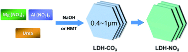
We present a facile synthesis of layered double hydroxides (LDHs) with submicrometer-sized platelets in lateral dimension, and their subsequent direct decarbonation. First, LDH–carbonate phase (LDH_CO3) with submicrometer-sizes in lateral dimension were controlled synthesized by a facile accelerated urea hydrolysis method. Then, using HNO3–NaNO3 mixed solution, the obtained LDHs with carbonate anions in the interlayer were directly decarbonated to their nitrate form (LDH_NO3). These results are important for the synthesis of novel nanomaterials with controlled morphology and will benefit their applications.
1. Introduction
Layered double hydroxides (LDHs), having unusual interlayer anion exchange capability, have received increasing attention in recent years. Structurally similar to the natural mineral of brucite, LDHs are intrinsically nanomaterials, composed of positively nanolayers with thickness around 0.48 nm and charge-balanced counter-anions with water molecules in the interlayer space.1 Since their morphology, structures, constituents and properties are highly tunable, LDHs have widely potential applications or been already applied in catalysts,2 adsorbent,3 thermal stabilizer4 or UV-shielding agent of polymer,5 enhanced oil recovery6 and hydrogel7 etc.
The synthesis of LDHs with controlled morphology is vital for their optimum performance and applications.8–18 Conventionally, LDHs are synthesized using a coprecipitation method by adding a sodium hydroxide (NaOH) solution to an aqueous mixture of metal salts.8–11 However, the resultant products always show ill-defined shape, and the single crystals are usually smaller than 400 nm in lateral size.10,11 Fortunately, a method called homogeneous precipitation12–15 was developed to obtain micron-scale (commonly 1–20 μm) hexagonal-shaped LDHs with carbonate as counter-anion (LDH_CO3). In sharp contrast, compared with the prevalent studies of small (<400 nm) and large (micron) LDH particles, the studies of synthesis of LDH crystals with lateral dimensions between 400 nm to 1 μm are rather limited, although they are highly desired in a variety of applications such as photo-functional materials. In order to obtain submicrometer-scale LDHs, high aging temperature (>150 °C) and long aging time,16 or organic solvent14 are always applied. Very recently, Zhu et al. achieved layered yttrium hydroxide (LYH) from ∼400 nm to ∼1 μm by 50% europium doping.17 However, this method cannot be expanded to other commonly used LDHs. Therefore, it still remains a challenge to conveniently prepare submicrometer-sized LDHs with controlled morphology.
Recently, deintercalation of carbonate ions, i.e. decarbonation, from LDH_CO3 using an acid–salt mixed solution was developed.19–21 In this way, LDH_CO3 crystals could be successfully exfoliated using the decarbonation and subsequent exfoliation in formamide. However, in this method, LDH_CO3 was first transformed to LDH_Cl (LDH with Cl− as the balanced anion), and then converted to LDH_NO3 via anion exchange between NO3− and Cl−. Two steps were adopted, although LDH_NO3 is probably the most frequently reported exchangeable LDHs and its anion exchange ability is much higher than LDH_Cl.9
Considering the slow nucleation procedure due to the slow hydrolysis of urea and the low degree of supersaturation during precipitation in urea method,22 it is reasonable to expect LDHs with obviously reduced lateral size by acceleration of the nucleation rate. Herein, we report an accelerated urea hydrolysis method by adding NaOH or hexamethylenetetramine (HMT) to obtain submicrometer-sized LDH_CO3 particles. Since the LDHs containing Mg and Al in the octahedral nanosheets (MgAl_LDHs) are the most extensively studied type, they were adopted in this investigation. Different synthesis parameters such as concentration and temperature were taken into account in order to control the process of LDH formation. The purity and the morphology of the resultant products were studied as well. Then, a direct decarbonation process of the submicron-sized LDHs was completed by a nitrate acid–sodium nitrate acid (HNO3–NaNO3) mixed solution method.
2. Reagents and methods
2.1. Reagents
All of the reagents, including hydrated magnesium nitrate (Mg(NO3)2·6H2O), hydrated aluminum nitrate (Al(NO3)3·9H2O), urea (purity >99.0%, Beijing Chemical Co., Ltd), NaOH and HMT (purity >99.0%, Shantouxilong Chemical Co., Ltd) are of analytical reagent pure (A.R.) grade. The water was distilled twice before use.
2.2. Preparation
2.2.1. Urea method. A typical synthetic process is described as follows. Mg(NO3)2, Al(NO3)3, and urea were dissolved in distilled water first, wherein the total concentration of metal ions ([Mg] + [Al]) was fixed at 0.15 M, and the molar ratios of Mg/Al and urea/([Mg] + [Al]) were 3 and 10, respectively. Then, the mixed solution was heated to and maintained at 90 or 100 °C under continuous stirring. The heating duration was around 29 h. Finally, the resultant precipitate was collected by filtration, washed and dried in a vacuum oven at 50 °C for 24 h.
2.2.2. Urea + NaOH method. The main difference between this method and the above urea method is the addition of NaOH aqueous solution. First, an aqueous solution of urea, Mg(NO3)2, and Al(NO3)3 were mixed at the Mg/Al/urea molar ratio of 3
:
1
:
40. After the mixture was heated to reflux at 100 °C for ca. 20 min, a translucent dispersion appeared due to formation of precipitate was added. Then, 50 mL of NaOH solution with concentration of 0.10 or 0.15 or 0.20 M was drop-wisely added into the mixture in 30 min. Totally, the time for the synthesis lasted for 28 h. The subsequent procedure was the same as the above urea method.
2.2.3. Urea + HMT method. In this method, HMT solution with concentration of 0.075 or 0.0375 M was added into the mixture of Mg(NO3)2 (0.075 M) and Al(NO3)3 (0.025 M) aqueous solutions. The molar ratio of Mg/Al/urea/HMT was 3
:
1
:
40
:
x (x = 1.5 or 3). The subsequent procedure was the same as the above urea method.
2.3. Direct decarbonation by HNO3–NaNO3
About 0.10 g of the powdery LDH_CO3 was added into a glass vessel containing of 100 mL HNO3–NaNO3 mixed solution, wherein the concentrations for the HNO3 and NaNO3 solutions were 0.005 and 5.0 M, respectively. Then, the glass vessel was purged with nitrogen gas, tightly capped, and sustained a subsequent ultrasonication treatment with the power of 100 W for 30 s. After that, the vessel was stored with constant shaking at 25 °C for 2 days. Finally, the product was filtrated, washed and dried under vacuum at 50 °C for 24 h.
2.4. Characterization
X-ray diffraction (XRD) patterns of the LDHs were collected using a Rigaku D/max 2400 diffractometer with Cu Kα radiation (λ = 0.15418 nm) at a scanning rate of 4° min−1 in the 2θ range of 3–70°. The size and morphology of the LDH layers were observed by a HITACHI S-4300 scanning electron microscopy (SEM). Its accelerating voltage was 15 kV. Before the measurements, the LDH powder was added into water and ultrasonicated for 10 min. Then, it was dripped onto a glass slide and air-dried at room temperature. Fourier transform infrared (FTIR) spectra were recorded on a Perkin-Elmer System 2000 FTIR spectrophotometer ranging from 4000 to 400 cm−1 at a nominal resolution of 4 cm−1.
3. Results and discussion
Theoretically, an accelerated nucleation rate is favorable to the growth of crystals with reduced size. Taking into account the slow nucleation procedure and the low degree of supersaturation during precipitation in homogeneous precipitation method, it is reasonable to expect LDHs with submicrometer-size in lateral dimension by acceleration of the nucleation rate. In this work, first, we prepared the micron-scale LDHs using the typical urea hydrolysis method for comparison. Then, an accelerated urea hydrolysis method using NaOH or HMT was proposed to accelerate the nucleation rate, aiming at achieving monodispersed LDHs with an average size between 400 nm and 1 μm. The concentration effect of NaOH or HMT solution was studied as well.
3.1. Synthesis of LDH_CO3 via urea method
Since the effects of various factors (including concentrations of total metal salts and urea, and reaction time) except hydrolysis temperature on LDH size have been extensively reported to synthesize micron-scale LDH_CO3 particles,14,15 here, the effect of hydrolysis temperature was studied. In Fig. 1, both XRD curves are essentially identical, which coincide with those of LDH_CO3 published in literatures.14,15 A series of intense and sharp diffraction peaks are clearly observed, suggesting the high crystallinity and three-dimensional order for the synthesized LDH_CO3 crystals. All of the peaks can be indexed as a rhombohedral structure. More importantly, no obvious XRD peaks of impurities appear, demonstrating the high purity of the synthesized products. The morphology of the LDH_CO3 particles was measured by SEM images. Large platelets with hexagonal shape can be clearly seen in Fig. 2. The dimensions of an average of individual LDH_CO3 particles synthesized at 90 and 100 °C are 3.24 ± 0.42 and 2.87 ± 0.47 μm, respectively. This implies that the increase of temperature can lead to a limited reduction of the particle size for the synthesized product. The main reason may lie in the limited acceleration of the nucleation rate with temperature increasing. Thus, the effect of temperature on the reduction of LDH size is rather limited for the typical urea hydrolysis method.
 |
| Fig. 1 XRD patterns of LDH_CO3 samples synthesized via urea hydrolysis method at (a) 90 or (b) 100 °C. | |
 |
| Fig. 2 SEM images for the LDH_CO3 particles synthesized by urea method at (a and b) 90 or (c and d) 100 °C. | |
3.2. Synthesis of LDH_CO3 via urea + NaOH method
Fig. 3 reveals the XRD patterns showing the effect of concentration of NaOH solution on the synthesized LDH_CO3 products. As clearly shown in Fig. 3(a), high NaOH concentration of 0.20 M inevitably resulted in various impurities including boehmite (AlO(OH)),15 hydromagnesite or hydrated magnesium carbonate hydroxide (HM),23 and magnesium oxide (MgO).24 At decreased concentration of NaOH solution of 0.15 M, the decrease of the relative intensities of the impurities suggests the reduction of the impurity content (Fig. 3(b)). Fortunately, no characteristic sharp peaks of impurities could be recognized in Fig. 3(c), demonstrating that pure phase has been successfully synthesized using the developed accelerated urea hydrolysis method of urea + NaOH at NaOH concentration of 0.10 M. Moreover, a series of intense and sharp diffraction peaks are clearly observed, suggesting the highly ordered structure and regular stacking in the synthesized LDH_CO3 crystals. All of the peaks can be indexed as a rhombohedra structure. The characteristic peak positions, the corresponding distances and the refined lattice parameters derived from the XRD patterns are illustrated in Table 1. The basal spacing of the synthesized LDH_CO3 particles is 0.76 nm, and the two weak diffraction peaks around 61.0° and 62.0° result from the characteristic reflections of (110) and (113) lattice planes. These XRD results are in consistency with previous investigations9–11 very well.
 |
| Fig. 3 XRD patterns of the LDH_CO3 particles synthesized via urea + NaOH method with NaOH concentration of (a) 0.20, (b) 0.15, or (c) 0.10 M. Symbols of triangles, circles and asterisk are possibly due to impurities of boehmite (AlO(OH)), hydromagnesite or hydrated magnesium carbonate hydroxide (HM), and magnesium oxide (MgO), respectively. | |
Table 1 Characteristic data derived from XRD patterns in Fig. 8 for the LDH_CO3 and LDH_NO3 particles
|
003 |
006 |
110 |
113 |
Lattice parameters |
2θ/° |
d/nm |
2θ/° |
d/nm |
2θ/° |
d/nm |
2θ/° |
d/nm |
a/nm |
c/nm |
LDH_CO3 |
Urea |
11.8 |
0.76 |
23.6 |
0.38 |
61.0 |
0.15 |
62.4 |
0.15 |
0.304 |
2.26 |
Urea + NaOH |
11.6 |
0.76 |
23.5 |
0.38 |
60.9 |
0.15 |
62.2 |
0.15 |
0.304 |
2.28 |
Urea + HMT |
11.7 |
0.76 |
23.5 |
0.38 |
60.9 |
0.15 |
62.2 |
0.15 |
0.304 |
2.27 |
LDH_NO3 |
Urea |
11.0 |
0.88 |
20.1 |
0.44 |
61.0 |
0.15 |
62.0 |
0.15 |
0.304 |
2.64 |
Urea + NaOH |
11.1 |
0.88 |
20.0 |
0.44 |
61.1 |
0.15 |
62.0 |
0.15 |
0.304 |
2.63 |
Urea + HMT |
11.1 |
0.88 |
20.1 |
0.44 |
61.0 |
0.15 |
61.9 |
0.15 |
0.304 |
2.63 |
The morphology of the synthesized pure LDH_CO3 particles was observed, and typical SEM images are shown in Fig. 4, wherein the concentration of NaOH solution is 0.10 M. Flowerlike texture composed of inclined platelets can be clearly observed. Calculated from at least 200 particles, the average lateral size of the synthesized LDH_CO3 particles was determined to be 0.82 ± 0.16 μm, smaller than the micron-scale LDH_CO3 samples prepared by typical urea hydrolysis method. Therefore, we conclude that the urea + NaOH method developed herein is effective to synthesis of pure LDH_CO3 particles with dramatically reduced lateral size of less than 1 μm.
 |
| Fig. 4 SEM images of the LDH_CO3 particles, prepared using urea + NaOH method with 0.10 M NaOH, with different magnification scale. | |
3.3. Synthesis of LDH_CO3 via urea + HMT method
As an alternative to the accelerated urea method of urea + NaOH, the LDH_CO3 samples were also prepared by adding HMT solution. Effect of further reduced alkali concentration (HMT concentration) was explored. Apparently, Fig. 5 confirms that pure LDH_CO3 particles were obtained with HMT concentration of 0.075 or 0.0375 M, demonstrated by the absence of XRD peaks of any impurities. As shown in Table 1, their peak positions and structural parameters are almost the same as those synthesized using the above urea and urea + NaOH methods.
 |
| Fig. 5 XRD patterns of the LDH_CO3 particles synthesized via urea + HMT method with HMT concentration of (a) 0.075 and (b) 0.0375 M. | |
On the other hand, the SEM images in Fig. 6 strongly support the HMT effect on reduction of the LDH particle size relative to those prepared by urea method. When the HMT concentration is 0.075 M, the as-prepared LDH_CO3 samples have an average particle size of 0.47 ± 0.11 μm (Fig. 6(a)). However, further decrease of HMT concentration led to an enlargement of lateral size for the synthesized LDH_CO3 particles. At HMT concentration of 0.0375 M, the resultant product exhibits an average size of 1.12 ± 0.30 μm.
 |
| Fig. 6 SEM images of the LDH_CO3 particles using urea + HMT method with HMT concentration of (a) 0.075 and (b) 0.0375 M. | |
Taking into account of the above results, therefore, we conclude that appropriate concentration of NaOH or HMT solution is very important to the synthesis of pure submicrometer-sized LDHs. High concentration resulted in the occurrence of impurities, while low concentration led to large-sized particles with lateral sizes larger than 1 μm.
3.4. Decarbonation of LDH_CO3 directly to LDH_NO3 using HNO3–NaNO3
To overcome the difficulty of anion exchange reaction of the interlayer CO32− anions in LDH_CO3, here, we present an HNO3–NaNO3 exchange method to directly convert the submicron-scale LDH_CO3 to LDH_NO3 without obvious particle morphology damage. Possible mechanism is proposed in the following. First, the nitrate acid is decomposed into its ion forms, and H+ is liberated. Then, the resulted H+ is combined with the interlayer CO32− to afford LDH with HCO3− as the balanced interlayer anion (LDH_HCO3). Finally, the interlayer HCO3− is exchanged with excess amount of NO3−, resulting in the product of LDH_NO3. Indeed, the pH value was monitored for the decarbonation process. After the ultrasonication treatment, the pH value of the system was about 3.5, and finally changed to around 5.0 after 2 days.
The direct decarbonation process for the submicron-scale LDH_CO3 is first demonstrated by FTIR spectra. Fig. 7 presents the FTIR spectra of the LDH_CO3 and the exchanged LDH_NO3. The broad absorption band between 3750 and 2700 cm−1 is due to stretching modes of the hydroxyl groups in the LDH platelets and the interlayer water molecules. Importantly, the distinction of the band at 1352 or 1384 cm−1 provides a convictive proof for the decarbonation process. All of the FTIR spectra of the three LDH_CO3 (Fig. 7(a)–(c)) have a characteristic band at 1352 cm−1 due to the stretching vibration mode of CO32−.10,14 After the HNO3–NaNO3 exchange process, this band disappeared with simultaneous appearance of a new band at 1384 cm−1 (Fig. 7(d)–(f)), characteristic of the stretching mode of NO3−.10,14,25 In addition, in the spectra of the exchanged LDH_NO3 particles, a new weak band at 827 cm−1 occurred due to the NO3− stretching vibration mode.25 And the stretching band at 860 cm−1 characteristic of CO32− (Fig. 7(a)–(c)) disappeared in the obtained LDH_NO3 samples (Fig. 7(d)–(f)). As for the absorption bands at 552 and 448 cm−1 in all spectra, they could be assigned to the M–O–M lattice vibrations of the octahedral sheets.11,14,26 These FTIR results confirm the success of the decarbonation process by the HNO3–NaNO3 mixed solution exchange.
 |
| Fig. 7 FTIR spectra of (a–c) the LDH_CO3 particles synthesized via (a) urea, (b) urea + NaOH and (c) urea + HMT method. (d–f) are the spectra of the LDH_NO3 particles after decarbonation treatment of the LDH_CO3 (a–c) by HNO3–NaNO3 mixed solution, respectively. | |
XRD results provide another proof of the interlayer anion exchange of CO32− by NO3−. Since CO32− anions have greater affinity to adjacent inorganic platelets, the corresponding LDH_CO3 patterns have high diffraction angles with small basing spacing relative to those of the LDH_NO3, as shown in Fig. 8. Table 1 illustrates the characteristic peak positions, corresponding distances, and the refined lattice parameters derived from the XRD patterns of LDH_CO3 and LDH_NO3 particles. The basal spacing (d003) of the three LDH_CO3 samples synthesized by urea, urea + NaOH or urea + HMT method are the same, 0.76 nm. After the decarbonation, the exchanged LDH_NO3 products had an obviously increased basal spacing of 0.88 nm. Note that no residues of the characteristic peaks for LDH_CO3 remain in Fig. 8(a)–(c), confirming the completely conversion of LDH_CO3 to LDH_NO3. In addition, all of the three curves exhibit two weak diffraction peaks around 61.0° and 62.0°, resulting from the reflections of individual platelets. After the decarbonation process, the values of the lattice parameter (a) remains unchanged to be 0.304 nm, suggesting that the crystalline ordered structure inside the individual LDH platelets was well preserved. In contrast, the c parameter increases greatly from around 2.27 nm to 2.63 nm due to the expansion of interlayer distance.
 |
| Fig. 8 XRD patterns of the LDH_NO3 particles after decarbonation treatment of the LDH_CO3 (synthesized via (a) urea, (b) urea + NaOH or (c) urea + HMT method) by HNO3–NaNO3 mixed solution, respectively. | |
Furthermore, the morphology of the exchanged LDH_NO3 particles was studied by SEM. Fig. 9 reveals that the shape of all of the LDH_NO3 particles still remains undamaged, being almost identical to the corresponding LDH_CO3 particles before the HNO3–NaNO3 exchange. And their average particle sizes were unchanged as well. However, the Mg/Al ratios decreased after the decarbonation for all of the submicron-scale LDHs. The Mg/Al molar ratios of the LDH_CO3 samples synthesized by urea, urea + NaOH and urea + HMT methods were 1.81, 1.84 and 1.94, respectively. After the direct decarbonation process using HNO3–NaNO3 solution, they decreased to 1.41, 1.45 and 1.54, respectively. The reason may be that the solubility of Mg(OH)2 is larger than that of Al(OH)3.
 |
| Fig. 9 SEM images of the LDH_NO3 particles after the HNO3–NaNO3 exchange process for the LDH_CO3 synthesized by (a) urea, (b) urea + NaOH, and (c) urea + HMT method, respectively. | |
4. Conclusions
We report a facile synthesis of LDHs with submicron-scale platelets in lateral dimension via accelerated urea hydrolysis method. By simply increasing of hydrolysis temperature in the typical urea method, submicrometer-sized LDHs cannot be obtained. In contrast, LDH–carbonate phase particles with lateral size dimension ranged from 400 nm to 1 μm were successfully achieved by an accelerated urea hydrolysis method developed herein. Appropriate concentration of NaOH or HMT solution is vital. High concentration resulted in various impurities, whereas low concentration led to large LDH particles. Subsequently, after being sustained a HNO3–NaNO3 mixed solution exchange process, the synthesized submicrometer-sized LDH_CO3 particles were directly decarbonated to the LDH_NO3 type without obvious morphology change. These results offer a facile way to prepare submicrometer-sized LDH particles, and may widen the applications of LDHs.
Acknowledgements
The authors are grateful to National Natural Science Foundation of China (51073162). G. Chen acknowledges the support of K. C. Wong Education Foundation, Hong Kong.
References
- T. Hibino and W. Jones, J. Mater. Chem., 2001, 11, 1321 RSC.
- O. D. Pavel, D. Tichit and I.-C. Marcu, Appl. Clay Sci., 2012, 61, 52 CrossRef CAS.
- Q. Wang, Y. Gao, J. Luo, Z. Zhong, A. Borgana, Z. Guo and D. O'Hare, RSC Adv., 2013, 3, 3414 RSC.
- J. Liu, G. Chen and J. Yang, Polymer, 2008, 49, 3923 CrossRef CAS.
- T. Cao, K. Xu, G. Chen and C.-Y. Guo, RSC Adv., 2013, 3, 6282 RSC.
- P. Fu, K. Xu, H. Song, G. Chen, J. Yang and Y. Niu, J. Mater. Chem., 2010, 20, 3869 RSC.
- Z. Hu and G. Chen, RSC Adv., 2013, 3, 12021 RSC.
- S. Miyata, Clays Clay Miner., 1975, 23, 369 CAS.
- S. Miyata and T. Kumura, Chem. Lett., 1973, 2, 843 CrossRef.
- Q. Wu, A. Olafsan, Ø. B. Vistad, J. Roots and P. Norby, J. Mater. Chem., 2005, 15, 4695 RSC.
- Z. Xu, G. Stevenson, C.-Q. Lu and G. Lu, J. Phys. Chem. B, 2006, 110, 16923 CrossRef CAS PubMed.
- U. Costantino, F. Marmottni, M. Nocchetti and R. Vivani, Eur. J. Inorg. Chem., 1998, 1439 CrossRef CAS.
- M. Ogawa and H. Kaiho, Langmuir, 2002, 18, 4240 CrossRef CAS.
- M. Adachi-Pagano, C. Forano and J.-P. Besse, J. Mater. Chem., 2003, 13, 1988 RSC.
- J.-M. Oh, S.-H. Hwang and J.-H. Choy, Solid State Ionics, 2002, 151, 285 CrossRef CAS.
- L. Albiston, K. R. Franklin, E. Lee and J. B. A. F. Smeulders, J. Mater. Chem., 1996, 6, 871 RSC.
- Q. Zhu, J. Li, C. Zhi, X. Li, X. Sun, Y. Sakka, D. Golberg and Y. Bando, Chem. Mater., 2010, 22, 4204 CrossRef CAS.
- Z. Zhang, G. Chen and K. Xu, Appl. Clay Sci., 2013, 72, 206 CrossRef CAS.
- N. Iyi, T. Matsumoto, Y. Kaneko and K. Kitamura, Chem. Mater., 2004, 16, 2926 CrossRef CAS.
- N. Iyi and T. Sasaki, Appl. Clay Sci., 2008, 42, 246 CrossRef CAS.
- K. Okamoto, T. Sasaki, T. Fujita and N. Iyi, J. Mater. Chem., 2006, 16, 1608 RSC.
- Z. Liu, R. Ma, M. Osada, N. Iyi, Y. Ebina, K. Takada and T. Sasaki, J. Am. Chem. Soc., 2006, 128, 4872 CrossRef CAS PubMed.
- T. Hibino and H. Ohya, Appl. Clay Sci., 2009, 45, 123 CrossRef CAS.
- Z. Xu and G. Q. Lu, Chem. Mater., 2005, 17, 1055 CrossRef CAS.
- Z. Xu and H. Zeng, Chem. Mater., 2001, 13, 4555 CrossRef CAS.
- D.-Y. Wang, F. R. Costa, A. Vyalikh, A. Leuteritz, U. Scheler, D. Jehnichen, U. Wagenknecht, L. Häussler and G. Heinrich, Chem. Mater., 2009, 21, 4490 CrossRef CAS.
|
This journal is © The Royal Society of Chemistry 2014 |
Click here to see how this site uses Cookies. View our privacy policy here.