Structural architecture and solubility of native and modified gliadin and glutenin proteins: non-crystalline molecular and atomic organization

Faiza Rasheed *a, William R. Newson a, Tomás S. Plivelic b, Ramune Kuktaite a, Mikael S. Hedenqvist c, Mikael Gällstedt d and Eva Johansson a
aDepartment, of Plant Breeding, The Swedish University of Agricultural Sciences, SE-230 53, Box 101, Alnarp, Sweden. E-mail: Faiza.Rasheed@slu.se; Tel: +46 40-41 5565 Tel: +46 73-53 31741
bMAX IV Laboratory, Lund University, Box 118, SE-221 00 Lund, Sweden
cKTH Royal Institute of Technology, School of Chemical Science and Engineering, Fibre and Polymer Technology, SE-10044 Stockholm, Sweden
dInnventia, Box 5604, SE-11486 Stockholm, Sweden

Received 2nd October 2013 , Accepted 4th November 2013

First published on 5th November 2013


Abstract

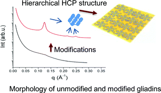
Wheat gluten (WG) and its components, gliadin and glutenin proteins, form the largest polymers in nature, which complicates the structural architecture of these proteins. Wheat gluten, gliadin and glutenin proteins in unmodified form showed few secondary structural features. Structural modification of these proteins using heat, pressure and the chemical chaperone glycerol resulted in a shift to organized structure. In modified gliadin, nano-structural molecular arrangements in the form of hexagonal closed packed (HCP) assemblies with lattice parameter of (58 Å) were obvious together with development of intermolecular disulphide bonds. Modification of glutenin resulted in highly polymerized structure with proteins linked not only by disulphide bonds, but also with other covalent and irreversible bonds, as well as the highest proportion of β-sheets. From a combination of experimental evidence and protein algorithms, we have proposed tertiary structure models of unmodified and modified gliadin and glutenin proteins. An increased understanding of gliadin and glutenin proteins structure and behavior are of utmost importance to understand the applicability of these proteins for various applications including plastic materials, foams, adhesives, films and coatings.

Introduction

Wheat gluten (WG) proteins form the largest and most complex protein network in nature due to the high molecular weight and heterogeneous origin of its proteins. WG is comprised of two component proteins, gliadin (monomeric) and glutenin (polymeric), with molecular weights ranging from 30 to 3000 kDa and these proteins possess a variety of secondary structures such as helices, sheets, random coils and turns.1,2 The understanding of structural conformation of a protein and arrangement of its secondary structural elements in a three dimensional array are often a pre-requisite for fundamental understanding of the biological role of that protein. Structural modifications and rearrangements of the proteins by energy and chemical inputs further increase the understanding of the biological and technical function of the protein. The basic structure of WG and its component proteins, as well as how the structure rearranges under different modifications is only partly understood, and needs to be further examined in order to predict the complete 3D structure.

Due to the complexity and amorphous nature of gluten proteins, attempts to crystallize these proteins in a normal room environment have not been successful, except under a microgravity environment.3 Up till now, the structural riddle of gluten component proteins has been evaluated using circular dichroism, infrared spectroscopy and X-ray scattering techniques, although the full picture has not been completed yet. Aggregation of the proteins has been evaluated using high performance liquid chromatography methods. The performed studies have shown that gliadin exhibited limited aggregation with mainly intramolecular disulphide bonds, while the glutenin is aggregated with intermolecular4 as well as intra-molecular1 disulphide linkages. Furthermore, the studies have indicated that the central repetitive domain of high molecular weight glutenin subunit (HMW-GS) have the form of beta turns arranged in a loose spiral structure. The N- and C-terminal domains are globular with few alpha helices.5,6 The gliadins are modelled as prolate ellipsoids (diameter ≈ 3.2 nm), when studied in solution with X-ray scattering.7 Some studies of gluten proteins have shown that these proteins have a higher ratio of intrinsically disordered regions as compared to structurally organized regions.8 The intrinsic disorder of gluten proteins is coherent with their biological role as seed storage proteins. The disordered proteins are suggested to become structurally organized only when bound to another molecule or owing to changes in the chemical or biochemical surroundings.9,10

Contrary to the intrinsically disordered nature of gluten proteins, previous studies have shown supramolecular assemblies in WG films modified with heat, pressure, shear and alkaline additives4,11 or urea.12 These studies presented a bidimensional hexagonal close packed (HCP) or tetragonal structure of aligned scattering objects. The mentioned studies have inspired us to further investigate the polymerization properties and structural conformation in individual gliadin and glutenin components and how the structure gets rearranged by changing the surrounding chemical environment of the protein. Heat above 75 °C combined with pressure is known to unfold and refold the gluten proteins and facilitate disulphide interchange reactions.12,13 This unfolding and refolding of proteins by heat and pressure can be further enhanced in the presence of additives e.g. glycerol, that brings proteins to a low energy state. The addition of glycerol may introduce important conformational changes by increasing molecular mobility of proteins as compared to the native form.

In addition to experimental data, some additional information to get more details into the gluten protein structural puzzle can be obtained through the use of protein structure prediction algorithms e.g. I-TASSER14,15 and Phyre2.16 For such predictions, the full length amino acid sequences of gluten proteins, reported previously, were utilized. These bioinformatics algorithms, based on fold recognition and homology modeling, give some insights into probable structure arrangement of proteins in a 3D array.

In the present study we evaluated the secondary and tertiary structures of gluten protein and polymerization behavior of its individual components i.e. gliadin and glutenin. Both, the gliadin and glutenin proteins were evaluated in their native state, and while the proteins were modified with glycerol, temperature and pressure. This study is also bridging experimental and bioinformatics based information to predict the tertiary (3D) structural conformations of WG, gliadin and glutenin proteins.

To our knowledge this is the first proposed detailed structure of individual gliadin and glutenin proteins and their possible modification.

Experimental procedures

Materials

Wheat gluten was generously supplied by Reppe AB, Lidköping, Sweden. According to the supplier, the gluten protein content was 77.7% of the dry weight (modified NMKL Nr 6, Kjeltec, N x 5.7, http://www.NMKL.org), the starch content was 5.8% of the dry weight (Ewers, polarimetric method) and the moisture content was 6.9% of the total weight (NMKL 23, 1991). Glycerol (99.5% purity) was kindly supplied by Karlshams Tefac AB, Sweden. Sodium dodecyl sulphate (SDS) and urea were purchased from Duchefa, Haarlem, Netherlands. NaH2PO4 was purchased from from J.T. Baker, Deventer, Netherlands. Propanol, trifluoroacetic acid (TFA), and isocratic grade acetonitrile were purchased from Merck, Germany. Gradient grade propanol was purchased from Honeywell, Germany. Dithiothreitol (DTT) was purchased from Saveen Werner AB, Sweden.

Methods

Extraction of gliadins and glutenin proteins. Sixteen grams of commercial WG powder was thoroughly mixed with 70% ethanol (200 ml). For homogenous mixing of gluten powder with ethanol, WG was added slowly from the top of sieve with continuous stirring of the mixture with magnetic stirrer. Then the mixture was kept on shaking for 30 min at 300 rpm at room temperature (25 °C) using a mechanical shaker (Hunkel Ika, Werk KS 500). After shaking, the mixture was centrifuged for 10 min at 12[thin space (1/6-em)]000 RCF (Beckman J2.21). Supernatant containing gliadin proteins was decanted into flasks. A rotary evaporator (Buchi) was used to evaporate ethanol from the supernatant in order to precipitate gliadins. Glutenin protein was separated out as a rubbery mass and was washed three times with Millipore water. After washing, the glutenin mass was cut into small pieces and both the gliadin precipitate and glutenin were frozen at −80 °C for two hours. After freezing, both the glutenin and gliadin were lyophilized (Edwards, Modulyo, freeze-drier, Edwards). Lyophilized glutenins and gliadin proteins were ground into powder using a laboratory mill (IKA, Yellow Line A10).
Protein modifications. The WG, gliadins and glutenin enriched proteins were modified by hot pressing for 10 minutes at 130 °C and 200 bar pressure. The proteins were modified without glycerol and also with the addition of 30% glycerol.
Size exclusion high performance liquid chromatography. The amount of soluble proteins was determined by a three step extraction procedure by size exclusion high performance liquid chromatography (SE-HPLC). Proteins were extracted in phosphate buffer containing 0.5% (wt/vol) SDS and 0.05 M NaH2PO4 (pH 6.9). Triplicates of each sample weighing (16.5 mg) were analyzed. Each sample was extracted serially in buffer; vortexing for 10 seconds followed by a shaking of five minutes at 2000 rpm on a laboratory shaker, 30 s ultrasonication at an indicated amplitude of 5 μm (Sanyo Soniprep). It was followed by a third extraction with 30 s + 60 s + 60 s ultrasonication with cooling to room temperature between sonications. In each case, the material was centrifuged at 16[thin space (1/6-em)]000 RCF for 30 minutes and the supernatant decanted into HPLC vials before the next extraction step.

Analysis was performed with an isocratic flow of 0.2 ml min−1 (50% acetonitrile, 0.1% TFA; 50% H2O, 0.1% TFA) using a Waters 2690 Separations Module and Waters 996 Photodiode Array Detector (Waters, Milford, USA). A 20 μl sample of supernatant was injected into a column consisting of; a prefilter and main column (SecurityGuard GFC 4000, Biosep-SEC-S 4000 300 mm × 4.5 mm, respectively, Phenomenex, Torrance USA). 3D absorption spectra were collected for 30 minutes and Millenium 32 software was used for analysis (Waters, version 4.00). Chromatograms were extracted at 210 nm and integrated into arbitrary fractions, polymeric proteins from 0 to 13.5 minutes and monomeric proteins from 13.5 to 28 minutes.

To calculate total extracted proteins (TEP), the pellet left after three extraction steps were dried, weighed, and analysed for N according to the Dumas method through volatilisation of N in a Carlo Erba N analyser.17 To calculate protein concentration, the N concentration was multiplied by a conversion factor of 5.7.

Reverse phase high performance liquid chromatography. Three 100 mg replicates of each sample were analyzed by RP-HPLC. Serial extraction was carried out using six different solvent systems (extraction buffers). The extraction buffers for each step were (1) 70% ethanol (2) 50% propanol (3) 50% propanol (4) 0.5% SDS, 50% propanol (5) 1% DTT, 50% propanol and (6) 1% DTT + 1% SDS, 6 M urea solution. The volume of buffer was kept constant i.e. 1 ml for each extraction. The first two extractions were done at room temperature. The 3rd, 4th, 5th extractions were done by heating the solution at 60 °C for 30 minutes and in 6th extraction, the solution was heated at 100 °C for five minutes in a dry oven.

At each extraction step, the samples were mixed with buffer and stirred at 2000 rpm for 30 minutes followed by a centrifugation at 12[thin space (1/6-em)]000 RCF for 30 minutes using Sorvall legend microcentrifuge. Supernatant from each extraction was kept for RP-HPLC analysis and pellet was mixed with next buffer.

The reverse phase chromatography was done by using a SUPELCO column (discovery bio wide pore C8, 5 μm 25 cm × 4.6 mm, catalog no 568323-4). A SUPELCO pre column (guard column, discovery bio wide pore C8, 5 μm, 2 cm × 4.0 mm) was used in combination with the main column. The solvent system for elution in RP-HPLC was based on two solvents; water (A) and gradient grade acetonitrile (B) containing 0.1% trifluoroacetic acid (TFA). The solvent flow rate was maintained at 0.8 ml min−1 and the temperature of column was maintained at 70 °C at a gradient flow of 28–72% for 1–30 minutes extraction time.

Fourier Transform Infrared Spectroscopy. FT-IR spectra (32 scans) were recorded on all WG, gliadin and glutenin proteins using a Spectrum 2000 FTIR spectrometer (Perkin-Elmer inc., USA). The spectrometer was equipped with a single reflection ATR accessory, golden gate from Speac Ltd. All the samples were dried in a desicator over a silica gel for at least 72 h before analysis.
Small angle X-ray scattering. Small Angle X-ray Scattering (SAXS) experiments were performed at the beamlines I711 and I911-4 of the MAX-lab Synchrotron in Lund, Sweden. A monochromatic beam of 1.2 and 0.91 Å wavelengths was used respectively. The range of the scattering vector, q= (4π/λ)sin(θ) (where 2θ is the scattering angle) was around 0.0085–0.35 Å−1 in both cases. Two dimensional scattering pictures were recorded using an area CCD detector (165 mm in diameter active area, from Marresearch, GmbH) using 5 or 1 min of exposure time. SAXS data were analyzed with the software bli9114.18 The curves are normalized by the integrated intensity incident on the sample during the exposure time, and corrected for sample absorption, parasitic scattering and background.
Wide angle X-ray scattering. Wide-angle X-ray scattering (WAXS) measurements were performed at the I911-2 beamline of the MAX-lab synchrotron. The wavelength employed was 1.04 Å and the sample to detector distance was 149.8 mm. Silicon powder was used as a calibration standard. Two dimensional images were registered with an area CCD detector (165 mm in diameter active area), exposing the samples for 1 min. Complementary WAXS measurements were performed at the I911-4 beamline with 0.91 Å wavelength and 290 mm sample–detector distance. The data were process with the software FIT2D.19 Parasitic scattering was subtracted for all diffractograms.
Bioinformatics algorithms used. Two protein structure prediction algorithms Phyre216 and I-TASSER14,15 were utilized to predict the three dimensional structure of unmodified gliadin and glutenin proteins. Domain analysis was done by simple modular architecture research (SMART) program.20

Results and discussion

Protein extractability

Monomeric and polymeric proteins (after SE-HPLC analyses) were present in all the unmodified protein samples and in the modified samples without glycerol (Fig. 1; samples described in Table 1). However, in all modified protein samples with glycerol no polymeric proteins were observed as shown in representative gliadin samples modified without and with glycerol (Fig. 1a and b). The lack of polymeric proteins in the extracts of modified samples with glycerol indicates stronger crosslinking of polymeric proteins in those samples. In summary, total extractable proteins (TEP); (analyzed indirectly by calculating the amount of protein left in the sample after three sequential extraction steps carried out for SE-HPLC analyses) was 81% in unmodified gliadin samples (Glia-UnM). A drastic reduction in TEP to 50% was seen in the modified gliadins without glycerol samples (Glia-0Gly), and the TEP was further reduced to 22% in samples of modified gliadins with 30% glycerol (70Glia–30Gly). In unmodified glutenin samples (Glut-UnM), TEP was 22% and the TEP was further decreased to 11% in samples of modified glutenin with glycerol (70Glut–30Gly) and without (Glut-0Gly) glycerol. Thus, by the modification of gliadin and glutenin proteins, solubility in SDS buffer even with sonication was reduced considerably and, especially the polymeric proteins were no longer extractable with the used buffers indicating an increased cross-linking.
image file: c3ra45522j-f1.tif
Fig. 1 SE-HPLC Analysis (a) chromatograms for three sequential extractions of Glia-0Gly (b) 70Glia–30Gly sample (c) total extracted proteins (TEP) (1st Ext. SDS buffer, 2nd Ext. SDS buffer + 30 s sonication, 3rd Ext. SDS buffer + 30 s + 60 s + 60 s sonication). Error bars on figures represent standard deviations.
Table 1 Protein types and modifications introduced
Category Protein types Preparation
Unmodified proteins Wheat Gluten (WG-UnM), Gliadin (Glia-UnM), Glutenin (Glut-UnM) Commercial WG, raw gliadin, raw glutenin
Modified proteins WG-0Glycerol (WG-0Gly), Glia-0Glycerol (Glia-0Gly), Glut-0Glycerol (Glut-0Gly) 130 °C, 200 bar, 10 min
70WG-30Glycerol (70WG-30Gly), 70Glia-30Glycerol (70Glia-30Gly), 70Glut-30Glycerol (70Glut-30Gly) Glycerol, 130 °C, 200 bar, 10 min


When wheat proteins are modified by the use of heat (130° C), pressure and additives, the proteins become more aggregated as compared to unmodified proteins as shown by the decrease in solubility in the present study and also in previous studies.21,22 Heating plays a role in unfolding of protein chains and re-aggregation via formation of new interchain disulphide and other covalent bonds.13,23,24 The present results show that the combination of heat, pressure and additives resulted in more hydrophobic sites being exposed, leading to intrachain bonds being rearranged to interchain crosslinks. The increased polymerization and molecular weight of polypeptide chains reduced the protein solubility in SDS buffer, and even sonication had little effect in increasing the protein solubility. The extremely reduced solubility of 70Glia–30Gly showed that the intramolecular crosslinks were heavily disrupted and new more stable network was formed by interchain disulphide reactions,25 and hydrogen bonding between the peptides.

Protein solubility in various solvents

The major part of the protein in the Glia-UnM was extracted with sequential 70% ethanol and 50% propanol (Fig. 2), indicating no interchain disulphide bonds in these samples. For the Glut-UnM, the main part of the protein was extracted with 6 M Urea, 1% DTT and 1% SDS, although some protein was also extracted with 70% ethanol (Fig. 2a). Thus, in the Glut-UnM, the main part of the protein is disulphide bonded, but some smaller aggregates are soluble in ethanol. In modified WG, gliadin and glutenin samples, protein solubility increased with increasing polarity of solvents and most of the proteins were extracted when the severe protein denaturants SDS, DTT and urea were added (Fig. 2b).
image file: c3ra45522j-f2.tif
Fig. 2 Protein solubility of unmodified and modified proteins based on RP-HPLC analysis from six sequential extraction steps (1; 70% ethanol, 2; 50% propanol, 3; 50% propanol, 60 °C 4; 50% propanol 0.5% SDS, 60 °C 5; 50% propanol 1% DTT, 60 °C 6; 1% DTT, 1% SDS, 6 M Urea solution, 100 °C).Error bars on figures represent standard deviations.

Thus, the RP-HPLC results confirmed the formation of intermolecular disulphide bonds in the 70Glia–30Gly, as the proteins were mainly soluble when DTT was added (Fig. 2b). These new interchain disulphide crosslinks are most likely entangled inside the hydrophobic core of the proteins, resulting in an inability to completely solubilize the samples by SDS and DTT, as demonstrated by the 4th and 5th extraction steps (Fig. 2b). At the 6th extraction step, when 6 M urea was added, very high amount of proteins were solubilized in the 70Glia–30Gly sample (Fig. 2a). The decrease in solubility with modification is due to the unfolding and refolding of proteins during heating and when using additives (glycerol). This unfolding of protein chains leads to the exposure of hydrophobic groups and facilitates the refolding with formation of new covalent and other bonds between polypeptide chains.4,21,26

The decrease in solubility in modified samples, especially in the 70Glut–30Gly samples even in solvents with denaturing agents e.g. SDS, urea and DTT, also indicate an increase in protein aggregation via covalent crosslinks other than disulphide bonds. The low solubility of 70Glut–30Gly even under reducing conditions revealed that proteins with 30% glycerol had formed non-disulphide covalent linkages in addition to interchain disulphide linkages. These alternative linkages might be due to β-elimination reactions during heating resulting in lanthionine (LAN) reactions27 or isopeptide bond formation28 in the 70Glut–30Gly sample.

Protein secondary structure prediction

Unmodified WG, gliadin and glutenin have a relatively disordered conformation of the secondary protein structure, as shown using the amide I region (1600–1700 cm−1) from Fourier Transform Infrared Spectroscopy (FT-IR; Fig. 3). In WG-UnM, broad shoulders were observed at 1650 and 1652 cm−1, indicating α-helix conformations (Fig. 3a). The Glia-UnM showed a high content of α-helix as demonstrated by a small peak at 1652 cm−1 (Fig. 3b). The Glut-UnM as shown in Fig. 3c had, compared to the other unmodified samples, a higher α-helix peak (1658 cm−1), but no peak or shoulder for β-sheets.
image file: c3ra45522j-f3.tif
Fig. 3 FT-IR spectra of unmoified and modified proteins (a) WG (b) gliadin (c) glutenin.

The occurrence of a broad shoulder (indicated by arrows; Fig. 3a) for 70WG–30Gly in the 1632 and 1625 cm−1 region showed a conversion of some of the α-helices into β-sheets, a more organized structural form. Reorganization and structural conversion of α-helices to beta sheets in WG with the additions of additives e.g. NaCl, KCl, and also with the addition of cysteins have been shown by Mejri et al.29

The modified gliadin samples, Glia-0Gly and 70Glia–30Gly, were structurally reorganized and showed more α-helix and β-sheets, as compared to Glia-UnM, indicated by broad shoulders at the specific regions (see arrows in Fig. 3b). Similar behavior is observed for Glut-0Gly and 70Glut–30Gly (Fig. 3c). The most aggregated structure with dominant secondary structures as β-sheets was observed for 70Glut–30Gly. A strong aggregation of the proteins and an increased number of intermolecular disulphide and other covalent and irreversible bonds in the protein samples with 30% glycerol was shown by SE and RP-HPLC. The FT-IR results also support the HPLC results, as disulphide cross-links have a greater tendency to result in β-sheets than in α-helix structures.30

Small angle X-ray scattering

The small angle X-ray scattering (SAXS) curves for unmodified WG, gliadin and glutenin proteins showed no distinct scattering features (Fig. 4a–c). The unmodified proteins showed an absence of specific structural arrangement at the molecular level, indicating aggregated proteins (without a specific structural arrangement). The Glia-0Gly showed only one peak with d-spacing of 41.25 Å (where d = 2π/q1 and q1 is the peak position). The 70Glia–30Gly showed the three distinct Bragg peaks with peak positional ratio of 1[thin space (1/6-em)]:[thin space (1/6-em)]√3[thin space (1/6-em)]:[thin space (1/6-em)]√4 indicating a hexagonal close packed (HCP) structure of the scattering objects. The interdomain distance “dI” (distance between the centers of two scattering objects) is calculated by the equation.
image file: c3ra45522j-t1.tif

image file: c3ra45522j-f4.tif
Fig. 4 SAXS profile of unmodified and modified proteins (a) WG (b) gliadin (c) glutenin.

The calculated interdomain distance in hexagonally arranged gliadin moieties is 58.36 Å (Fig. 4b). The hexagonal structure of 70Glia–30Gly proteins shows that the gliadin proteins undergo thermodynamically favorable interactions at high temperature (130 °C), pressure and addition of glycerol, and thereby attain a specific geometrical arrangement at the supramolecular level. This arrangement in 70Glia–30Gly gives an insight into the tertiary structure of the modified gliadins. We are speculating that the found arrangements are related to the increased aggregation and disulphide bond formation in the molecules as was found from the HPLC results, as well as a greater amount of secondary structures of proteins, stabilized by cooperative hydrogen bonds (FT-IR results).

Glut-0Gly showed a scattering curve similar to those of the unmodified samples. In the 70Glut–30Gly sample one broad peak was observed with d-spacing of 70.10 Å. This peak might be related to the aggregated structure of the proteins in these samples with the high amounts of disulphide and other irreversible bonds, leading to extremely low solubility of the proteins and high amount of β-sheets, as shown by HPLC and FT-IR. Thus, no Bragg peaks were observed in glutenin samples, meaning that an ordered structure is dependent on protein composition. The modified 70WG-30Gly showed a broad peak with interdomain distance 77.6 Å. It is also observed a small, low intensity peak close to the main peak (Fig. 4a; indicated by arrow). It resembles the first Bragg peak of the HCP structure observed in gliadins, although additional experimental evidence is needed to confirm such assumption.

Wide angle X-ray scattering

The WG, gliadin and glutenin proteins were analyzed at the atomic level with wide angle X-ray scattering (WAXS). Two broad peaks with characteristic d-values, d1 and d2, were seen in all the unmodified and modified WG, gliadin and glutenin proteins (Fig. 5a). The d1 and d2 spacings for all WG samples were 9.9 and 4.5 Å, respectively. For gliadin, the d1 and d2 values were 9.7 and 4.5 Å for both unmodified and modified proteins. Also glutenin proteins showed similar values for all samples i.e. 9.8 and 4.5 Å. The 4.5 Å distance is characteristic of the average backbone distance within the α-helix structure of wheat gluten proteins. The larger d-spacing corresponds to the inter-helix distance.7 Despite the fact that the average d-values did not change significantly among the diffractograms, the effect of glycerol on the inter-helix distance was very clear comparing proteins without and with glycerol. The d1 peak intensity was reduced and the width was increased in all cases (Fig. 5b)
image file: c3ra45522j-f5.tif
Fig. 5 (a) Glycerol modified proteins: the two main peaks with characteristic distances d1and d2 are indicated with arrows (b) changes in d1 peaks width in modified proteins without and with glycerol.

Ab initio structure modeling of gliadins and glutenins proteins

In order to visualize the tertiary structure of WG proteins, experimental results were utilized in combination with computational modeling applying reported amino acid sequences (see Table 2) for individual α/β-, γ- and ω-gliadins,31–33 LMW-GS34 and HMW-GS.35 Both the bioinformatics based algorithms, I-TASSER (fold recognition) and Phyre2 (homology modeling), showed in this study similar results as to secondary and tertiary structure prediction and these results supported basically the experimental results for unmodified protein samples. In general, the confidence of prediction was low for all the evaluated proteins as most part of the sequence lack protein data bank (PDB) homologues and prediction was mainly ab initio modeling. For all unmodified proteins, the prediction modeling showed mostly disordered regions with some α-helices in certain regions of the proteins. A somewhat higher proportion of α-helix was found in omega gliadins as compared to the other investigated proteins. The few β-sheets predicted were mainly found in the HMW–glutenins. The predicted tertiary and quaternary structure for unmodified gliadins (α/β-, γ-, and ω-gliadins as monomer) and glutenins respectively, are shown in Fig. 6.
Table 2 Protein sequences (Fasta format) used to predict structure
>sp|P18573|GDA9_WHEAT alpha/beta-gliadin MM1 OS = Triticum aestivum MKTFLILALLAIVATTARIAVRVPVPQLQPQNPSQQQPQEQVPLVQQQQFPGQQQPFPPQQPYPQPQPFPSQQPYLQLQPFPQPQLPYPQPQLPYPQPQLPYPQPQPFRPQQPYPQSQPQYSQPQQPISQQQQQQQQQQQQKQQQQQQQQILQQILQQQLIPCRDVVLQQHSIAYGSSQVLQQSTYQLVQQLCCQQLWQIPEQSRCQAIHNVVHAIILHQQQQQQQQQQQQPLSQVSFQQPQQQYPSGQGSFQPSQQNPQAQGSVQPQQLPQFEEIRNLALETLPAMCNVYIPPYCTIAPVGIFGTN
>sp|P04729|GDB1_WHEAT gamma-gliadin B–I OS = Triticum aestivum37 MKTFLVFALIAVVATSAIAQMETSCISGLERPWQQQPLPPQQSFSQQPPFSQQQQQPLPQQPSFSQQQPPFSQQQPILSQQPPFSQQQQPVLPQQSPFSQQQQLVLPPQQQQQQLVQQQIPIVQPSVLQQLNPCKVFLQQQCSPVAMPQRLARSQMWQQSSCHVMQQQCCQQLQQIPEQSRYEAIRAIIYSIILQEQQQGFVQPQQQQPQQSGQGVSQSQQQSQQQLGQCSFQQPQQQLGQQPQQQQQQQVLQGTFLQPHQIAHLEAVTSIALRTLPTMCSVNVPLYSATTSVPFGVGTGVGAY
>gi|73912496|dbj|BAE20328.1| omega-5 gliadin OS = Triticum aestivum MKTFIIFVLLAMAMNIASASRLLSPRGKELHTPQEQFPQQQQFPQPQQFPQQQIPQQHQIPQQPQQFPQQQQFLQQQQIPQQQIPQQHQIPQQPQQFPQQQQFPQQHQSPQQQFPQQQFPQQKLPQQEFPQQQISQQPQQLPQQQQIPQQPQQFLQQQQFPQQQPPQQHQFPQQQLPQQQQIPQQQQIPQQPQQIPQQQQIPQQPQQFPQQQFPQQQFPQQQFPQQEFPQQQQFPQQQIARQPQQLPQQQQIPQQPQQFPQQQQFPQQQSPQQQQFPQQQFPQQQQLPQKQFPQPQQIPQQQQIPQQPQQFPQQQFPQQQQFPQQQEFPQQQFPQQQFHQQQLPQQQFPQQQFPQQQFPQQQQFPQQQQLTQQQFPRPQQSPEQQQFPQQQFPQQPPQQFPQQQFPIPYPPQQSEEPSPYQQYPQQQPSGSDVISISGL
>sp|P10386|GLTB_WHEAT glutenin, low molecular weight subunit 1D1 OS = Triticum aestivum MKTFLVFALLAVAATSAIAQMETRCIPGLERPWQQQPLPPQQTFPQQPLFSQQQQQQLFPQQPSFSQQQPPFWQQQPPFSQQQPILPQQPPFSQQQQLVLPQQPPFSQQQQPVLPPQQSPFPQQQQQHQQLVQQQIPVVQPSILQQLNPCKVFLQQQCSPVAMPQRLARSQMLQQSSCHVMQQQCCQQLPQIPQQSRYEAIRAIIYSIILQEQQQVQGSIQSQQQQPQQLGQCVSQPQQQSQQQLGQQPQQQQLAQGTFLQPHQIAQLEVMTSIALRILPTMCSVNVPLYRTTTSVPFGVGTGVGAY
>sp|P10388|GLT5_WHEAT glutenin, high molecular weight subunit DX5 OS = Triticum aestivum MAKRLVLFVAVVVALVALTVAEGEASEQLQCERELQELQERELKACQQVMDQQLRDISPECHPVVVSPVAGQYEQQIVVPPKGGSFYPGETTPPQQLQQRIFWGIPALLKRYYPSVTCPQQVSYYPGQASPQRPGQGQQPGQGQQGYYPTSPQQPGQWQQPEQGQPRYYPTSPQQSGQLQQPAQGQQPGQGQQGQQPGQGQPGYYPTSSQLQPGQLQQPAQGQQGQQPGQAQQGQQPGQGQQPGQGQQGQQPGQGQQPGQGQQGQQLGQGQQGYYPTSLQQSGQGQPGYYPTSLQQLGQGQSGYYPTSPQQPGQGQQPGQLQQPAQGQQPGQGQQGQQPGQGQQGQQPGQGQQPGQGQPGYYPTSPQQSGQGQPGYYPTSSQQPTQSQQPGQGQQGQQVGQGQQAQQPGQGQQPGQGQPGYYPTSPQQSGQGQPGYYLTSPQQSGQGQQPGQLQQSAQGQKGQQPGQGQQPGQGQQGQQPGQGQQGQQPGQGQPGYYPTSPQQSGQGQQPGQWQQPGQGQPGYYPTSPLQPGQGQPGYDPTSPQQPGQGQQPGQLQQPAQGQQGQQLAQGQQGQQPAQVQQGQRPAQGQQGQQPGQGQQGQQLGQGQQGQQPGQGQQGQQPAQGQQGQQPGQGQQGQQPGQGQQGQQPGQGQQPGQGQPWYYPTSPQESGQGQQPGQWQQPGQGQPGYYLTSPLQLGQGQQGYYPTSLQQPGQGQQPGQWQQSGQGQHWYYPTSPQLSGQGQRPGQWLQPGQGQQGYYPTSPQQPGQGQQLGQWLQPGQGQQGYYPTSLQQTGQGQQSGQGQQGYYSSYHVSVEHQAASLKVAKAQQLAAQLPAMCRLEGGDALSASQ


image file: c3ra45522j-f6.tif
Fig. 6 Predicted and proposed models of proteins (a) predicted model of unmodified gliadin (α/β-, γ- and ω) and proposed structure for glycerol modified gliadin (b) predicted model of unmodified glutenin (LMW-, HMW-GS) and proposed structure for glycerol modified glutenin. (Sequences used to predict unmodified gliadin and glutenin structures are shown in Table 2).

Domain analysis of unmodified proteins by simple modular architecture research (SMART) developed by Letunic et al.20 predicted most regions of proteins as low complexity region without any specific domain. The α/β-gliadin region, which was rich in glutamine (Q) and leucine (L), showed coiled-coil domains. The AAI (alpha amylase inhibitor/plant lipid transfer protein/seed storage protein) domain identified in α/β, γ-gliadin and LMW-GS was predominantly in cystein-rich C-terminal regions. In addition to seed storage proteins, the AAI domain is identified in plant lipid transfer proteins and trypsin–alpha amylase inhibitors.36 The structural feature of the AAI domain is a four-helical bundle in a right-handed superhelix with a folded leaf topology.37 This topology is stabilized by cysteine bonds and only occurs in cysteine rich regions of the gliadin proteins. The AAI domain is also known from foam-forming soluble proteins and has also previously been identified in wheat gluten proteins.38

Tertiary structure prediction for gliadins and glutenins

The present study has clearly pinpointed vast opportunities to modify disordered plant proteins into more organized structural features. Especially, the gliadins were modified so that they formed HCP structures by addition of energy in the form of heat and pressure, and by addition of glycerol. HCP structures have previously been reported from certain metals and from some synthetic polymers.39,40 Furthermore, HCP structures have been observed in WG samples with thermo-mechanical energy added in the form of heat, pressure and with chemical energy through additives e.g., NH4OH and urea.11,12,23 Energy addition is known to unfold and refold the gluten proteins and facilitate disulphide interchange reactions,23 as was also the case in this study, shown by the solubility results. Addition of both forms of energy created low energy interactions between proteins and glycerol, which was sufficient to create structural hierarchical order of the proteins.

Glycerol is known as a chemical chaperone,41 and chaperons (molecular chaperones) are small proteins known to contribute to protein folding in vivo.42 The specific function of the molecular chaperons is to work as house keepers in the cell i.e. identify and correct the misfoldings/mislocalization in proteins. The mechanism and signaling pathways have been studied and identified for several chaperones and misfolded proteins.43,44 The chemical chaperones in vitro are on their way to being explored for mechanistic approach but their function is very much understood and these are widely used in therapeutic studies.45 Glycerol together with other polyols is well known to stabilize the proteins from heat-induced denaturation and in high concentrations accelerate the folding and polymerization of proteins, although specific contribution to protein folding is not fully understood.45 Glycerol acted as an in vitro chemical chaperon in the gliadin sample, thereby contributing to the HCP structure by folding the protein aggregates in a space saving manner. The tertiary structure of the modified gliadin (70Glia–30Gly) can be summarized from our results as being strongly aggregated by disulphide bonds, and thereby with low solubility. A suggested model of the 70Glia–30Gly with HCP structure is shown in Fig. 6a. The scattering objects are of the average size of gliadin protein (45–70 Å). So, we suppose that the scattering objects are originating from amorphous three-dimensional arrangement of individual gliadin proteins, stabilized by low energy interactions and covalent bonds. The tertiary structure of gliadin proteins is further reinforced by α-helices and β-sheets, probably arranged as ‘crystal lattice’ forms,46 mainly in N and C-terminal regions. The proposed model for 70Glia–30Gly sample (Fig. 6a) is that the scattering area in the gliadin is the hydrophobic core, which is denser than the hydrophilic part of the proteins. The scattering object in Fig. 6 is shown to have aggregated/compact hydrophobic core of proteins with some structural part, with a major part of random loops and coils, and thread-like structure.

Also, in the WG sample (70WG–30Gly), indications of a possible, though incomplete, HCP structure were present. However, to obtain a complete hierarchically arranged HCP structure in WG, chemical additives e.g. NH4OH or urea are needed, in order to contribute to full unfolding and refolding of the proteins, as has been shown by Kuktaite et al., (2011, 2012).11,12 The occurrence of hexagonal structure in gliadins in rather ‘native’ state has not been reported previously, when comparing to chemical environment e.g. with NH4OH and urea, in case of WG. Organized protein structures are of great importance both in terms of understanding their biological and chemical functions, but not least, due to the improved properties when utilizing proteins in the biomedical and biophysical sectors. Several investigations have recently demonstrated that hierarchical structural assemblies e.g. HCP in metals, have potential to improve the properties of materials that could be utilized in biomedical and biophysical material applications.47 Keeping in mind the importance of HCP structural arrangements for the metallic material industry, increased understanding of mechanism for organized structures, e.g. hexagonal structure in proteins, may open new areas of protein bio-nano applications in bio-based or biomaterial industries. The functional importance of HCP structure in protein-based materials containing NH4OH has been described with enhanced strength of materials.

Highest level of protein aggregation and polymerization from the HPLC and FT-IR data was found in the 70Glut–30Gly, and in this sample also glycerol was contributing to protein folding. Despite this, there was a lack of specific geometrical features and high ordered structures in the 70Glut–30Gly. This might be due to the presence of intermolecular disulphide bonds and strong aggregation of glutenins already before modification, leading to incomplete unfolding. The modification of glutenins also led to additional intermolecular bonds beside the disulphide bonds, a conclusion that can be drawn from the decrease of total solubility of the glutenins from the RP-HPLC analyses. Alternative bonds that have been described previously in WG are the formation of LAN through β-elimination and isopeptide bonds formation.27,48 Especially LAN formation has been attributed to high temperature and alkali conditions.48 However, both types of bonds are irreversible than disulphide bonds, and might be responsible for steric hindrance so that HCP structures are not able to form in the 70Glut–30Gly sample. The broad peak from SAXS analyses of the 70Glut–30Gly might be the result of the extremely complex and aggregated, although disordered structure of the protein. An attempt to predict three dimensional structure conformation of 70Glut–30Gly is shown in Fig. 6b. This proposed structure showed the highly polymerized and undifferentiated glutenin proteins comprised of many LMW-GS and HMW-GS.

Conclusion

To conclude, this study depicts that WG and its subsequent component proteins are dynamic structural entities i.e. possess different structural elements under the effect of different modifiers. These facts make it possible to establish the large potentials to modify plant proteins into specific structures to modulate the functional properties of proteins for various applications. The results have been summarized in Fig. 6 by showing the proposed structural models for gliadin and glutenin proteins derived from various experimental evidences. The HCP structure in gliadins in a so-called “native” state without the use of chemical additives has not been reported previously in any study. In order to hierarchically arrange proteins into space saving HCP structures, a complete unfolding and refolding is necessary, primarily with the formation of intermolecular disulphide bonds together with the presence of chemical chaperone. Here we showed that glycerol might act as a chemical chaperone to create ordered and space saving structures by facilitating the protein folding and enhanced polymerization of plant proteins.

Competing financial interests

Author declares no competing financial interests.

Acknowledgements

Maria-Luisa Prieto-Linde and Dr Ali Hafeez Malik are thanked for their kind technical assistance in RP-HPLC and Nitrogen determination analysis, respectively. This work was supported by TC4F, VINNOVA and Bioraffinaderi Öresund. Max-lab beamline stations, I711 and I911 are acknowledged for the provision of beam time.

References

  1. P. R. Shewry and A. S. Tatham, J. Cereal Sci., 1997, 25, 207–227 CrossRef CAS.
  2. B. Lagrain, B. Goderis, K. Brijs and J. A. Delcour, Biomacromolecules, 2010, 11, 533–541 CrossRef CAS PubMed.
  3. S. Aibara, J. Cryst. Growth, 1995, 155, 247–253 CrossRef CAS.
  4. N. H. Ullsten, S. W. Cho, G. Spencer, M. Gallstedt, E. Johansson and M. S. Hedenqvist, Biomacromolecules, 2009, 10, 479–488 CrossRef CAS PubMed.
  5. A. S. Tatham, P. R. Shewry and B. J. Miflin, FEBS Lett., 1984, 177, 205–208 CrossRef CAS.
  6. J. M. Field, A. S. Tatham and P. R. Shewry, J. Biochem., 1987, 247, 215–221 CAS.
  7. N. H. Thomson, M. J. Miles, Y. Popineau, J. Harries, P. Shewry and A. S. Tatham, Biochim. Biophys. Acta, Protein Struct. Mol. Enzymol., 1999, 1430, 359–366 CrossRef CAS.
  8. E. W. Blanch, D. D. Kasarda, L. Hecht, K. Nielsen and L. D. Barron, Biochemistry, 2003, 42, 5665–5673 CrossRef CAS PubMed.
  9. A. K. Dunker, C. J. Brown, J. D. Lawson, L. M. Iakoucheva and Z. Obradovic, Biochemistry, 2002, 41, 6573–6582 CrossRef CAS.
  10. V. N. Uversky, Protein Sci., 2002, 11, 739–756 CrossRef CAS PubMed.
  11. R. Kuktaite, T. s. S. Plivelic, Y. Cerenius, M. S. Hedenqvist, M. Gällstedt, S. Marttila, R. Ignell, Y. Popineau, O. Tranquet, P. R. Shewry and E. Johansson, Biomacromolecules, 2011, 12, 1438–1448 CrossRef CAS PubMed.
  12. R. Kuktaite, T. S. Plivelic, H. Ture, M. S. Hedenqvist, M. Gallstedt, S. Marttila and E. Johansson, RSC Adv., 2012, 2, 11908–11914 RSC.
  13. J. D. Schofield, R. C. Bottomley, M. F. Timms and M. R. Booth, J. Cereal Sci., 1983, 1, 241–253 CrossRef CAS.
  14. Y. Zhang, BMC Bioinf., 2008, 9 DOI:10.1186/1471-2105-9-40.
  15. A. Roy, A. Kucukural and Y. Zhang, Nat. Protoc., 2010, 5, 725–738 CrossRef CAS PubMed.
  16. L. A. S. Kelley and M. J. E. Sternberg, Nat. Protoc., 2009, 4, 363–371 CrossRef CAS PubMed.
  17. A. J. Andersson, E. Johansson and P. Oscarson, Plant Soil, 2005, 269, 1135–1139 CrossRef.
  18. A. C. Labrador, Y. Cerenius, C. Svensson, K. Theodor and T. S. Plivelic, J. Phys.: Conf. Ser., 2013, 425, 072019 CrossRef.
  19. A. P. S. Hammersley, S. O. Svensson, M. Hanfland, A. N. Fitch and D. Hausermann, High Pressure Res., 1996, 14, 235–248 CrossRef.
  20. I. Letunic, T. Doerks and P. Bork, Nucleic Acids Res., 2011, 40, D302–D305 CrossRef PubMed.
  21. M. Gallstedt, A. Mattozzi, E. Johansson and M. S. Hedenqvist, Biomacromolecules, 2004, 5, 2020–2028 CrossRef PubMed.
  22. N. H. Ullsten, M. Gallstedt, E. Johansson, A. Graslund and M. S. Hedenqvist, Biomacromolecules, 2006, 7, 771–776 CrossRef CAS PubMed.
  23. E. Johansson, A. H. Malik, A. Hussain, F. Rasheed, W. R. Newson, T. Plivelic, M. S. Hedenqvist, M. Gällstedt and R. Kuktaite, Cereal Chem., 2013, 90, 367–376 CrossRef CAS.
  24. H. Wieser, Acta Paediatr., 1996, 412, 3–9 CrossRef CAS.
  25. R. Kieffer, F. Schurer, P. Köhler and H. Wieser, J. Cereal Sci., 2007, 45, 285–292 CrossRef CAS PubMed.
  26. V. M. Micard, M.-H. Morel, J. Bonicel and S. Guilbert, Polymer, 2001, 42, 477–485 CrossRef CAS.
  27. J. A. Gerrard, Trends Food Sci. Technol., 2002, 13, 391–399 CrossRef CAS.
  28. M. Petitot, C. Brossard, C. Barron, C. Larré, M.-H. Morel and V. Micard, Food Chem., 2009, 116, 401–412 CrossRef CAS PubMed.
  29. M. Mejri, B. Roge, A. Bensouissi, F. Michels and M. Mathlouthi, Food Chem., 2005, 92, 7–15 CrossRef CAS PubMed.
  30. A. De Simone, R. Berisio, A. Zagari and L. Vitagliano, J. Pept. Sci., 2006, 12, 740–747 CrossRef CAS PubMed.
  31. F. García-Maroto, C. Maraña, F. García-Olmedo and P. Carbonero, Plant Mol. Biol., 1990, 14, 867–868 CrossRef.
  32. T. W. C. Okita, V. Cheesbrough and C. D. Reeves, J. Biol. Chem., 1985, 260, 8203–8213 CAS.
  33. H. Matsuo, K. Kohno and E. Morita, FEBS J., 2005, 272, 4431–4438 CrossRef CAS PubMed.
  34. V. Colot, D. Bartels, R. Thompson and R. Flavell, Mol. Gen. Genet., 1989, 216, 81–90 CrossRef CAS.
  35. Y. Q. Gu, J. Salse, D. Coleman-Derr, A. Dupin, C. Crossman, G. R. Lazo, N. X. Huo, H. Belcram, C. Ravel, G. Charmet, M. Charles, O. D. Anderson and B. Chalhoub, Genetics, 2006, 174, 1493–1504 CrossRef CAS PubMed.
  36. K.-F. Lin, Y.-N. Liu, S.-T. D. Hsu, D. Samuel, C.-S. Cheng, A. M. Bonvin and P.-C. Lyu, Biochemistry, 2005, 44, 5703–5712 CrossRef CAS PubMed.
  37. D. N. Nair, V. Singh, Y. Yamaguchi and D. D. Singh, Planta, 2012, 236, 1499–1505 CrossRef CAS PubMed.
  38. L. J. Salt, J. A. Robertson, J. A. Jenkins, F. Mulholland and E. N. C. Mills, Proteomics, 2005, 5, 1612–1623 CrossRef CAS PubMed.
  39. M. Maurer, J. C. Ousset, M. F. Ravet and M. Piecuch, Europhys. Lett., 1989, 9, 803–808 CrossRef CAS.
  40. Y. H. Ma, L. Han, K. Miyasaka, P. Oleynikov, S. N. Che and O. Terasaki, Chem. Mater., 2013, 25, 2184–2191 CrossRef CAS.
  41. S. Diamant, N. Eliahu, D. Rosenthal and P. Goloubinoff, J. Biol. Chem., 2001, 276, 39586–39591 CrossRef CAS PubMed.
  42. W. J. Welch and C. R. Brown, Cell Stress Chaperones, 1996, 1, 109–115 CrossRef CAS.
  43. M. P. Mayer and B. Bukau, Cell. Mol. Life Sci., 2005, 62, 670–684 CrossRef CAS PubMed.
  44. W. Wang, B. Vinocur, O. Shoseyov and A. Altman, Trends Plant Sci., 2004, 9, 244–252 CrossRef CAS PubMed.
  45. D. H. Perlmutter, Pediatr. Res., 2002, 52, 832–836 CrossRef PubMed.
  46. J. M. Kroshta, in Protein-based films and coatings, ed. A. Gennadios, CRC Press, 2002, pp1–32 Search PubMed.
  47. Y. Li, T. Sasaki, Y. Shimizu and N. Koshizaki, J. Am. Chem. Soc., 2008, 130, 14755–14762 CrossRef CAS PubMed.
  48. I. Rombouts, B. Lagrain, M. Brunnbauer, P. Koehler, K. Brijs and J. A. Delcour, J. Agric. Food Chem., 2011, 59, 1236–1243 CrossRef CAS PubMed.

This journal is © The Royal Society of Chemistry 2014
Click here to see how this site uses Cookies. View our privacy policy here.