Piotr J.
Glazer
a,
Peter
Verbrugghe
c,
Kehinde
Adesanya
b,
Paul
Herijgers
c,
Peter
Dubruel
b and
Eduardo
Mendes
*a
aDelft University of Technology, Department of Chemical Engineering, Julianalaan 136, 2628 BL Delft, The Netherlands. E-mail: e.mendes@tudelft.nl
bUniversity of Ghent, Krijgslaan 281, 9000 Ghent, Belgium
cKatholieke Universiteit Leuven, Herestraat 49, 3000 Leuven, Belgium
First published on 25th November 2013
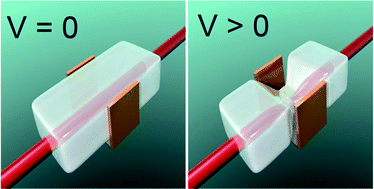
The electro-responsiveness in blood plasma and in blood mimicking fluids of methacrylic acid modified Pluronic (P127) hydrogel is investigated. It is observed that the hydrogel's response to an applied potential, in all buffer solutions, is very high, and comparable in amplitude and time response to classic polyelectrolyte gels. The highest actuation amplitude is achieved in PBS, which is directly related to the largest current value obtained for that buffer. The electro-actuation achieved in blood plasma is comparable to that in KREBS and KCl buffers. The good performance in blood plasma is attributed to the low protein adhesion to hydrogel surface. Preliminary biocompatibility studies demonstrate that the investigated hydrogel can be considered biocompatible.
The production of artificial biocompatible materials, which can autonomously execute desired work in vivo in a controllable way in response to the smallest change in surrounding environment, however, still remains a big challenge. Among the most promising candidates largely claimed in the literature, are polyelectrolyte hydrogels, that is, polymer hydrogels with charged groups incorporated into their macromolecular network.8–12 Because of their chemical structure that couples swelling degree to electrostatic interactions, they are capable of volumetric and/or mechanical changes in the response to external stimuli such as electric field, pH change or specific salt concentration variations. Gel-based materials have already been proposed as artificial muscles,13,14 stimuli responsive valves15 and active drug delivery systems.16 It is important to stress that hydrogels miniaturization improves the response speed significantly: electro-actuation induced volume change times below second are achievable, as we recently demonstrated with micrometer-sized electro-responsive hydrogel based cilia.17,18 This renders polyelectrolyte hydrogels ideal candidates for biomedical applications as electrical stimuli are usually easy to implement inside the body.
It was recently demonstrated that Pluronic/methacrylic acid sodium salt (PLMANa) hydrogel exhibit radial deswelling in KCl and KREBS solutions when surrounded by interdigitated electrodes. This electrically controlled swelling state of the gel creates possible applications such as blood vessel occlusion.19 The importance of electrically controlled shrinkage lies in a fact that it gives the physician time needed to approach the treatment location with the hydrogel prior to the swelling. In order to verify whether this system could be implemented in vivo we study the electro-responsiveness of Pluronic/methacrylic acid sodium salt hydrogel in blood plasma and compare it to blood-mimicking buffers (KREBS, PBS and KCl solutions). As we focus on the response properties of the gel, we use here a more appropriate geometry for this study than those potentially used in real applications. In order to produce hydrogel-based implantable devices with appropriate mechanical properties and short response times when actuating in vivo, some comprehensive tests in-vitro are first required. In addition, as the methacrylic acid is not FDA approved, preliminary biocompatibility evaluation of this new Pluronic/methacrylic acid based system is also reported below (biocompatibility data is presented in ESI†).
When the polymerization is complete the hydrogel is removed from the container and washed in an excess of demineralised water to remove any residual material. The hydrogel is then placed in MQ water, replaced twice a day, for a period of 4–5 days. The concentration of the mobile ions inside the hydrogel, that neutralize MANa, is not equal to that in the outer solution, creating an osmotic pressure difference. Those mobile ions tend to reduce the concentration gradient trying to diffuse to the outer solution. However, electro-neutrality condition prevents them from leaving the hydrogel and the osmotic pressure difference instead pumps water in, causing swelling of the hydrogel. The time associated with the hydrogel's swelling process is measured in days (as illustrated in Fig. 1). As the hydrogel volume increases, the density of charged groups is reduced, therefore, also decreasing the osmotic pressure difference. Each time the water is replaced, the hydrogel reaches its final volume as a balance between polymer matrix-solvent affinity, network elasticity which resists expansion, and charged groups-mobile ions interactions.20 The hydrogel can increase its own weight in MQ up to 42 times before reaching the equilibrium swelling. Directly before performing electro-actuation experiments, the hydrogel is cut into rectangular beams. For consistency between the actuation experiments described here, all hydrogels are allowed to first reach swelling equilibrium in MQ. Then, actuation experiments are carried out in aqueous electrolytes or blood plasma that contain varying concentrations of salt. It is important to stress that the hydrogel has a different equilibrium swelling when placed in a solution with a given salt concentration. However, the diffusion-driven de-swelling kinetics occur on a time scale of hours. On the contrary, the actuation experiments in the presence of electric field are carried out in the matter of minutes, immediately after the hydrogel is placed in a given salt solution. The difference of time scales for those two phenomena ensures that the hydrogel is in a similar state for measurements carried out in different buffers.
For the electro-actuation measurements, a Flat Bed Electrophoresis Unit MULTI (Carl Roth GMBH) is used. This unit consists of a glass tank and two platinum wires mounted in movable frames that serve as electrodes. The glass tank, together with the electrodes, is placed on a cooling unit that removes excess heat from the system during experiments. In addition, the cooling unit's surface is marked with a grid pattern that allows to precisely measure the hydrogel's shape variations during electro-actuation. The distance between the two platinum electrodes is set to 10 cm and the hydrogel is placed in the tank center – mid-way between the electrodes. An electric potential difference of 15 V is applied between the electrodes. The experiment is conducted in steady voltage mode, therefore whatever current is necessary to maintain this voltage drop is provided by the power supply. When a potential above 1.2 V is applied across aqueous solutions water hydrolysis takes place.14 Because the applied potential is much larger than 1.2 V, the variations in the electrolysis potential are relatively small and therefore the electric field is approximately constant in all the experiments. This allows us to compare the influence of different blood-mimicking solutions on hydrogel electro-responsiveness. The mentioned electrode/gel configuration was chosen to easily visualize the electro-responsiveness. In case of bio-medical application like blood vessel occlusion, the typical size of the hydrogel is of the order of millimetres. As a consequence of the hydrogel size reduction, the voltage/current required for electro-actuation is considerably decreased.11
During electro-actuation the shape of the hydrogel is monitored, every 30 seconds, using a Nikon Coolpix 4500 camera mounted above the electrophoresis unit. The images are then processed with the ImageJ software and the hydrogel curvature against time is extracted.
In order to prepare Krebs–Ringer Bicarbonate Buffer (KREBS) 9.5 grams of powder (Sigma Aldrich, K4002) are dissolved in 1 litre of demineralised water. To prepare Phosphate buffered saline (PBS) 5 tablets (Sigma Aldrich, P4417) are dissolved in 1 litre of demineralised water (unless mentioned otherwise). The blood plasma is obtained from the Red Cross (Belgium).
The biocompatibility tests and procedures are described in ESI.†
It is therefore crucial, if one intend to develop any realistic implantable electro-responsive gel system, to consider the electro-responsiveness of polyelectrolyte gels in environments that mimics biological conditions in terms of ionic concentrations, tonicity and ionic motilities (blood plasma). Furthermore, the gels have to be biocompatible and must not promote protein clotting when placed in blood. Below, we investigate all these aspects for a Pluronic based polyelectrolyte gels.
As mentioned above, the electro-responsiveness of the polyelectrolyte hydrogel is directly related to the difference in ionic concentrations between the hydrogel and its surrounding medium.21–23 One possible way to control this parameter is to vary the hydrogel's swelling ratio in pure water, which is in turn related to the ratio between PL and MANa used during synthesis. However, in some cases poor mechanical properties (hydrogel becomes very brittle at high swelling ratios) are observed. It is therefore important to find a balance between an acceptable mechanical strength, fast electro-actuation and material integrity in order to envisage a biomedical application.
The swelling process in a hydrogel is diffusion driven and propagates from the outer boundary of the hydrogel towards the hydrogel's centre. When the PLMANa hydrogel is placed directly in a large excess of MQ water the swelling process is very abrupt. Due to the high osmotic pressure difference between hydrogel and surrounding solution, the hydrogel is subject to large stress gradients that may create cracks on the hydrogel surface affecting sample integrity. To prevent this event, a step-wise swelling method was developed. In this method the hydrogel is first pre-swollen in a small liquid volume (50 ml) for 8 hours, during that time the hydrogel doubles its mass, and then the water volume is increased to 500 mL and repeatedly replaced (at least twice a day). This approach improves the hydrogel's mechanical properties and prevents hydrogel breaking during swelling. An example of the hydrogel swollen with the pre-swelling step (pre-swelling period marked with blue rectangle) and the hydrogel relative mass change in time is illustrated in Fig. 1. Absorbing water, the PLMANa hydrogel increases its own mass around 42 times after 4 days that correlates to an enormous volume expansion. This significant network expansion is of great importance in terms of possible blood vessel occlusion as already stressed. However, if one think of a medical application of this material it should also be possible to supress the swelling with external stimuli. Therefore the hydrogels electro-responsiveness is validated as described below.
During swelling, the PLMANa hydrogel reaches equilibrium with MQ water. When the hydrogel is then placed in a given buffer and an electric potential is applied trough the solution, ion accumulation and depletion regions, localized initially at the buffer/hydrogel boundaries, propagate into the hydrogel. The local change in the ion concentration at the hydrogel boundaries that induces significant gel shrinkage at anode facing side of the gel causes electro-actuation.21,22 As a result, the difference in ionic concentrations between the outer solution and the hydrogel equilibrated in MQ, which is directly related to the swelling ratio (eqn (1)), where M is the hydrogel mass and Mprep hydrogel mass at preparation state should have a significant influence on the electro-responsiveness of PLMANa gel.
![]() | (1) |
Therefore, different samples of PLMANa gels, swollen in MQ to 761% (Qw% = 7), 1372% (Qw% = 13), 2175% (Qw% = 21) and 4285% (Qw% = 42) weight increase, were placed in KREBS, PBS, KCl and blood plasma buffers and their electro-responsiveness was measured. The schematic illustration of the experiment, electro-responsiveness characterization procedure and graphical illustration of the ion accumulation/depletion mechanism responsible for actuation are illustrated in Fig. 2.
![]() | ||
Fig. 2 Schematic illustration of the experimental geometry. The rod-like hydrogel placed between two electrodes (a) before and (b) after two minutes under applied electric potential. The curvature characterization procedure is represented by the black arc of radius R. (c) Graphical interpretation of the enrichment/depletion mechanism (darker and lighter colours at the hydrogel/solution boundaries represent accumulation and depletion of ions at the anode and cathode side respectively).21 |
For each swelling degree and each solution used, the hydrogel curvature (1/R) against time was extracted (Fig. 3).
From inspection of Fig. 3, it is clear that even at low swelling degrees the gel is electro-responsive. In addition an increase in the electro-actuation speed with the increase of swelling degree is observed due to the increase in difference between ionic strength inside the gel and embedding solution. The actuation of the hydrogel swollen 42 times (Qw% = 42) is almost twice as big as for the hydrogel swollen only 7 times. The Qw% = 13 and Qw% = 21 gels have comparable electro-actuation speed, for all buffers except KREBS. The electro-actuation of Qw% = 7 gel is high and comparable with that of hydrolysed polyacrylamide hydrogels.21 A typical example of an actuating PLMANa hydrogel, embedded in blood plasma with the electric potential applied, is illustrated in Fig. 4.
It is important to stress that the mechanical properties of the hydrogel, which determine the ease of hydrogel handling and manipulation, are much better when the hydrogel's Qw% is 7 or 13 than when it is 21 or 42 times. The hydrogels swollen 7 and 13 times are relatively easy to handle and flexible. It is worth mentioning, that the step-wise swelling method produces hydrogels that are uniformly swollen, without any visual defects and flexible during actuation. The storage modulus of the Qw% = 42 hydrogel is around 2 kPa.
From Fig. 3 we notice that the electro-response of the hydrogel in blood plasma is similar to that in 0.1 M KCl and in KREBS solution. It is therefore important to investigate to what extend the buffer used determines the electro-actuation speed. In Fig. 5 the hydrogel curvature versus time for different solutions is extracted.
The highest electro-responsiveness is achieved when the hydrogel is embedded in PBS solution. This can be related to the current values obtained for the investigated solutions. As the power supply works in the constant voltage mode, the high current value indicates significant difference, in terms of ionic concentrations, between the gel (equilibrated in MQ water) and buffer used. Indeed, the current value measured during actuation was highest for PBS (92 mA after 2 minutes) clearly indicating that the ionic concentrations play an important role in the gel response. The current values after 2 minutes for KREBS and KCl were 82 mA and 70 mA respectively. In case of blood plasma the value obtained was 60 mA.
The speed and amplitude response of actuation in blood plasma is similar to those in blood-mimicking fluids. This suggests that interaction between the gel and proteins present in blood are not playing a decisive role and we can anticipate as experimentally confirmed (see ESI for more details†) that protein adhesion to the gel surface is low.
In order to answer the question whether PLMANa hydrogel system can be considered as material for biomedical applications some preliminary biocompatibility tests like cell toxicity, haemolytic and protein adhesion were conducted (see ESI for more details†). From the obtained results we can conclude that PLMANa gel shows no cytotoxic nor haemolytic effects and is relatively resistant to protein adhesion. All these features are of crucial importance in biomedical applications. Currently more extensive biocompatibility and in vivo studies of this promising material are being conducted.
Footnote |
† Electronic supplementary information (ESI) available. See DOI: 10.1039/c3ra45395b |
This journal is © The Royal Society of Chemistry 2014 |