Sunil
Pandey†
*,
Ashmi
Mewada†
,
Mukeshchand
Thakur†
,
Sreenath
Pillai
,
Roopa
Dharmatti
,
Chinmay
Phadke
and
Madhuri
Sharon
*
N.S.N. Research Center for Nanotechnology and Bio-Nanotechnology, Ambernath, MS, India. E-mail: gurus.spandey@gmail.com; sharonmadhuri@gmail.com; Tel: +91 9004024937
First published on 14th October 2013
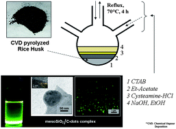
Due to the abundance of silica and carbon in rice (Oryza sativa) husk (RH), we exploited it for the synthesis of a mesoporous silica oxide micro-particles (meso-SiO2)/C-dot complex for biological imaging using a novel hot injection method commonly used for semiconductor quantum dots. Carbon dots (C-dots) with a high degree of green fluorescence were observed under UV-light they were found to be embedded to a significant extent in meso-SiO2 as confirmed by transmission electron microscopy (TEM) and energy dispersive spectroscopy (EDS). The mesoporous nature of the complex was confirmed using N2 adsorption–desorption measurements. Surface functionalization was studied using Fourier transform infrared spectroscopy (FTIR). The synthesized meso-SiO2/C-dots complex was used for labeling yeast cells since the fungi closely represents eukaryotic organisms. Furthermore, the meso-SiO2/C-dot complex was found to be highly bio-compatible for Vero cells. This study might help in the further utilization of the complex for the theranostic application of simultaneous cellular imaging and drug delivery.
One of the most interesting outcomes of the work is the one pot synthesis of C-dots protected with meso-SiO2 micro-particles. In an earlier attempt to synthesize C-dots entrapped in meso-SiO2 in order to enhance the homogeneous size distribution, silica oxide nanoparticles were prepared separately followed by their attachment to synthesized C-dots and surface protection using polyethylene glycol.13 In order to prepare meso-SiO2 capped C-dots, we selected rice husk as an ideal precursor because of the very high content of silica present in it (>67%). We also developed a hot injection method for the preparation of the above complex at 200 °C using a three neck flask. This method was found to be extremely reproducible and yielded high quality C-dots.
i. Rice husk, being a natural material act as a highly biocompatible precursor for a C-dot synthesis.
ii. Involvement of toxic chemical constituents such as tetraethyl orthosilicate (TEOS) is avoided.
iii. The process is rapid hence ideal for the commercial synthesis of C-dots.
iv. Mesoporous silica oxide along with C-dots can act as a versatile drug delivery vehicle with a high drug loading capacity.
Pyrolysis of RH before the synthesis of C-dots was done to solubilize the major carbon content in ethyl acetate, and particularly, the high amount of silica present in the RHA (Table 1). During carbonization of RH the carbonaceous materials get oxygenated, which is an important prerequisite for C-dot synthesis.14 A major part of the silica gets separated during the reflux due to its high solubility in ethyl acetate. After injection of 5 M NaOH along with EtOH, there was a transformation in the color from black to pale yellow, a common color marker for C-dot synthesis.7,11,12 In the reaction vessel, due to a slightly alkaline environment (pH 6.4) and the presence of surface passivation agents such as ethanol, the synthesis of highly fluorescent C-dots was catalysed. An alkaline environment is found to be highly favorable for the synthesis of C-dots.15 Simultaneously, the formation of mesoporous silica oxide is helped by the cationic surfactant CTAB. Simultaneous formation of C-dots can take place, which can attach to meso-SiO2 by adsorption, or covalent bond formation (Fig. 4b) alternatively cysteamine hydrochloride in a lower concentration could act as a bridge between mesoporous silica and the as synthesized C-dots. Due to the above chemical reactions a C-dots@meso-SiO2 conjugate is formed (Fig. 1). Another important observation was the presence of a silvery white suspension along with C-dots indicating the formation of meso-SiO2.13 This silvery yellow solution was first purified by dialysis against distilled water for 12 h. A clear water soluble suspension was obtained after dialysis, which exhibited a strong green color under UV light (λ = 260 nm) as depicted in Fig. 2. Aqueous solubility and bright fluorescence are two important attributes of C-dots, which are not present in other forms of carbon.15 With a certain level of disagreement, the inception of fluorescence in C-dots is believed to be due to the presence of energy traps.1 Quantum confinement is another mechanism considered to be responsible for the above phenomenon. Moreover, the association of SP3 hybridized carbons and oxygen plays a very critical role in the inception of fluorescence in carbon nanoparticles such as C-dots.16
| Sr. No. | Constituents | Percentage (%) |
|---|---|---|
| 1. | Silicon dioxide (SiO2) | 81.33 |
| 2. | Calcium oxide (CaO) | 0.32 |
| 3. | Ferric oxide (Fe2O3) | 0.98 |
| 4. | Aluminium oxide (Al2O3) | 0.18 |
| 5. | Magnesium oxide (MgO) | 0.11 |
| 6. | Titanium oxide (TiO2) | 0.07 |
| 7. | Carbon (C) | 8.62 |
| 8. | Loss of Ignition (L.O.I) | 10.11 |
![]() | ||
| Fig. 1 Schematic showing CTAB assisted synthesis of meso-SiO2 and attachment of C-dots to form the meso-SiO2/C-dot complex. | ||
Fig. 2 depicts UV-Vis spectroscopy of as prepared C-dots along with meso-SiO2. A sharp peak at 260 nm and a broad hump at 972 nm indicate the formation of C-dots in the solution. The peak at 260 nm arises due to the π → π* electron transition of C
C associated with C-dots17 and another peak at 972 nm is due to the presence of silica oxide or the meso-SiO2/C-dot complex. Due to significant absorption in the near infrared (NIR) region, this complex can also be used for photothermal therapy of solid tumors and/or NIR induced drug release, thus establishing a fundamental platform for chemo-photothermal therapy. Absence of background absorbance in the visible region indicates the absence of other carbonaceous materials, which usually absorb at higher wavelengths.18,19 The inset of the Fig. 2 presents photoluminescence spectra of the meso-SiO2/C-dot complex at 464, 535 and 552 nm after excitations at 250, 350 and 450 nm respectively. There is a continuous red shift and enhancement in intensity of the peak with an increasing excitation wavelength. Excitation dependent emission is a signature marker of C-dots.
![]() | ||
| Fig. 2 UV-Vis spectroscopy of meso-SiO2/C-dots synthesised using rice husk. Inset shows photoluminescence spectra excited at (a) 250 (b) 350 and (c) 450 nm. | ||
The most revealing feature of the present research is the synthesis of meso-SiO2 capped on the surface by C-dots and/or that the C-dots were found to be trapped inside the meso-SiO2. The TEM image displayed in Fig. 3a shows meso-SiO2/C-dots along with free C-dots and meso-SiO2 in the background. Fig. 3b shows mesoporous silica micro-particles along with an enlarged view showing the presence of tiny C-dots ∼10 nm in size on the surface. The inset shows an enlarged image of the entrapped C-dots with a lattice fringe width of 0.3 nm and the porous nature of the complex is seen in a HRTEM image (Fig. 3c). X-Ray diffraction (XRD) (Fig. 3d) reveals a characteristic peak at 2θ = 26.29° and a weak peak at 2θ = 43.67°, which are assigned to the (002) and (101) diffraction patterns of graphitic carbon respectively. The peak at 26.29° corresponds to an interlayer spacing of ∼3.77 Å, which is higher than that between the (002) planes in bulk graphite i.e. 3.44 Å.20 The Raman spectrum of RH derived C-dots is shown in Fig. 3e. The G-band observed at 1594.89 cm−1 with respect to the more intense peak of the D-band at 1331.52 cm−1 indicates the presence of messy carbon nanomaterials in the form of C-dots.21 Energy dispersive spectrometry (EDS) (Fig. 3f) shows that the major components are silicon (Si) and carbon (C) in the elemental composition of the meso-SiO2/C-dot complex. The prominence of silicon and carbon explains the association of C-dots and silicon oxide. Another peak corresponding to elemental oxygen (O) is present, which is particularly important for the fluorescent properties of C-dots. Sodium (Na) and bromide (Br) come from CTAB and traces of Cl come from Cys-HCl, which are used during the synthesis procedure. Fig. 3g shows a fluorescent image of the meso-SiO2/C-dot complex under UV excitation when viewed under a fluorescent microscope. Bright green complexes were seen because of the florescence emerging from the C-dots embedded in the meso-SiO2/C-dot complex.
In order to confirm the mesoporous nature of SiO2, a N2 adsorption–desorption measurement was performed. It showed type IV behavior (Fig. 4a). The pore size of the meso-SiO2 was found to be ∼30–40 nm as confirmed by the Brunauer–Emmett–Teller (BET) method. Surface functionalization studies were performed using FTIR. Fig. 4b shows FTIR spectra of the meso-SiO2/C-dot complex. Typical IR bands were obtained around 916–1094 cm−1, indicating symmetric stretching vibrations of Si–O–Si at 916 cm−1 and asymmetric stretching vibrations at 1094 cm−1. The weak band at 594 cm−1 was attributed to –CH2 alkane bending. The band at 1411 cm−1 is possibly due to aromatic –NO2 groups on surface passivized C-dots. Other surface passivation was observed at 1652 cm−1 arising from the C
O stretching vibrations. A relatively intense IR band of a typical –CH stretching vibration from the C-dots was observed around 2952 cm−1. An intense wide band was observed at 3355 cm−1, which indicates typical surface passivation by –OH stretching onto C-dots and that typical silanol (Si-OH) groups were formed during the formation of the meso-SiO2/C-dot complex.
An important use for blended meso-SiO2/C-dots was found to be yeast cell imaging as shown in Fig. 5. C-dots are more advantageous over conventional imaging materials such as gold nanoparticles due to high background scattering from biological cells.21 Semiconductor quantum dots (QDs), due to presence of highly toxic metals such as arsenic, lead and cadmium have limited applications in biological imaging.22
The cytotoxicity results (Fig. 6) demonstrate that C-dots blended with meso-SiO2 were non-toxic to Vero cells, thus providing an indication of the potential advantages of C-dots in imaging over conventional semiconductor QDs. These C-dots after administration into the body undergo rapid renal clearance and are quickly metabolised, unlike other fluorescent probes including QDs.23 At all of the concentrations of the complex (0.1–0.5 mg ml−1), the percentage viability of the cells were found to be >95% indicating exceptional bio-compatibility of the synthesised complex. In drug delivery applications, both mesoporous silica and C-dots individually can act as drug delivery vehicles.24,25 However, with the meso-SiO2/C-dot complex, a high drug loading efficiency can be achieved and simultaneous bio-imaging could be performed. Hence, the meso-SiO2/C-dot complex has a potential nano-theranostic application. A stronger photoluminescence property could be obtained with meso-SiO2/C-dots compared to C-dots alone.26
![]() | ||
| Fig. 6 Percentage cell viability plot showing effect of the meso-SiO2/C-dot complex on Vero cell lines. | ||
Footnote |
| † Authors have equal contribution in this work. |
| This journal is © The Royal Society of Chemistry 2014 |