DOI:
10.1039/C3RA45142A
(Paper)
RSC Adv., 2014,
4, 9457-9462
Synthesis and enhanced microwave properties of uniform hollow Fe nanospheres and their core–shell silica nanocomposites
Received
19th September 2013
, Accepted 23rd January 2014
First published on 23rd January 2014
Abstract
Monodispersed hollow magnetite nanospheres with diameter of about 462 nm and a shell thickness of approximately 90 nm were successfully synthesized through a simple solvothermal process in ethylene glycol (EG) in the presence of urea. The electron and transmission microscopy images showed that the obtained magnetite spheres are composed of small particles and confirmed their hollow interior. Furthermore, by annealing in an H2 atmosphere, the crystalline structure is converted from fcc (Fe3O4) to bcc (Fe) which has been confirmed by X-ray diffraction (XRD) results. The Ms value of reduced samples (hollow Fe nanospheres) increases to 198 emu g−1 which is mostly equal to that of bulk Fe. Relevant hollow Fe/SiO2 core–shell nanospheres were prepared through a modified Stöber process. The enhanced microwave attenuation can be achieved for this core–shell nanospheres which is attributed to the improved electromagnetic impedance match and strong and broadband natural resonance at high frequency for the metal nanospheres.
1. Introduction
With the accelerated developments of wireless communications and high-frequency circuit devices in the gigahertz range, electromagnetic interference (EMI) has become a serious problem.1–3 Much research interest has been focused in this area to fabricate high-efficiency electromagnetic wave attenuation (EMA) materials with broad attenuation frequency, high attenuation capacity, light weight, good thermal stability.4–6 The traditional EMA materials, such as ferrites and metal powders, have some disadvantages. For instance, they usually have high density and encounter the high eddy current loss at the high frequency for the metal powders. As compared to the traditional ferrites and metal powders, the ferrite and metal nanospheres with hollow structure have some outstanding advantages, such as low density, high specific surface area and low eddy current loss. Therefore, it is expectable that hollow ferrite and metal nanospheres can be used as new EMA materials with light weight and high attenuation properties. Recently, several efforts have been made toward the development of EMA materials by employing hollow ferrite nanospheres as fillers, such as magnetite and Zn ferrite hollow nanospheres.7,8 However, as compared with ferrites, Fe powder should be the better candidate for microwave attenuation purpose because of its high saturation magnetization, high Curie temperature, higher complex permeability and higher working frequency.9
In this study, monodisperse hollow magnetite nanospheres were prepared by a simple solvothermal process with the assistance of urea and ethylene glycol. The formation mechanism is mainly based on oriented nanocrystals aggregation and subsequent local Ostwald ripening process.10 Afterward, hollow Fe nanospheres were obtained by hydrogen-thermally reducing prepared hollow magnetite nanospheres. Furthermore, as we know, metallic particles have the high conductive value which may result in the poor impedance matching between the materials and free space and thus worsen their attenuation properties.11,12 In order to overcome this problem, an insulating SiO2 layer were applied on the prepared hollow Fe nanospheres. The microstructure, morphology and magnetic properties of prepared hollow magnetite, Fe and SiO2 coated Fe nanospheres were investigated. The EMA performance of composites containing hollow Fe and SiO2 coated Fe nanospheres as fillers was evaluated according to the transmission line theory.
2. Experimental procedure
2.1 Synthesis of monodispersed hollow magnetite nanospheres
Ferric chloride hexahydrate (FeCl3·6H2O), urea (H2NCONH2) and Ethylene glycol (EG) were purchased from Sigma-Aldrich Co. LLC. And all reagents are analytically pure and used as-received without further purification. The autoclaves for solvothermal reaction were purchased from Parr Instrument Company. The procedure for the preparation of hollow magnetite nanospheres was based on our previous study.13 In a typical experiment, 3 mmol FeCl3·6H2O and 0.03 mol urea were dissolved in 30 ml ethylene glycol (EG) under continuous magnetic stirring to form a clear solution. Subsequently, the mixture was transferred to a 50 ml Teflon-lined autoclave and maintained at 200 °C for 10 to 16 h. After reaction, the autoclave was cooled to ambient temperature and the products were obtained by filtering and washing with distilled water and ethyl alcohol for several times followed by drying in a vacuum oven at 60 °C for 2 h. The detailed procedure can be illustrated as in Fig. 1.
 |
| | Fig. 1 Fabrication process of the hollow magnetite (Fe3O4) spheres. | |
2.2 Synthesis of hollow Fe nanospheres
A portion of the as-prepared hollow Fe3O4 nanospheres was reduced in a hydrogen atmosphere at 400 °C for 120 min. After that, reduced products were cooled in H2 atmosphere to room temperature naturally in the furnace.
2.3 Synthesis of hollow Fe/SiO2 core–shell nanocomposites
Silica coating of hollow Fe nanospheres was performed through a modified Stöber process by using tetraethyl orthosilicate (TEOS, Si(OC2H5)4) (95%, Sigma-Aldrich), absolute ethanol (Fisher Scientific Co.,), and ammonium hydroxide solution (NH4OH, 28%, Sigma-Aldrich) as SiO2 precursor.14 In a typical coating process, hollow Fe nanospheres (0.6 g) were dispersed in ethanol (200 ml) and ultrasonicated in order to break down agglomerates. In the next step, ammonium hydroxide solution (2.5 ml, 28%) and 1 ml of TEOS were added to the mixture to initiate the deposition of silica shells on the hollow Fe nanoparticles. Thereafter, the suspension was mechanical stirred at 300 rpm for 2 h to complete the reaction at room temperature. The Fe/SiO2 particles were then washed with ethanol two times and then dried at 60 °C for 1 h in a vacuum oven.
2.4 Characterization
The morphologies of the prepared hollow Fe3O4, Fe and SiO2 coated Fe nanospheres were examined using a JSM 6701F field emission scanning electron microscope (FE-SEM) at an accelerating voltage of 20 kV and a JOEL JEM 2100F transmission electron microscope (TEM) at an accelerating voltage of 150 kV. Particle crystalline structures of the prepared Fe3O4, Fe and SiO2 coated Fe hollow nanoparticles were examined by using an X-ray diffractometer (XRD, Rigaku Ultima IV) with Cu Kα radiation (λ = 1.54056 Å and scan rate of 2° min−1). Static magnetic properties of the hollow Fe3O4, Fe and SiO2 coated Fe nanospheres at room temperature were measured by using a vibrating sample magnetometer (VSM, ADE Magnetics EV-9). Silicon resin composites were prepared by homogeneously mixing 60 wt% of the prepared hollow Fe3O4, Fe and SiO2 coated Fe nanospheres with silicon resin and pressed into toroidal-shaped samples with an outer diameter of 7.00 mm and an inner diameter of 3.04 mm. Complex permeability and permittivity of the composites were measured by using an Agilent VNA (Vector Network Analyzer) HP8722D with a reflection-through-line calibration, over 0.05–8.55 GHz, using a set of 7 mm coaxial air-line with length of 49.96 mm. Frequency dependence of reflection loss (RL) of the composites was estimated from their complex permittivity (εr = εr′ − jεr′′) and permeability (μr = μr′ − jμr′′) according to the following equations:| |
 | (1) |
| |
 | (2) |
where Zin is impedance of the composites backed by a ground plane, Z0 is the intrinsic impedance of free space, c is the velocity of light in free space, t is thickness of the attenuation material, ƒ is the frequency of the incident EM wave.
3. Results and discussion
3.1 FESEM and TEM images
The time-dependent shape evolution of Fe3O4 nanospheres was studied. In a control experiment, the reaction temperature was maintained at 200 °C and the reaction time was changed from 10 h to 16 h. Fig. 2 presents the morphology of nanospheres for different solvothermal treatment times. For a heating time between 10 and 12 h, the obtained samples are composed of uniform and compact nanospheres and some small holes on the nanospheres could be observed which implied the appearance of hollow structure of the nanospheres. If the heating time was prolonged to 14 to 16 h, most of the nanospheres were broken or collapsed and turned into the cracked structures. The reason for this transition is still not clear and needs some further investigation.
 |
| | Fig. 2 SEM images of the hollow magnetite nanospheres obtained for different reaction time: (a) 10 h, (b) 12 h, (c) 14 h, (d) 16 h. | |
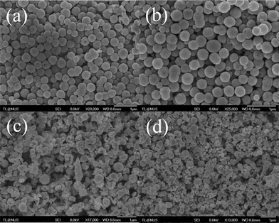
As the optimal reaction condition has been confirmed to be 200 °C for 12 h, all the samples hereafter were prepared under this reaction condition. The morphology of obtained and reduced products was investigated by SEM and TEM as shown in Fig. 3. From the low magnified images as shown in Fig. 3(a) and (c), it reveals that the products consist of a large quantity of uniform nano-structured spheres without agglomeration. The high magnified images shown in Fig. 3(b) exhibits detailed surface morphology of the obtained Fe3O4 nanospheres, indicating that the spheres are composed of some much smaller particles. The detailed surface morphology of the reduced products is shown in Fig. 3(d). It is noteworthy, from the SEM image, all the small individual nanoparticles which were aggregated together to form Fe3O4 hollow spheres during the solvothermal process are disappeared and the surface of reduced particles becomes more uniform which is attributed to the combination of small nanoparticles into one piece during the annealing process. Meanwhile, the increase in the number of cracked particles could be observed for the samples after reduction treatment which may be due to the shrinkage and the loss of oxygen atoms during the sintering process. The histogram of the particle size distribution given in Fig. 3(g) and (h) showed that, for both prepared and reduced samples, the size distribution is narrow and the average particle sizes are mostly 462 nm and 409 nm for prepared and reduced samples, respectively. The decrease in the average particle size also proved the shrinkage during the reduction process. In addition, the cracked spheres which can be observed in Fig. 3(e) confirms that the spheres have a hollow interior. The hollow structure is further investigated by the TEM image as shown in Fig. 3(f). The intensive contrast between the black margin and the bright center of the particles confirms the existence of hollow structures in the resulting spheres which is consistent with the SEM observation. The insert in Fig. 3(f) shows the close view of a Fe3O4 hollow sphere and the wall thickness can be estimated to be around 90 to 100 nm.
 |
| | Fig. 3 (a)–(d) SEM images of the hollow magnetite and Fe nanospheres. (e) SEM image of some cracked magnetite nanospheres. (f) TEM image of prepared magnetite nanospheres. The insert in (f) shows a high-magnification TEM image of a typical magnetite hollow sphere. (g) Particle size distribution of magnetite nanospheres. (h) Particle size distribution of Fe nanospheres. | |
The SEM images of the obtained Fe hollow spheres with SiO2 coating are shown in Fig. 4(a) and (b). Compared with the Fe hollow spheres, the SiO2 coated samples exhibit a more regular spherical shape with a smooth surface which is due to the deposition and growth of the silica layer. The histogram of the particle size distribution given in Fig. 4(d) indicates an increase in the particle size for the samples after SiO2 coating and the average particle size is around 516 nm. Based on the average particle sizes of the Fe hollow spheres without and with SiO2 coating, the average thickness of the silica layer can be estimated to be ∼54 nm. Fig. 4(c) shows the TEM image of reduced products coating with a layer of SiO2. It is obvious that the original nanospheres are coated with a relatively uniform SiO2 layer and the thickness of the silica layer can be determined to be around 50 nm which is consistent with the value estimated from the average particle sizes.
 |
| | Fig. 4 (a) and (b) SEM and (c) TEM images of the Fe hollow spheres after SiO2 coating. (d) Particle size distribution of the Fe hollow spheres after SiO2 coating. | |
3.2 XRD patterns
Fig. 5a shows the typical XRD pattern of the prepared Fe3O4 hollow spheres. All reflection peaks and positions can be indexed to the cubic-structured Fe3O4 phase (JCPDS 85-1436) and well-resolved peaks reveal good crystallinity of these nanospheres. No impurity phase can be detected, indicating that the products prepared by using this method are in a single phase. Calculations using the Debye–Scherrer formula for the strongest peak (311) showed the grain sizes of 46 nm. This result confirms the fact that the hollow nanospheres of ∼462 nm in size are made up of smaller building blocks of nanoparticles, which is consistent with that determined by SEM in Fig. 3b. Fig. 5b shows the XRD peaks of the reduced samples. The result indicates that the samples annealed under H2 atmosphere at temperature of 400 °C for 2 h were converted from Fe3O4 with fcc structure to metallic Fe with bcc structure. The XRD pattern for the reduced samples coating with the SiO2 layer is shown in Fig. 5c. No characteristic peaks beside the Fe peaks can be detected and the weak and broad peak at the low angle should be attributed to the SiO2 layer with amorphous structure.
 |
| | Fig. 5 XRD patterns of magnetite hollow spheres (a), Fe hollow spheres (b) and Fe hollow spheres with SiO2 coating (c). | |
3.3 Static magnetic properties
Fig. 6 shows the magnetization hysteresis curves of hollow Fe3O4, Fe and SiO2 coated Fe nanospheres at room temperature. As shown in purple line in Fig. 6, from the hysteresis loops of the sample, it is found that the magnetite spheres exhibit ferromagnetic behavior. The saturation magnetization value (Ms) of the synthesized Fe3O4 hollow nanospheres is about 86.7 emu g−1 which is close to the theoretical value of bulk Fe3O4 (∼92 emu g−1). The coercivity (Hc) value is around 60 Oe. However, the Ms value of reduced samples (Fe nanospheres) increases to 198 emu g−1 which is mostly equal to that of bulk Fe. It also can prove that most of the prepared Fe3O4 has reduced to Fe. Meanwhile, the coercivity (Hc) value increases to 129 Oe. After coating a layer of SiO2 on the Fe nanospheres, the value of Ms decreases from 198 emu g−1 (original Fe nanospheres) to 156 emu g−1 (SiO2 coated Fe nanospheres). This decrease in magnetism is attributed mainly to the decrease in weight ratio of Fe in the particles.
 |
| | Fig. 6 The hysteresis loops of hollow magnetite, Fe and SiO2 coated Fe nanospheres measured at room temperature. | |
3.4 Dynamic magnetic properties
The real part (μ′) and the imaginary part (μ′′) of the relative permeability are shown as a function of frequency for the silicon resin composites with 60 wt% of hollow Fe3O4, Fe, and SiO2 coated Fe nanospheres as fillers in Fig. 7. For the Fe3O4 composites, the μ′ value increases from 2.4 to 2.8 firstly and then decreases to 0.8 with the frequency increasing to 8.55 GHz. The μ′′ exhibits a broad peak in a wide frequency range and the maximum value is around 1.2, however, the resonance frequency is around 2.8 GHz. For the Fe composites, the μ0′ value is still around 2.4, but the μ′ value is higher than the Fe3O4 composites throughout the whole frequency range and the resonance frequency shifts from 2.8 GHz to 6 GHz as compared with Fe3O4 composites. For the SiO2 coated Fe composites, the result indicates that both μ′ and μ′′ values decrease. The decrease in permeability can be attributed to the reduction of saturation magnetization after coating nonmagnetic SiO2.
 |
| | Fig. 7 Complex permeability curves of the silicon resin composites filled with 60 wt% of hollow magnetite, Fe, SiO2 coated Fe nanospheres. | |
Fig. 8 shows the real part (ε′) and imaginary part (ε′′) of relative permittivity for the silicon resin composites with 60 wt% of hollow Fe3O4, Fe and SiO2 coated Fe nanospheres as fillers. As shown in the figure, for the Fe3O4 composites, both real part (ε′) and imaginary part (ε′′) of relative permittivity are almost constant in the whole frequency range with the values of ε′ = ∼12.2 and ε′′ = ∼0.5. However, for the Fe composites, ε′ value increases to 68 at 0.05 GHz as compared to the Fe3O4 composites and then decrease to a certain value in the high-frequency range. The large permittivity is mainly determined by the increased conductivity after reducing the oxide to metal and thus the electrical charge of the particles is more easily polarized. However, the ε′ value of composites with hollow Fe nanospheres as fillers is still lower than that of the composites with other kinds of metallic fillers.12 That can be attributed to the special hollow structure and the air inside of the nanospheres which can partly cut off the polarization between particles. The ε′ and ε′′ values of SiO2 coated Fe composites substantially decrease and almost keep constant in the measured range, in which the magnitudes of ε′ and ε′′ are around 17 and 1, respectively. The increase of resistivity and the decrease of space-charge polarization after coating SiO2 nanoshell mainly contribute to the lower permittivity.
 |
| | Fig. 8 Complex permittivity curves of the silicon resin composites filled with 60 wt% of hollow magnetite, Fe, SiO2 coated Fe nanospheres. | |
3.5 Refection loss (RL)
Fig. 9 and 10 show the typical relationship between reflection loss (RL) and frequency for the silicon resin composites with 60 wt% hollow Fe and SiO2 coated Fe nanospheres, respectively. The minimum RL value shifts toward the lower frequency region with the increase in attenuation material thickness. The RL values of composites derived from the hollow Fe nanospheres always show the quite narrow band EM wave attenuation and for the samples with thickness of 3 to 4 mm, the RL value even cannot research −10 dB. However, with the same thickness, the composites made with the SiO2 coated hollow Fe nanospheres can readily achieve RL value of −10 dB. RL value of −20 dB at about 2.8 GHz is reached by the composite with a thickness of 5 mm. It is very clearly showed that composites made of hollow Fe nanospheres with a layer of insulated SiO2 coating have the enhanced EM wave attenuation properties as compared to the samples without the coating. As far as we know, in order to achieve the low RL values, two key factors need to be met. Firstly, make sure the incident microwave can transmit into the materials with minimum reflection and furthermore the materials can effectively attenuate the microwave that has transmitted into them. In order to let the incident microwave transmit into the materials instead of reflecting back to the space, the material should have an impedance matching with that of free space, thus the material should have close values of permittivity and permeability.15 As the permittivity value of the Fe composites is much higher than its permeability value, so most of the incident microwave cannot transmit into the material. As a result, microwave attenuation is weak. Comparatively, the difference between permittivity and permeability of samples after coating is smaller. In other words, it has a better impedance match and thus has higher microwave attenuation.
 |
| | Fig. 9 Dependences of reflection loss (RL) on frequency for the silicon resin composites filled with 60 wt% of hollow Fe nanospheres. | |
 |
| | Fig. 10 Dependences of reflection loss (RL) on frequency for the silicon resin composites filled with 60 wt% of hollow Fe nanospheres with SiO2 coating. | |
4. Conclusions
In conclusion, monodisperse Fe3O4 ferrite hollow nanospheres with the diameter approximately 462 nm were successfully synthesized through a simple solvothermal method. The TEM image and some of the cracked particles in the SEM images have confirmed the interior hollow structure of the nanoparticles. Relevant hollow Fe nanospheres can be directly obtained through a simple hydrogen reduction process. The XRD curves have confirmed that crystalline structure converted from fcc to bcc which indicates that the Fe3O4 has been reduced to Fe after being annealed under H2. Their core–shell silica nanocomposites were prepared using a modified Stöber process. As compared to the Fe nanospheres without SiO2 coating, the permittivity of hollow Fe nanospheres with 50 nm SiO2 shell decreases dramatically, while permeability decreases slightly. Because of the improved electromagnetic impedance match due to the decrease of permittivity after SiO2 coating and large magnetic loss due to strong and broadband natural resonance at high frequency for metallic nanospheres, the SiO2 coated Fe silicon resin composites show the enhanced microwave attenuation. The RL value exceeding −20 dB for SiO2 coated Fe composites is obtained with thickness of 5 mm.
References
- Y. L. Yang, M. C. Gupta, K. L. Dudley and R. W. Lawrence, Adv. Mater., 2005, 17, 1999 CrossRef CAS.
- V. B. Bregar, IEEE Trans. Magn., 2004, 40, 1679 CrossRef CAS.
- J. Joo and C. Y. Lee, J. Appl. Phys., 2000, 88, 513 CrossRef CAS PubMed.
- Z. H. Yang, Z. W. Li, L. Liu and L. B. Kong, J. Magn. Magn. Mater., 2012, 324, 3144 CrossRef CAS PubMed.
- Z. W. Li, Z. H. Yang and L. B. Kong, J. Appl. Phys., 2011, 110, 063907 CrossRef PubMed.
- X. A. Li, B. Zhang, C. H. Ju, X. J. Han, Y. C. Du and P. Xu, J. Phys. Chem. C, 2011, 115, 12350 CAS.
- F. L. Wang, J. R. Liu, J. Kong, Z. J. Zhang, X. Z. Wang, M. Itoh and K. I. Machida, J. Mater. Chem., 2011, 21, 4314 RSC.
- A. G. Yan, X. H. Liu, R. Yi, R. R. Shi, N. Zhang and G. Z. Qiu, J. Phys. Chem. C, 2008, 112, 8558 CAS.
- L. Z. Wu, J. Ding, H. B. Jiang, C. P. Neo, L. F. Chen and C. K. Ong, J. Appl. Phys., 2006, 99, 083905 CrossRef PubMed.
- P. Hu, L. J. Yu, A. H. Zuo, C. Y. Guo and F. L. Yuan, J. Phys. Chem. C, 2009, 113, 900 CAS.
- Y. X. Gong, L. Zhen, J. T. Jiang, C. Y. Xu and W. Z. Shao, J. Magn. Magn. Mater., 2009, 321, 3702 CrossRef CAS PubMed.
- Z. H. Yang, Z. W. Li, Y. H. Yang, L. Liu and L. B. Kong, J. Magn. Magn. Mater., 2013, 331, 232 CrossRef CAS PubMed.
- X. H. Lin, G. B. Ji, Y. S. Liu, Q. H. Huang, Z. H. Yang and Y. W. Du, CrystEngComm, 2012, 14, 8658 RSC.
- J. Ge and Y. Yi, Adv. Mater., 2008, 20, 3485 CrossRef CAS.
- L. G. Yan, J. B. Wang, X. H. Han, Y. Ren, Q. F. Liu and F. S. Li, Nanotechnology, 2010, 21, 095708 CrossRef PubMed.
|
| This journal is © The Royal Society of Chemistry 2014 |
Click here to see how this site uses Cookies. View our privacy policy here. 


