Namita Kumaria,
Nilanjan Deya and
Santanu Bhattacharya*ab
aDepartment of Organic Chemistry, Indian Institute of Science, JNCASR, Bangalore 560 012, India. E-mail: sb@orgchem.iisc.ernet.in
bChemical Biology Unit, JNCASR, Bangalore 560 064, India
First published on 18th November 2013
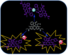
Cation sensing properties of the three positional isomers of rhodamine based sensors (1–3) are studied in water. The sensors differ only in the position of pyridine's nitrogen. The chemosensor 1, with pyridine nitrogen at ortho-position, showed a selective colorimetric detection of Cu(II) ions in water, at physiological pH 7.4 and also in medium containing BSA (bovine serum albumin) and blood serum. Notably the compound 2 and 3, with pyridine end located at meta- and para-positions did not show any color change with Cu(II) ions, although both the compounds showed turn-on change both in color and fluorescence with Hg(II) ions specifically. All the probes showed ratiometric changes with the specific metal ions. The changing position of nitrogen also changed the complexation pattern of the sensors with the metal ions. Probe 1 showed 2
:
1 complexation with Cu(II), whereas 2 and 3 showed 1
:
1 complexation with Hg(II) ions. The mechanism investigation showed that the change in color upon addition of metal ions is due to the ring-opening of the spirolactam ring of the probes. Cu(II) interacted with ligand 1 through a three-point interaction mode comprising carbonyl oxygen, amido nitrogen and pyridine nitrogen end. But in case of 2 and 3, Hg2+ only interacted through pyridine nitrogen ends. Quantitative estimation of Cu2+ and Hg2+ in complex biological media such as bovine albumin protein (BSA) and human blood serum were performed using these sensors. Rapid on-site detection as well as discrimination of these toxic ions was demonstrated using easily prepared portable test-strips.
Mercury ion is another highly toxic transition metal ion. It is carcinogenic and highly toxic.1 The standard for the maximum allowable level of inorganic Hg in drinking water is only 2 ppb according to the United States Environmental Protection Agency (EPA).11,12
Rhodamine based sensors have gained much popularity in the last few years due to their strikingly different properties in the closed spirolactam ring and open-ring form. In the closed spirolactam form, the central quaternary carbon of lactone ring breaks the π-conjugation of xanthene moiety and renders nearly colorless and non-fluorescent entity. On the other hand, the opening of this spirolactam ring extended the π-conjugation which leads enhancement of color and fluorescence emission in the visible region. Exploiting this idea in 1997, Czarnik et al. first reported Rhodamine based chemosensor.13 Following this work, the spirolactam-ring opening phenomenon has been utilized by many groups and a number of reports have appeared in literature for the detection of various ions based on rhodamine as signalling moiety.14,15
Many rhodamine based sensors have been reported for the detection of the Cu2+ ion13,16–24 as well as for the Hg2+ ion.14,15,25–33 Some of the reports have shown the detection both Cu2+ and Hg2+ ion.34–36 But most of the reports have shown the detection only in aqueous–organic mixture. So far very few instances are known which report the sensing in complete water or in aqueous–organic medium with water as a major solvent.37–42 The response of rhodamine based sensors are known to depend upon the choice of solvent system and nature of spacer between binding site and signalling unit.14,15 But till date no effort has been made to investigate the effect of different positional isomers on metal ion selective binding ability of rhodamine based sensors.
Earlier we have developed sensors and functional assemblies for the detection of various analytes.43–47 In this work we have investigated the sensing properties of the three positional isomers based on rhodamine as signalling moiety (1–3). The three isomers differ in position of nitrogen end of the pyridine moiety. The probe 1 has been reported earlier for the detection Hg2+ and Cu2+ ion in mixtures of different aqueous–organic medium.48,49 However, no effort has been made to explore the properties of this sensor in pure water and also in biological samples. The properties of isomeric 2 and 3 are however not known in any medium as they have not even been investigated earlier. Notably by changing the position of the nitrogen atom in the sensors, it has completely changed the sensing properties of these probes. The sensor 1, containing pyridine nitrogen at the ortho-position showed a visible colorimetric and ratiometric detection of Cu2+ ion in natural water as well as in buffer solution at physiological pH 7.4. In contrast, the other two probes 2 and 3 possessing pyridine nitrogen at meta- and para-position respectively did not show any evidence of interaction with Cu2+ ion. However, both of these compounds showed selective interaction with Hg2+ ion in water. Both 2 and 3 showed visible colorimetric and ratiometric change with the Hg2+ ion. Also these two sensors showed ‘turn-on’ detection of the Hg2+ ion under fluorescence spectroscopy.
Addition of other cations (Ag+, Cd2+, Co2+, Fe2+, Fe3+, Hg2+, Mg2+, Mn2+, Ni2+, Pb2+ and Zn2+) did not induce any change in visible color. The UV-vis absorption spectra of 1 were recorded individually upon addition of various cations. The absorption spectrum of 1 showed two bands with the absorption maxima at 244 and 310 nm. The addition of Cu2+ ion to the solution of 1 resulted in the appearance of a new band with the absorption maximum at 530 nm with a concomitant decrease in the absorbance at 310 nm (Fig. 1). The addition of other cations however, did not show any such change in the absorption spectra. Further the UV-vis titration was performed upon gradual addition of Cu2+ ion. The titration showed progressive increases in the absorption maxima at 530 nm with concomitant decreases at 310 nm (Fig. 2). The titration curve showed two isosbestic points at 340 and 255 nm, indicating the existence of an equilibrium between 1 and 1-Cu2+ complex (Fig. 2). Further when the absorbance ratio at 529 and 308 nm was plotted with the addition of Cu2+ ion, a linear response with the added Cu2+ ion was observed (inset of Fig. 2). This showed that the probe 1 can be used as a ratiometric sensor for the detection of Cu2+ ion in pure water.
![]() | ||
| Fig. 2 UV-vis titration of 1 (10 μM) in water with Cu2+ (0 to 30 μM) [inset: ratiometric plot with added equiv. of Cu2+ at the absorbance ratio of 529 and 308 nm]. | ||
Further sensing property of 1 towards various cations was investigated at physiological pH 7.4 (Fig. S3, ESI†). At this pH also, the probe 1 remained colorless with the two bands with absorption maxima at 241 and 310 nm. Upon addition of Cu2+ ion, the probe 1 showed the emergence of a new band at 530 nm as was observed in pure water. The selectivity of the sensor was checked by adding various other cations to the probe solution. It did not show any change with other cations, which indicated that it remains selective towards the Cu2+ ion. Further the plot of absorbance at 530 nm with various cations showed that the addition of Cu2+ ion resulted in a 5-fold increase in the absorbance compared to other cations (Fig. S3b, ESI†). The titration was performed with Cu2+ ion which showed a linear increase in the absorbance at 530 nm against the added Cu2+ ion (Fig. S3d, ESI†).
Thus the sensor 1 showed the ‘turn-on’ detection of Cu2+ in water without any interference from the other cations.
The selectivity was checked with the addition of other cations. Importantly none of the two probes showed any change with the addition of other cations (Fig. 3). Both the probes showed >10-fold enhancement in the absorbance at 532 nm with the addition of Hg2+ compared to the addition of other cations. Further the titration of both the sensors 2 and 3 were performed with the gradual addition of Hg2+ ion. Both 2 and 3 maintained isosbestic points in titration with the Hg2+ ions (Fig. S4, ESI†). This showed the formation of equilibrium between each of 2 or 3 and their corresponding metal complexes. The titration resulted in the gradual increase in absorbance at 532 nm with the concomitant decrease at ∼310 nm. Both sensors showed ratiometric changes with the added Hg2+ ion (Fig. S4, ESI†).
Further the applicability of the sensor for the detection of Hg2+ multiple times was checked using EDTA. Both the sensors showed complete reversal of the probe's absorbance with the addition of EDTA and showed that the sensors (2 and 3) can be used multiple times for the detection of the Hg2+ ion (Fig. S5b, ESI†).
Further the pH dependence of 2 and 3 were also checked for the detection of Hg2+ ion. Both the sensors showed effective detection of Hg2+ ion above pH 5. But above pH 7, the change in the absorbance due to the addition of Hg2+ was very little (Fig. S6b and c, ESI†). Thus the effective range of pH for the Hg2+ ion detection using 2 and 3 is from pH 5 to pH 7.
The addition of other cations did not show any change in the fluorescence intensity. The titrations were performed with the gradual addition of Hg2+ ion to both 2 and 3. The addition of Hg2+ resulted in an increase in the emission intensity at 560 nm (Fig. S8, ESI†). Thus both 2 and 3 can be used as turn-on sensors for the detection of Hg2+ ion in water.
:
1 interaction between 1 and Cu2+ (Fig. S9a, ESI†). The binding constant was calculated with the help of Benesi–Hildebrand equation for 2
:
1 stoichiometry (Fig. S9b†). 1 showed a very strong binding with Cu2+ with a binding constant of log
K = 8.54 ± 0.01.
Further we determined the stoichiometry of interaction of both 2 and 3 with the Hg2+ ion. Both the probes showed 1
:
1 stoichiometry with Hg2+ ion (Fig. S10a, ESI†). The binding constants were calculated for both 1 and 2 with Hg2+ ion using Benesi–Hildebrand equation for 1
:
1 stoichiometry (Fig. S10b, ESI†). Both the probes 2 and 3 showed good binding towards the Hg2+ ion with binding constant of log
K = 3.7 ± 0.01 and 3.91 ± 0.01 respectively.
Furthermore we have also calculated the selectivity coefficients of all the three probes towards the metal ions in terms of the relative enhancement in absorbance of probe (Table 1, and Fig. S11, ESI†).
| Metal ion | Compound 1 | Compound 2 | Compound 3 |
|---|---|---|---|
| a Based on the relative enhancement in absorbance of probe upon addition of metal ions. | |||
| Ag+ | 5.55 | 9.31 | 6.09 |
| Cd2+ | 2.63 | 7.59 | 5.19 |
| Co2+ | 7.81 | 8.70 | 10.72 |
| Cu2+ | 100.00 | 8.70 | 8.47 |
| Hg2+ | 7.07 | 100.00 | 100.00 |
| Mg2+ | 5.75 | 4.90 | 3.11 |
| Mn2+ | 5.55 | 7.32 | 5.96 |
| Ni2+ | 8.14 | 8.57 | 4.10 |
| Pb2+ | 7.64 | 7.96 | 8.07 |
| Zn2+ | 8.19 | 4.91 | 10.46 |
| Fe2+ | 7.65 | 6.23 | 5.89 |
| Fe3+ | 6.82 | 6.92 | 5.83 |
:
2 metal complex of 1 with Cu2+ (Fig. S12, ESI†). Similarly ESI mass spectra of 2 + Hg2+ and 3 + Hg2+ also exhibited sharp peaks with proper isotopic distribution corresponding to 1
:
1 complexes (Fig. S13 and 14, ESI†).
To further examine the mode of interaction between the probe and the metal ion, we have performed 1H NMR titration of the sensors with the metal ions. The titration of 1 could not be performed with Cu2+ ion, as the addition of Cu2+ ion to the solution of 1 completely quenched all the peaks, due to the paramagnetic nature of the Cu2+ ion.
Further both 2 and 3 showed similar interactions and stoichiometry with Hg2+ ion. Taking into account of this we performed 1H NMR titration of 2 with Hg2+ ion to explore the mechanism of complexation (Fig. 5). Upon addition of Hg2+ to the probe solution, the pyridine protons (Hb, Hc, Hd and He) experienced greater extent of downfield shift compared to the protons present near the vicinity of carbonyl group (Hf). This indicated that the Hg2+ interacted with probes through pyridine nitrogen end rather than carbonyl of spirolactam ring. The resonance signal of the imine proton (Hj) showed a downfield shift of 0.06 ppm and broadened. This indicates the involvement of imine proton in opening of the spirolactam ring.
To confirm this further, we have recorded the FT-IR spectra of 2 both in absence and presence of Hg2+ ion. The addition of Hg2+ did not show any change in carbonyl (C
O) stretching frequency upon complexation with Hg2+ (Fig. S15, ESI†). The non-involvement of carbonyl oxygen in coordination with Hg2+ has also been reported by Duan et al.48
Furthermore for Cu2+ ion to interact with rhodamine based probes, three-point interaction mode has been reported54 comprising the oxygen atom of carbonyl, nitrogen atom of amido, and another coordinating atom of adjacent ligand. Czarnik et al. have shown the interaction of Cu2+ towards the carbonyl oxygen and amido nitrogen of the rhodamine based probe.13 In addition Zhang et al. and Lee et al. have independently demonstrated that the third coordination site required for the binding with Cu2+ in rhodamine type probes can be pyridine or picolyl nitrogen.54,55
Now, as the compound 1 possesses a rigid C
N bond, the tridentate ligand model may not be favorable for the binding with Cu2+. That could be the reason for Cu2+ induced 1
:
2 complexation which enhances the flexibility in the system to achieve appropriate geometry. To check the involvement of carbonyl oxygen (C
O) in the complexation, the FT-IR spectra were recorded for both 1 and 1-Cu2+. The addition of Cu2+ resulted into ∼49 cm−1 shift in C
O stretching frequency (Fig. S16, ESI†). This was further confirmed by the synthesizing analogous molecule 4, where the instead of pyridine, phenyl ring was substituted (see ESI† for details). This compound did not show any change with Cu2+ ions. This confirmed the involvement of pyridine's nitrogen in interaction with Cu2+ ion.
Apart from the experimental evidences, DFT calculations were performed with all the probes using B3LYP/6-31G* basis set. All possible configurations of the probes were made and screened for the low energy conformers by semi-empirical PM3 method. Then most stable conformers were selected by B3LYP/6-31G* method. The distance between carbonyl oxygen and pyridine nitrogen ends were found to be 2.85, 3.84, 4.64 Å for probes 1, 2, and 3 respectively (Fig. 6). The presence of a rigid backbone with distant pyridine ends made probes 2 and 3 incapable of providing suitable geometry for Cu2+ even with 1
:
2 complexation. Instead they can easily form linear complex with Hg2+ through pyridine ends. Thus in 1, the pyridine nitrogen which is in close vicinity with the carbonyl oxygen, enables it to form more flexible 1
:
2 complexes with Cu2+ to fulfill the required tridentate mode of ligand geometry. This was further supported from energy minimized structures (using B3LYP/LANL2DZ method) of the corresponding metal complexes (Fig. S17, ESI†). The structures of metal complexes showed that Cu2+ form complex with 1 comprising carbonyl oxygen, amido nitrogen and pyridine nitrogen end. However, with 2 and 3, Hg2+ could only interact through pyridine's nitrogen ends.
Based on the above a plausible mechanism of interaction may be proposed as shown in Scheme 2.
We have employed these probes to detect Cu2+ or Hg2+ ions from diverse sources such as tap water, sea water and swimming pool. Emission spectra of probes 1 and 3 were recorded immediately upon addition of different amount of metal ions to these water samples. Addition of Cu2+ induced significant quenching in emission intensity of 1 in all the three water samples. Emission intensity varied linearly with the added Cu2+ ion (0 to 150 ppb), which made the system suitable for the estimation of Cu2+ in real life water samples (Fig. S18, ESI†). As both 2 and 3 showed similar changes with Hg2+ ion, we herein chose 3 as it showed greater increase in emission intensity and better quantum yield. The addition of Hg2+ resulted in increase in the emission intensity of 3 as expected. In all the three water samples significant fluorescence changes were observed with a good linear response (Fig. S19, ESI†) upon addition of Hg2+ (0 to 40 ppb). Significantly low level of metal ion could be detected using these probes which ensured the effective analysis of Cu2+ or Hg2+ in natural water sources without any potential interference. The estimated detection limits have been tabulated in Table 2. This shows that the probes may indeed be used for the detection of above mentioned metal ions in real water samples.
| System | Linear response (r2) | Detection limit (in ppb) | |
|---|---|---|---|
| a Sources.b Bangalore city water supply.c From a nearby swimming pool.d From Arabian sea. | |||
| 1 with Cu2+ | In tap waterb | 0.979 | 6.7 ± 0.02 |
| In pool waterc | 0.973 | 4.0 ± 0.04 | |
| In sea waterd | 0.981 | 5.8 ± 0.03 | |
| 3 with Hg2+ | In tap waterb | 0.996 | 5.2 ± 0.03 |
| In pool waterc | 0.980 | 5.6 ± 0.02 | |
| In sea waterd | 0.998 | 2.5 ± 0.03 | |
![]() | ||
| Fig. 7 Photographs of the test strips made from 1 and 3 (100 μM) for the detection of Cu2+ and Hg2+ in water both under day light and under UV lamp (365 nm). | ||
A comparison of the applicability and analytical part of the present probes with some of the previous reports in terms of their water solubility, detection limit and specific application is shown in Tables 3 and 4 below. Thus these probes can detect both the metals ions Cu2+ and Hg2+ in water. Accordingly we have used the present probes to check their applicability for the detection of above ions in certain biological media.
| Ref. | Medium | Detection limit (nM) | Application probed |
|---|---|---|---|
| a J. Am. Chem. Soc., 1997, 119, 7386.b Org. Lett., 2006, 8, 2863.c Org. Lett., 2009, 11, 4442.d Org. Lett., 2010, 12, 3852.e Analyst, 2009, 134, 1826.f Org. Biomol. Chem., 2010, 8, 5277.g Sens. Actuators, B, 2009, 141, 506.h Chem.–Eur. J., 2008, 14, 6892.i Sens. Actuators, B, 2009, 135, 625.j Present work. | |||
| a | HEPES–CH3CN (4 : 1), Incubation time = 2 min |
— | — |
| b | Tris–CH3CN (1 : 1) |
25 | — |
| c | HEPES–CH3CN (6 : 4) |
— | — |
| c | HEPES–CH3CN (1 : 1) |
— | — |
| d | PBS buffer, Incubation time = 10 min | 37 | Cell imaging |
| e | MeOH–Water (8 : 2) |
3 | — |
| f | EtOH–water (8 : 1) |
150 | — |
| g | CH3CN–water (3 : 7), Incubation time = 1 min |
50 | Cell imaging |
| h | CH3CN–water (3 : 7) |
40 | In tap water |
| i | Water, Incubation time = 7 min | 200 | — |
| Probe 1j | Water (DMSO < 0.5%) | 63 | In real life water samples and also using test strips |
| Ref. | Medium | Detection limit (nM) | Application probed |
|---|---|---|---|
| a Org. Lett., 2010, 12, 476.b J. Org. Chem., 2008, 73, 8571.c Chem. Commun., 2009, 4417.d Org. Biomol. Chem., 2010, 8, 4143.e J. Org. Chem., 2009, 74, 2167.f Org. Biomol. Chem., 2010, 8, 3618.g Org. Biomol. Chem., 2010, 8, 4819.h Org. Lett., 2006, 8, 859.i Anal. Chim. Acta, 2010, 663, 85.j Present work. | |||
| a | EtOH–water (1 : 1) |
4.5 | Cell imaging |
| b | CH3CN | — | — |
| c | 20% EtOH–H2O | — | Cell imaging |
| d | MeCN–water (95 : 5) |
10 | Cell imaging |
| e | MeCN–water (15 : 85) |
— | — |
| f | 2.5% CH3CN in water | 27.5 | Cell imaging |
| g | Aqueous ethanol | 1.7 | Cell imaging |
| h | Aqueous solution of pH 3.4 | 100 | — |
| i | EtOH–water (1 : 1) |
40 | Detection shown in tap and river water samples |
| Probe 2j | Water (DMSO 0.5%) | 47 | Detection shown in real life water samples and also using test strips |
| Probe 3j | 20 | ||
Even in this highly concentrated BSA medium (0.1 mg mL−1), the sensor 1 showed effective detection of Cu2+ (Fig. S20a, ESI†). A linear change in the absorbance at 562 nm was observed upon the addition of Cu2+ (Fig. S20b, ESI†). Similarly, titration of 3 with Hg2+ in presence of BSA exhibited “turn-on” response. A linear change in emission at 560 nm band clearly indicated high sensitivity of the probe towards Hg2+ even in presence of BSA (Fig. S21, ESI†). On the other hand a complex system, human blood serum contains different electrolytes, antibodies, antigens, and hormones. We further investigated the efficiency of the probes towards the detection of Cu2+ and Hg2+ ions in high serum condition (10%). In this condition also, the sensitivity of the probes found equally good to detect metal ions (Fig. S22, ESI†). The control experiments were also performed to ensure that the probe molecules did not interact with the BSA or the components of human blood serum.
:
2 fashion, whereas 2 and 3 specifically interact with Hg2+ in 1
:
1 stoichiometry. In all the cases the spectral changes were observed due to reversible spirolactam ring opening. Compound 1 was found to interact with Cu2+ through a well known tridentate ligand model comprising carbonyl oxygen, amido nitrogen and pyridine nitrogen. However, in 2 and 3 pyridine nitrogen ends were only involved in linear complexation with Hg2+. The stoichiometry and mode of interaction were found to be dependent on both the flexibility of ligands and nature of the metal ions investigated. The sensitivity of metal ion recognition by probe 1 and 3 was also investigated in the presence of excess of plasma protein (BSA) and human blood serum. The detection as well as discrimination of these (Cu2+ and Hg2+) toxic metal ions was also achieved in different natural water sources at ppb level. In addition we have also demonstrated the rapid on-site detection of these ions using portable test strips.
The fluorescence quantum yield was calculated by using rhodamine 6G (Φ = 0.94 in EtOH) as a reference.50 And the quantum yield is calculated using the equation
| Φunk = Φstd[(Iunk/Aunk)/(Istd/Astd)](ηunk/ηstd)2 |
Footnote |
| † Electronic supplementary information (ESI) available: Synthetic procedures, experimental details, additional spectroscopic data. See DOI: 10.1039/c3ra45054f |
| This journal is © The Royal Society of Chemistry 2014 |