Nanocomposites of monodisperse nanoparticles embedded in high-K oxide matrices – a general preparation strategy

Simona Kubickovaa, Jiri Plocekb, Alice Mantlikovaa and Jana Vejpravova*a
aDepartment of Magnetic Nanosystems, Institute of Physics of the ASCR, v.v.i., Na Slovance 2, 18221 Prague, Czech Republic. E-mail: vejpravo@fzu.cz; Tel: +420 266 05 2325
bInstitute of Inorganic Chemistry of the ASCR, v.v.i., 250 68 Rez, Czech Republic

Received 2nd September 2013 , Accepted 2nd December 2013

First published on 10th December 2013


Abstract

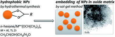
We present a general approach, which enables preparation of multifunctional nanocomposites of monodisperse nanoparticles embedded in oxide matrices. The two-step route has been successfully applied to nanocomposites composed of CoFe2O4 nanoparticles embedded in high-K oxide matrices (ZrO2, Al2O3 and TiO2). First, hydrophobic CoFe2O4 nanoparticles were produced by hydrothermal synthesis and then their incorporation in the oxide matrix was completed by the sol–gel method using the corresponding alcoxides. The as-prepared samples were subjected to annealing at temperatures ranging from 200 to 700 °C, and characterized in detail by the Powder X-Ray Diffraction (PXRD), Energy Dispersive Analysis (EDX), Mössbauer Spectroscopy (MS) and magnetic measurements. The particle size does not change with the annealing temperature, while the amorphous matrices crystallize at temperatures above 400 °C. At much higher annealing temperatures, partial decomposition of the CoFe2O4 occurs accompanied by formation of additional phases. The magnetic measurements also confirmed presence and stability of the uniform CoFe2O4 nanoparticles in the matrices. Thus the proposed method allows preparation of new types of nanocomposites constituted of uniform nanoparticles of the desired type (magnetic, luminescent etc.) embedded in the favored oxide matrix.


1. Introduction

Recently, nanocomposites of nanoparticles embedded in a functional matrix attracted considerable attention due to large application potential in various fields like biomedicine, catalysis, data storage and smart sensing.1–3 The functional matrix is chosen according to its required intrinsic physical, chemical and mechanical properties; however, it plays several important roles in the nanocomposites as well. It reduces contact of the embedded nanoparticles hence their interactions, enhances their stability due to minimized contact with the environment and avoids their agglomeration during thermal treatment leading to better control of their final size.

The most frequent material of the matrix used in nanocomposites is the amorphous silicon dioxide, which can be easily obtained by several variants of the sol–gel method.4,5 It is an excellent insulator, with low level of electronic defects, great transparency and possibility of surface functionalization by various functional groups. It can be easily etched and patterned down to a nanometer scale.6 By a simple modification of the sol–gel route using nitrates or chlorides as a source of the corresponding cations, nanoparticles of transition metal oxides can be grown in the porous silica matrix by controlled annealing.7 The particle size is driven by the annealing temperature due to the diffusion of the ions in the matrix, so the higher the annealing temperature, the larger particle size.

However, for many applications, a well-defined and uniform particle size is essential. Therefore, additional heat treatment is a limiting factor as the variation of the particle size is limited by the concentration of the cations, their diffusion coefficients and solubility in the silica matrix. Moreover, the in-matrix grown particles have a significant size distribution. At higher temperatures, the matrix transforms to crystalline form, which typically leads to formation of high-temperature phases. Another point is that the modified sol–gel method is limited to nanoparticles of simple oxides with low solubility of the cations in the matrix, which also prevents rapid formation of higher oxide phases even at much lower temperatures.

To avoid the above mentioned difficulties, we have introduced a novel two-step procedure, which can be applied to preparation of a large spectrum of nanocomposites, where the common sol–gel derived approach fails. In the first step, hydrophobic nanoparticles are prepared by a hydrothermal method. In the second step, the nanoparticles are embedded in an oxide matrix formed by hydrolysis of the corresponding alcoxides, followed by gelation and mild drying. This procedure can be applied to many types of nanoparticles such as magnetic (iron oxides, spinel ferrites), quantum dots, luminescent, photoactive (TiO2), up-conversion (Ln-doped NaYF4), SERS active (Au, Ag) etc. in combination with different matrices.

In our study, we have tested the proposed method on preparation of the nanocomposites of CoFe2O4 nanoparticles integrated in selected oxide matrices (TiO2, Al2O3 and ZrO2), diverse to the prevalent SiO2. The CoFe2O4 nanoparticles are well-known for their convenient magnetic properties (saturation magnetization 80 A m2 kg−1, coercivity 2 T at low temperatures), also their hydrothermal preparation is mastered.8–10 The ZrO2, TiO2, and Al2O3 oxides show many attractive properties, like high dielectric constant and optical transparency, and their preparation by the sol–gel method is rather routine.11

The TiO2 is a photoactive material, that is mainly used as an antibacterial agent, a component of self-cleaning surfaces or water and air purification technologies based on the photocatalysis.3,12 The CoFe2O4/TiO2 nanocomposites have been now intensively studied due to their advantage of magnetic separation from the liquid phase after the purification process.13,14

The Al2O3 exhibits promising catalytic properties15 and has been widely studied as a candidate for surface passivation of the solar cells.16 The Al2O3 matrix is mostly used as a passive spacer to avoid the particle agglomeration and to control the inter-particle interactions.17,18 ZrO2 is known as a diluted magnetic semiconductor (DMS).19,20 To our best knowledge there are no reports on the CoFe2O4/Al2O3 and CoFe2O4/ZrO2 nanocomposites yet. Therefore we also address the most appropriate conditions for preparation of the CoFe2O4/Al2O3 and CoFe2O4/ZrO2 nanocomposites by the general two-step route.

All samples were characterized in detail by the Powder X-Ray Diffraction (PXRD) and Transmission Electron Microscopy (TEM) in order to observe the phase composition, particle size and dispersion of the nanoparticles in the matrix. The Mössbauer Spectroscopy (MS) was performed for deeper studies of the iron-containing phases in the samples. Finally, the magnetic measurements were carried out to confirm the presence of the CoFe2O4 particles in the composites and to determine the CoFe2O4-to-oxide matrix ratio. All results were compared with those of the bare CoFe2O4 nanoparticles. We demonstrate that the uniform CoFe2O4 nanoparticles can be homogeneously embedded in the high-K oxide matrices without change of their diameter, size distribution and magnetic response.

2. Experimental details

2.1. Preparation

The two-step synthesis was used to prepare the nanocomposites of CoFe2O4 embedded in various oxide matrices. The hydrophobic CoFe2O4 nanoparticles were prepared by the hydrothermal synthesis10 and subsequently incorporated in the matrices, as is schematically shown in Fig. 1.
image file: c3ra44840a-f1.tif
Fig. 1 (Top panel) Scheme of the general two-step route including preparation of monodisperse nanoparticles by the hydrothermal method (step 1) and their incorporation in the oxide matrix by the sol–gel route (step 2). (Bottom panel) TEM image of the CoFe2O4 nanoparticles prepared by hydrothermal synthesis (on the left) together with the HR-TEM image of the powder form of the Co_free sample.
2.1.1. Preparation of CoFe2O4 nanoparticles. The 4.0 g of sodium hydroxide was dissoluted in mixture of water (20 ml) and ethanol (100 ml) and then 38.10 ml of oleic acid was added to the mixture (solution 1A). The 2.91 g of cobalt(II) nitrate hexahydrate and 8.08 g of iron(III) nitrate nonahydrate were dissolved in 180 ml of water (solution 1B). Solution 1A was stirred intensively at room temperature and added drop-wise to the solution 1B. The final mixture was stirred and sonicated at room temperature for 30 min (at 40 W transferred power). Afterwards, the mixture was heated at 180 °C for 10 h under autogeneous pressure in autoclave. The clear fraction was removed and the black-brown fraction was precipitated by addition of ethanol, decanted in magnetic field and dispersed in n-hexane using ultrasonic bath. This purification was repeated for four times. Concentration of the cobalt ferrite in the prepared ferrofluid was determined gravimetrically and by Thermogravimetry-Differential Thermal Analysis (TG-DTA). The reference (matrix-free) sample, labeled as Co_free was obtained by drying the ferrofluid at 100 °C.
2.1.2. Preparation of the nanocomposites – incorporation into matrices. The 0.22 mmol of cobalt ferrite nanoparticles in hexane was diluted with dry n-hexane to required amount (12.5 ml for Al2O3, 14.9 ml for TiO2 and 9.8 ml for ZrO2). The 3 mmol of aluminium isopropoxide, titanium n-butoxide and zirconium n-butoxide was added to the solution, respectively and stirred thoroughly. The mixtures were then stirred intensively and 2 ml solution of isopropylalcohol in water was added in ratio of 200[thin space (1/6-em)]:[thin space (1/6-em)]1 (v/v), 50[thin space (1/6-em)]:[thin space (1/6-em)]1 (v/v) and 20[thin space (1/6-em)]:[thin space (1/6-em)]1 (v/v) for the Al2O3, TiO2 and ZrO2 matrices, respectively. The final mixture was left to stay at 40 °C for 5 days. All gels were finally treated thermally in vacuum within two steps. First, the gel was heated to 200 °C (0.2 °C min−1), keeping the gel at 200 °C for 2 hours, and finally annealed at 300, 400, 500, 600, or 700 °C with the step of 1 °C min−1 keeping the product at the final temperature for 2 hours. The samples were labeled accordingly to its matrix and the annealing temperature, respectively, as M_T, where M = Al, Ti, Zr and T = 200–700 °C.

2.2. Characterization methods

All samples were characterized using the PXRD at room temperature by the Bragg Brentano geometry on the Philips X'Pert PRO MPD X-ray diffraction system equipped with the X'Celerator detector using Cu-anode (CuKα; λ = 1.5418 Å) or Co-anode (CoKα; λ = 1.7889 Å). The phase composition, particle size (using the isotropic approximation) and lattice parameters of the selected samples were obtained by Rietveld refinement implemented in the FullProf software.21

Concentration of the nanoparticles in the ferrofluid was determined gravimetrically by Thermogravimetry-Differential Thermal Analysis (TG-DTA) using a TG-DTA device by SETARAM. The TEM and High-Resolution TEM (HR-TEM) using HRTEM JEOL JEM 3010 was performed in order to study the morphology of the prepared nanocomposites and determination of the particle size of the CoFe2O4 nanoparticles. The samples were also studied by the Energy Dispersive X-ray Spectroscopy (EDX) analysis using scanning electron microscope MIRA 3 LMH by Tescan with the accelerating potential of 15 kV.

The MS measurement was done in the transmission mode with 57Co diffused into Rh matrix as the source moving with constant acceleration. The spectrometer (Wissel, Germany) was calibrated by standard α-Fe foil and the isomer shift is related to this standard at 293 K. The resulting parameters were determined in the NORMOS program.

The magnetic measurements were carried out using the SQUID magnetometer (MPMS7XL, Quantum Design). The zero field-cooled (ZFC) and field-cooled (FC) curves were recorded as follows: at first, the sample was cooled down to 10 K, then the magnetic field of 0.01 T was applied and the temperature dependence of the magnetization was measured up to 400 K (ZFC curve). Afterwards, the sample was cooled down to 10 K in the applied field and the FC curve was obtained. The magnetization isotherms (field dependence of the magnetization) were measured in the range of ±7 T, at selected temperatures.

3. Results and discussion

3.1. Phase analysis

The PXRD was carried out to obtain the phase composition of the samples and to determine the temperature of crystallization of the matrix. The results are summarized in Table 1, together with the phase composition determined by MS. Generally, the increasing annealing temperature caused decomposition of the CoFe2O4 nanoparticles leading to the creation of parasitic phases and crystallization of the matrix above 500 °C. The presence of the amorphous matrix at lower annealing temperature (200–400 °C) is manifested by the broad maxima at diffraction angle about 22° and increased intensity of the background. The particle diameter, dXRD determined as the size of the coherently diffracting domain of the CoFe2O4 nanoparticle is in the range of 5.5–7.0 nm and does not change significantly with the increasing annealing temperature. The overlapped reflections of the crystalline matrices and CoFe2O4 phase disallowed the precise determination of the particle size of the CoFe2O4 nanoparticles in multiphase samples.
Table 1 The phase composition of all samples determined by PXRD and MS. The matrix is either amorphous or crystalline, where (C) and (M) denote cubic and monoclinic structure, respectively. The content of individual crystalline phases in the samples is described as follows: * 5–30%, ** 30–60%, *** >60%
Sample Matrix PXRD Phases PXRD Phases MS
Al_200 Amorphous CoFe2O4*** CoFe2O4***
Al_300 Amorphous CoFe2O4*** CoFe2O4***
Al_400 Amorphous CoFe2O4***, AlO(OH)* CoFe2O4***
Al_500 Amorphous CoFe2O4***, CoAl2O4* CoFe2O4***
Al_600 Amorphous CoFe2O4**, CoAl2O4** CoFe2O4***
Al_700 Amorphous CoFe2O4*, CoAl2O4*, FeAl2O4** CoFe2O4***, FeAl2O4*
Ti_200 Amorphous CoFe2O4*** CoFe2O4***
Ti_300 Amorphous CoFe2O4*** CoFe2O4***, Fe2TiO4*
Ti_400 Amorphous CoFe2O4*** CoFe2O4/Fe3+*, Fe2TiO4***
Ti_500 TiO2 anatase***, TiO2 rutile* CoFe2O4*, FeTiO3* CoFe2O4/Fe3+*, Fe2TiO4***, FeTiO3*
Ti_600 TiO2 anatase**, TiO2 rutile* CoFe2O4*, FeTiO3* CoFe2O4/Fe3+**, Fe2TiO4**, FeTiO3*
Zr_200 Part. ZrO2 (C)** CoFe2O4** CoFe2O4***
Zr_300 Part. ZrO2 (C)** CoFe2O4** CoFe2O4***
Zr_400 Part. ZrO2 (C)** CoFe2O4*
Zr_500 ZrO2 (C)** CoFe2O4** Fe3+ ions***
Zr_600 ZrO2 (C)** CoFe2O4** Fe3+ ions***
Zr_700 ZrO2 (C)**, ZrO2 (M)* CoFe2O4**
Co_free N/A CoFe2O4*** CoFe2O4***


The EDX reveals the CoFe2O4/matrix weight ratio for the samples containing only CoFe2O4 phase and matrix. The values are summarized in Table 2 together with the theoretical ratio and the ratio calculated from the values of the saturation magnetization, Ms as is discussed further.

Table 2 Comparison of the weight content of CoFe2O4 in the matrix determined by EDX analysis, WEDX and magnetic measurements at 10 and 300 K, WM10, WM300 respectively with the theoretical one, WT
Sample WT (wt%) WEDX (wt%) WM10 (wt%) WM300 (wt%)
Al_200 25.0 30.4 20.7 20.8
Al_600 25.0 7.1
Ti_200 17.0 15.5 15.5 14.6
Ti_600 17.0 5.2
Zr_200 12.0 13.7 13.8 12.5
Zr_600 12.0 1.5


The TEM images were captured to obtain the morphology of the nanocomposites. The particle diameter determined by TEM, dTEM is in good agreement with the dXRD for all evaluated samples. The representative TEM images are shown in Fig. 4, 6 and 8, respectively. The MS was performed to confirm the presence of CoFe2O4 and exclude presence of the other iron phases, for the samples for that the PXRD measurements were not sufficient for the analysis due to the overlapped reflections. MS can clearly distinguish between the Fe2+ and Fe3+ ions due to the total different isomer shift as will be presented further, thus clearly indicates presence of other phases than CoFe2O4.

The crystal structure determined by PXRD and MS of individual nanocomposites will be now discussed in more details followed by the results of magnetic measurements.

3.1.1. The matrix-free sample, Co_free. The diffraction pattern displays only the reflections corresponding to the spinel structure with the lattice parameter a = 8.40 Å, that is in good agreement with the CoFe2O4 phase (PDF4 databasis, card no. 00-022-1086). The particle size determined using the Rietveld refinement is 5.5 ± 0.5 nm. The TEM image (Fig. 1) shows well-crystalline particles with dTEM ≈ 6 nm.

The MS spectra reveals that the most of the particles are in the blocked state, as is observed by the presence of sextet with asymmetric absorption peaks corresponding to the small particle size with non-zero size distribution. Therefore, the convolution of the Gaussian distribution function and the Lorentzian profile function was used to refine the spectra. The smaller values of the hyperfine fields (46.6 T for the Oh-sites; 41.3 T for the Td-sites) are consistent with the small particle size, dXRD = 5.5 nm. The isomer shift, δ ≈ 0.33 mm s−1 and zero quadrupole shift are attributed to the ferrite spinel structure. The doublet and the singlet correspond to the particles in superparamagnetic (SPM) regime and close to the blocking temperature, respectively. The PXRD patterns together with the MS spectra and magnetic measurements are depicted in Fig. 2.


image file: c3ra44840a-f2.tif
Fig. 2 (Top panel) The PXRD pattern of the Co_free sample with the refined pattern (full black line) on the left together with the MS spectra performed at room temperature on the right. The open circles mark the Bragg reflections of the CoFe2O4 phase. (Bottom panel) The ZFC–FC curves at 0.01 T and the magnetization isotherms measured at selected temperatures.
3.1.2. CoFe2O4/TiO2 nanocomposites. The diffraction patterns of the Ti_200, Ti_300 and Ti_400 samples depicted in Fig. 3 exhibit only reflections corresponding to the spinel structure with the lattice parameters, a = 8.38 Å, that is in good agreement with the reference sample, Co_free. The lower intensities of the CoFe2O4 reflections with the increasing annealing temperature are observed, suggesting the decomposition of the CoFe2O4 to additional phases. This observation has been further confirmed by the MS, where the doublet with high δ = 0.98 mm s−1 corresponding to the Fe2+ ions is detected in all samples except Ti_200 (Fig. 3). The high quadrupole splitting (ΔEq = 2.15 mm s−1) can be ascribed to the Fe2TiO4 phase that crystallizes in the spinel structure,22 therefore can be hardly resolved in PXRD due to the overlapped reflections with the CoFe2O4 phase.
image file: c3ra44840a-f3.tif
Fig. 3 The PXRD patterns of the CoFe2O4/TiO2 series on the left. The MS spectra of selected samples performed at room temperature on the right.

The crystalline matrix is detected for the samples annealed at 500 °C and higher temperatures, predominantly in anatase form. The asymmetry of the anatase reflections indicates presence of the rutile with tendency to increase the amount of the rutile at the expense of the anatase upon further annealing of the sample at 600 °C, which is connected with the increase of the size of the crystallites of the matrix.23,24 The reflection located at 38° can be attributed to the FeTiO3 phase, as is consistent with the results obtained by MS, where another doublet appears for the Ti_500 and Ti_600 samples with δ = 1.1 mm s−1, ΔEq = 0.58 mm s−1 corresponding to the FeTiO3 phase.25

The TEM image depicted in Fig. 4 of the Ti_200 sample shows crystalline nanoparticles (see HR-TEM image) with low size distribution dispersed in amorphous matrix. The particle size, dTEM = 6 nm is in good agreement with the dXRD.


image file: c3ra44840a-f4.tif
Fig. 4 (Top panel) The TEM image of the Ti_200 sample on the left with the HR-TEM image on the right. (Bottom panel) The ZFC–FC curves at 0.01 T for the Ti_200 sample and the Ti_600 sample in the inset on the left. The magnetization isotherms at 10 K for both samples on the right.
3.1.3. CoFe2O4/Al2O3 nanocomposites. The diffraction patterns together with the MS spectra are depicted in Fig. 5. Clearly resolved peaks corresponding to the spinel structure are observed at low temperatures (200, 300 °C) with the lattice parameters around a = 8.38 Å, which is in good agreement with the reference sample. The matrix remains amorphous through the whole series, as is consistent with A. Corrias et al.26 who observed the crystallization of the Al2O3 matrix at temperatures higher than 700 °C. Annealing of the nanocomposite at 400 °C led to the occurrence of pseudoboehmite phase AlO(OH) manifested by broad reflections at 28°, 49° and 54°.27 The presence of AlO(OH) is supported by the TEM observation (see Fig. 6), where the needle-shape aggregates typical for this phase is observed for the Al_400 sample.26
image file: c3ra44840a-f5.tif
Fig. 5 The diffraction patterns of the CoFe2O4/Al2O3 series on the left. The MS spectra of the selected samples performed at room temperature on the right.

image file: c3ra44840a-f6.tif
Fig. 6 (Top panel) The TEM images of the Al_200 and Al_400 samples, respectively. (Bottom panel) The ZFC–FC curves at 0.01 T for the Al_200 sample and the Al_600 sample in the inset on the left. Magnetization isotherms at 10 K for both samples on the right.

Increase of the annealing temperature to 500 °C has resulted into the asymmetry of the peak profiles, that can be elucidated by the presence of the parasitic CoAl2O4 and FeAl2O4 phases, that also crystallize in spinel structure with slightly different lattice parameter (a = 8.104 Å and 8.156 Å respectively, PDF4 databasis, cards no. 00-044-0160; 01-086-2320). The shift of the peaks to the higher angles for the Al_700 sample is clearly observable and points to the dominant presence of these spinel structures. The CoAl2O4 and FeAl2O4 cannot be unambiguously resolved by the PXRD, due to the nearly similar lattice parameters. However, the presence of FeAl2O4 has been detected by MS (δ = 0.99 mm s−1, ΔEq = 1.7 mm s−1).28 For all samples except the Al_700 sample, only the Fe3+ ions (δ = 0.35 mm s−1) corresponding to the CoFe2O4 phase have been detected by the MS.

3.1.4. CoFe2O4/ZrO2 nanocomposites. The samples annealed at lower temperatures (200–400 °C) exhibit diffraction peaks matching with the reflections of the spinel structure with lattice parameter a = 8.39 Å, that corresponds to the CoFe2O4 phase. The broad peak around 30° can be attributed to the partially crystalline cubic ZrO2 matrix (see Fig. 7). This peak gets narrower with the increasing temperatures, suggesting the continuous crystallization of the matrix. The MS exhibit similar profile as for the matrix-free sample confirming the presence of the CoFe2O4 nanoparticles (see Fig. 7). The crystalline particle with size around 5.5 nm can be seen in HR-TEM image in Fig. 8.
image file: c3ra44840a-f7.tif
Fig. 7 The diffraction patterns of the CoFe2O4/ZrO2 series on the left. The MS spectra of the selected samples performed at room temperature on the right.

image file: c3ra44840a-f8.tif
Fig. 8 (Top panel) The HR-TEM images of the Zr_200 and Zr_600 samples. (Bottom panel) The ZFC–FC curves at 0.01 T for the Zr_200 sample and the Zr_600 sample in the inset on the left. The magnetization isotherms at 10 K for both samples on the right.

The rapid change of the diffraction pattern is observed at 500 °C, where the matrix is well crystalline, predominantly in the cubic form, partially transferring to the monoclinic form at higher temperature (700 °C). The crystalline matrix is also observed by HR-TEM (Fig. 8) for the Zr_600 sample. The CoFe2O4 phase cannot be properly distinguished by the PXRD due to the overlapped reflections with the ZrO2 phases. The MS spectra contain only the doublet with δ = 0.36 mm s−1 corresponding to the Fe3+ ions and ΔEq = 1.07 mm s−1 which is higher than for the CoFe2O4 phase (usually ΔEq = 0.76 mm s−1 (ref. 29)). The presence of the CoFe2O4 in the Zr_600 sample has been elided by magnetic measurements, as is shown further. Therefore, the observed doublet in MS can be interpreted as the Fe3+ ions dispersed in crystalline matrix.

3.2. Magnetic measurements

3.2.1. General remarks. The magnetic measurements were carried out in order to unambiguously confirm presence of the CoFe2O4 in form of nanosized particles, through the typical high coercivity at low temperatures. The two selected samples from each series were measured, one with the amorphous matrix annealed at 200 °C, one with the crystalline matrix annealed at 600 °C. The ZFC–FC measurements were performed and character of the curves has been compared with the matrix-free sample. The necking of the magnetization isotherms that affected the determination of precise value of the coercivity, H10c is caused by the insufficient fixation of the sample. Therefore, estimation of the H10c was done by approximating the necked part by linear term. The values of saturation magnetization determined after subtraction of the paramagnetic contributions were used to determine the weight content of the CoFe2O4 particles in the matrices (Table 2). The resulted characteristic parameters are summarized in Table 3. The ZFC–FC curve of the Co_free sample shows the blocking temperature, TB, around 270 K with saturation of the FC curve pointing to strong interparticle interactions.30 The magnetization isotherms exhibit large H10c around 1 T typical for the CoFe2O4 phase (see Fig. 2 and Table 3).
Table 3 Magnetic parameters: blocking temperature, TB; saturation magnetization at 10 and 300 K, M10s, M300s and coercivity at 10 K, H10c
Sample TB (K) M10s (A m2 kg−1) M300s (A m2 kg−1) H10c (mT)
Al_200 ≈260 12.0 10.0 690
Al_600 ≈295 7.0 3.4 620
Ti_200 ≈265 9.0 7.0 720
Ti_600 >400 4.0 2.5 100
Zr_200 ≈295 8.0 6.0 740
Zr_600 >400 1.5 0.7 50
Co_free ≈270 58.0 48.0 1000


3.2.2. Nanocomposites. All three samples annealed at 200 °C (Ti_200, Al_200 and Zr_200) exhibit similar ZFC–FC curves as the matrix-free sample (see Fig. 4, 6 and 8) with the nearly similar TB pointing at the presence of the CoFe2O4 nanoparticles, that has been further confirmed by high coercivity at 10 K (Table 3). No coercivity is observed at 300 K, H300c suggesting the particles are in superparamagnetic regime. The weight contents of the CoFe2O4 in matrix calculated from Ms are in good agreement with the theoretical ones.

The ZFC–FC curves of the Zr_600 and Ti_600 samples do not match with the Co_free sample at all. The non-saturation of the FC curve of the Ti_600 sample indicates the weakening of the interparticle interactions, that can be explained by decomposition of the CoFe2O4 particles into smaller fragments (ions, clusters) in the matrix or by increase of the distances between the particles in the matrix or due to the phase transformation of the part of the CoFe2O4 particles. The mostly linear FC curve of the Zr_600 sample points at the strong interactions within the particles suggesting the formation of aggregates. The absence of the CoFe2O4 phase has been further confirmed by very low H10c and Ms.

On the other hand, the presence of CoFe2O4 has been unambiguously confirmed in the Al_600 sample by the high H10c and similar course of the ZFC–FC curve as for the Co_free sample. The unsaturation of the low temperature part of the FC curve points at the weakening of interparticle interactions that can be proved by the presence of weak magnetic phases CoAl2O4 and FeAl2O4, detected by PXRD (see previous discussion) and also indicated by the decrease of the Ms.

4. Conclusions

We have developed a general approach, which enables preparation of nanocomposites of monodisperse nanoparticles in amorphous or crystalline oxide matrix. The proposed method was successfully applied to preparation of nanocomposites of monodisperse CoFe2O4 nanoparticles embedded in high-K oxide matrices – TiO2, Al2O3 and ZrO2, respectively. In the first step, the uniform nanoparticles were prepared by the hydrothermal synthesis and subsequently, they were incorporated into the matrices using the sol–gel method. The detailed characterization by Powder X-Ray Diffraction, electron microscopy, Mössbauer Spectroscopy and magnetic measurements confirmed presence of the CoFe2O4 nanoparticles without change of the particle size with the increasing annealing temperature up to the onset of crystallization of the matrices (above 400 °C). The preparation of a nanocomposite with a crystalline matrix is also possible using suitable annealing conditions. The results clearly demonstrated that nanocomposites with well-defined particles embedded in various oxide matrices can be easily obtained by the two-step preparation method. The universal principle of the route – embedding the hydrophobic nanoparticles in the matrix using the corresponding alcoxides – opens possibilities to fabricate variety of multicomponent systems, which cannot be obtained by the common methods, mainly due to absence of soluble salts, interaction of the components during synthesis or requirement of a narrow particle size distribution.

Acknowledgements

The authors thank for financial support of the Czech Science Foundation, the project no. P108/10/1250 and by long-term research plan of the Ministry of Education of the Czech Republic no. MSM0021620857. Magnetic measurements were performed in MLTL (http://mltl.eu/), which is supported within the program of Czech Research Infrastructures (project no. LM2011025). Work of S. Kubickova is also supported by SVV-2013-267307.

References

  1. S. Komarneni, J. Mater. Chem., 1992, 2(12), 1219–1230 RSC.
  2. P. Granitzer and K. Rumpf, Materials, 2011, 4, 908–928 CrossRef.
  3. A. Fujishima and K. Honda, Nature, 1972, 238, 37–38 CrossRef CAS.
  4. H. Dislich, J. Non-Cryst. Solids, 1986, 80, 115–121 CrossRef CAS.
  5. K. A. Vorotilov, E. V. Orlova and V. I. Petrovsky, Thin Solid Films, 1992, 209, 188–194 CrossRef CAS.
  6. J. Robertson, Eur. Phys. J.: Appl. Phys., 2004, 28, 265–291 CrossRef CAS.
  7. D. Niznansky, N. Viart and J.-L. Rehspringer, J. Sol-Gel Sci. Technol., 1997, 8, 615–618 CAS.
  8. G. A. Sawatzky, F. V. der Woude and A. H. Morrish, Phys. Rev., 1969, 187, 747–757 CrossRef CAS.
  9. A. Hutlova, D. Niznansky, J.-L. Rehspringer, C. Estournes and M. Kurmoo, Adv. Mater., 2003, 15, 1622–1625 CrossRef CAS.
  10. A. Repko, D. Niznansky and J. P. Vejpravova, J. Nanopart. Res., 2011, 13, 5021–5031 CrossRef CAS.
  11. H. Shimizu and T. Nishide, Characterization of Sol-Gel-Derived and Crystallized HfO2, ZrO2, ZrO2–Y2O3 Thin Films on Si(001) Wafers with High Dielectric Constant, 13. chapter in Advances in Crystallization Processes, ed Dr Yitzhak Mastai, In-Tech, 2012 Search PubMed.
  12. A. Zaleska, Recent Pat. Eng., 2008, 2, 157–164 CrossRef CAS.
  13. C. Leng, J. Wei, Z. Liu, R. Xiong, C. Pan and J. Shi, J. Nanopart. Res., 2013, 15, 1643(1)–1643(11) CrossRef.
  14. H. Li, Y. Zhang, S. Wang, Q. Wu and C. Liu, J. Hazard. Mater., 2009, 169, 1045–1053 CrossRef CAS PubMed.
  15. M. A. Karakassides, D. Gournis, A. B. Bourlinos, P. N. Trikalitis and T. Bakas, J. Mater. Chem., 2003, 13, 871–876 RSC.
  16. Y. Zhao, C. Zhou, X. Zhang, P. Zhang, Y. Dou, W. Wang, X. Cao, B. Wang, Y. Tang and S. Zhou, Nanoscale Res. Lett., 2013, 8, 114(1)–114(7) Search PubMed.
  17. O. Petracic, X. Chen, S. Bedanta, W. Kleemann, S. Sahoo, S. Cardoso and P. P. Freitas, J. Magn. Magn. Mater., 2006, 300, 192–197 CrossRef CAS PubMed.
  18. J.-Z. Kong, Y.-P. Gong, X.-F. Li, A.-D. Li, J.-L. Zhang, Q.-Y. Yan and D. Wu, J. Mater. Chem., 2011, 21, 5046–5050 RSC.
  19. S. Ostanin, A. Ernst, L. M. Sandratskii, P. Bruno, M. Dane, I. D. Hughes, J. B. Staunton, W. Hergert, I. Mertig and J. Kudrnovsky, Phys. Rev. Lett., 2007, 98, 016101(1)–016101(4) CrossRef.
  20. N. H. Hong, C.-K. Park, A. T. Raghavender, O. Ciftja, N. S. Bingham, M. H. Phan and H. Srikanth, J. Appl. Phys., 2012, 111, 07C302(1)–07C302(3) Search PubMed.
  21. J. Rodriguez-Carvajal, An introduction to the program FullProf, 2000 Search PubMed.
  22. K. Ono, L. Chandler and A. Ito, J. Phys. Soc. Jpn., 1968, 25, 174–176 CrossRef CAS.
  23. Q. Zhang, L. Gao and J. Guo, Appl. Catal., B, 2000, 26, 207–215 CrossRef CAS.
  24. V. Tyrpekl, J. P. Vejpravova, A. G. Roca, N. Murafa, L. Szatmary and D. Niznansky, Appl. Surf. Sci., 2011, 257, 4844–4848 CrossRef CAS PubMed.
  25. S. L. Ruby and G. Shirane, Phys. Rev., 1961, 123, 1239–1240 CrossRef CAS.
  26. A. Corrias, M. F. Casula, A. Falqui and G. Paschina, Chem. Mater., 2004, 16, 3130–3138 CrossRef CAS.
  27. K. I. Shefer, S. V. Cherepanova, E. M. Moroz, E. Y. Gerasimov and S. V. Tsybulya, J. Struct. Chem., 2010, 51, 132–141 CrossRef CAS PubMed.
  28. C. M. Yagnik and H. B. Mathur, J. Phys. C: Solid State Phys., 1968, 1, 469–472 Search PubMed.
  29. S. R. Ahmed, S. B. Ogale, G. C. Papaefthymiou, R. Ramesh and P. Kofinas, Appl. Phys. Lett., 2002, 80, 1616–1618 CrossRef CAS PubMed.
  30. B. Bittova, J. P. Vejpravova, M. P. Morales, A. G. Roca and A. Mantlikova, J. Magn. Magn. Mater., 2012, 324, 1182–1188 CrossRef CAS PubMed.

This journal is © The Royal Society of Chemistry 2014
Click here to see how this site uses Cookies. View our privacy policy here.