Investigation of antimicrobial PEG-poly(amino acid)s

Frankie Costanza a, Shruti Padhee a, Haifan Wu a, Yan Wang b, Jesse Revenis a, Chuanhai Cao c, Qi Li *b and Jianfeng Cai *a
aDepartment of Chemistry, University of South Florida, 4202 E. Fowler Ave, Tampa, FL 33620, USA. E-mail: jianfengcai@usf.edu
bDepartment of Medical Oncology, Shuguang Hospital, Shanghai University of Traditional Chinese Medicine, Shanghai, 201203, China. E-mail: lzwf@hotmail.com
cCollege of Pharmacy, University of South Florida, 4202 E. Fowler Ave, Tampa, FL 33620, USA

Received 14th August 2013 , Accepted 4th November 2013

First published on 6th November 2013


Abstract

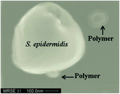
There has been significant interest in the development of antimicrobial cationic polymers due to their low manufacture cost and ease of synthesis compared to small antimicrobial peptides (AMPs). These polymers are designed to mimic amphiphilic structures of AMPs which can disrupt negatively charged bacterial membranes, and can therefore lead to potential antibiotic agents to fight emerging drug resistance. However, the reports of biodegradable antimicrobial polymer nanoparticles are rare. Herein we report the development of antimicrobial PEG-poly(amino acid)s. Some of these multi-block PEG-poly(amino acid)s form defined nanoparticles in solution, and display potent and broad-spectrum antimicrobial activity. Fluorescence and SEM studies show that these polymers are likely to kill bacteria by disrupting bacterial membranes. As these polymers are biodegradable and easy to scale up, they may provide an attractive approach for the development of antibiotic agents.


Introduction

The extensive use of antibiotics in recent decades for medical and agricultural purposes has elicited the widely known issue of bacterial resistance.1 As such, the development of new antimicrobial agents with novel mechanisms has been a constant area of research. This includes the need for new antibacterial materials to prevent contamination in food packaging, storage, medical supplies, and drinking water,2–4 in addition to drug discovery. One approach to circumvent resistance could be the development of antimicrobial peptides (AMPs).1,5,6 Cationic AMPs exist in most organisms and are the first line of natural defense against infections. Although they possess different secondary structures or sometimes even random structures in solution, they are believed to adopt an amphipathic conformation when interacting with negatively charged bacterial membranes, and subsequently disrupt the function of bacterial membranes.5 As this biophysical interaction lacks defined bacterial targets, this mechanism of action is not expected to develop resistance as easily as traditional antibiotics. Based on such recognition, many classes of AMP mimetics have been developed.7–14 These small peptidomimetics are capable of mimicking the function and mechanism of AMPs, and are shown to have potent and broad spectrum antimicrobial activity. However, their step-by-step preparation is tedious, and they are generally not economical to produce in substantial quantities, limiting the potential of widespread use as affordable biomaterials in the future.

Recently, there have been some advances in polymer antimicrobial agents such as polymers based on methacrylates,15,16 norborenes,17,18 amidoamines,19 and others.3,20–23 These polymers contain both hydrophobic and cationic components, which are critical for the disruption of bacterial membranes. Many of them have also been shown to kill both Gram-positive and Gram-negative bacteria, including clinically relevant pathogens. In addition, they are generally prepared through a one-pot polymerization, allowing the generation of products in large amounts. As such, there is promising potential for these synthetic materials to be used as antimicrobial agents to treat bacterial infections. However, the development of biodegradable antimicrobial nanoparticles is very rare. Poly(amino acid)s,23 synthesized by ring-opening polymerization, show attractive features for further exploration. They have been widely used for drug delivery and other biomedical applications.24–27 However, their antimicrobial development is rare.23 In this study, we designed, synthesized and studied the antimicrobial activity of biodegradable polymer nanoparticles – PEGylated amphiphilic poly(amino acids). The PEG is introduced to the polymers because PEG (polyethylene glycol) is essentially non-toxic and biocompatible.28–31 Meanwhile, PEGylation also promotes the formation of nanoparticles. Additionally, PEG is widely available, inexpensive, highly water soluble and easily modifiable by size and end group functionality. It has also been used in the development of nanoparticles with a hydrophilic corona which would enhance the stability of the micelle and increase circulation time.32,33 Another attractive aspect of using PEG in a block type antimicrobial nanoparticle is that it has non-adhesive antifouling properties which can prevent protein and cell adhesion.30 Another report shows that PEG does not reduce activity to bacteria, and may potentially decrease toxicity to human cells.19 As such, we also expected that introduction of a PEG segment to poly(amino acid)s will promote the formation of nanoparticles, which may facilitate bacterial membrane disruption, and therefore possess improved antimicrobial activity.

Experimental section

Materials and methods

Synthesis of polymer P3. (The other polymers were prepared by a similar method). 0.4 g of PEG–NH2 was dissolved in 10 mL of anhydrous dioxane and purged with N2. Separately 0.49 g (20 eq.) of Z-lysine NCA was dissolved in 5 mL anhydrous dioxane, passed through a 2 micron filter to remove any decomposed NCA, thoroughly purged with N2 and then added via syringe. The reaction was then allowed to proceed for 3 days under an active N2 atmosphere. Then 10 equivalents of Z-Lysine NCA and 15 equivalents of Phenylalanine NCA were dissolved in 5 mL anhydrous dioxane, passed through a 2 micron filter, purged, and added via syringe. Polymerization was allowed to continue for another 3 days. The clear solution was then precipitated into ether and the product was collected via filtration.
Antimicrobial activity. The bacterial strains used for testing the efficacy of polymers were multi-drug resistant S. epidermidis (RP62A), B. subtilis (BR151), K. pnuemoniae (ATCC 13383) and multi-drug resistant P. aeruginosa (ATCC 27853). The antimicrobial activities of the polymers developed were determined in sterile 96-well plates by the 2-fold serial-dilution method. Bacterial cells were grown overnight at 37 °C in 5 mL medium after which a bacterial suspension of approximately 106 CFU mL−1 in Luria broth or trypticase soy was prepared ensuring that the bacterial cells were in the mid-logarithmic phase. Aliquots of 50 μL bacterial suspension were added to 50 μL of medium containing different concentrations of polymers for a total volume of 100 μL in each well. The 96-well plates were incubated at 37 °C for about 20 h. The Biotek microplate reader was used to measure the optical density (OD) at a wavelength of 600 nm after about 20 h. The experiments were carried out as three independent biological replicates, each in duplicate. The lowest concentration at which complete inhibition of bacterial growth is observed is defined as the minimum inhibitory concentration (MIC).
Hemolytic activity. Freshly drawn human red blood cells (hRBC's) were used for the assay. The blood sample was washed with PBS buffer several times and centrifuged at 700 g for 10 min until a clear supernatant was observed. The hRBC's were re-suspended in 1× PBS to get a 5% v/v suspension which was used to perform the assay. 50 μL of different polymers solutions were added to sterile 96-well plates. Then 50 μL of 5% v/v hRBC solution was added to make up a total volume of 100 μL in each well. The 0% hemolysis point and 100% hemolysis point were determined in 1× PBS and 0.2% Triton-X-100 respectively. The 96 well plate was incubated at 37 °C for 1 h and centrifuged at 3500 rpm for 10 min. The supernatant (30 μL) was then diluted with 100 μL of 1×PBS and hemoglobin was detected by measuring the optical density at 360 nm by Biotek microtiter plate reader (Type: Synergy HT).
%Hemolysis = (Abssample − AbsPBS)/(AbsTriton − AbsPBS) × 100
Fluorescence microscopy. A double staining method with DAPI (4′,6-diamidino-2-phenylindole dihydrochloride, Sigma, >98%) and PI (propidium iodide, Sigma) as fluorophores was used to visualize and differentiate the viable from the dead B. subtilis cells. DAPI being a double stranded DNA binding dye, stains all bacterial cells irrespective of their viability. Ethidium derivatives such as propidium iodide (PI) is capable of passing through only damaged cell membranes and intercalates with the nucleic acids of injured and dead cells to form a bright red fluorescent complex. The bacterial cells were first stained with PI and then with DAPI. The bacterial cells were grown until they reached mid-logarithmic phase and then ∼2 × 10³ cells were incubated with 100 μg mL−1 polymer P5 for 4 h. Then the cells were pelleted by centrifugation at 3000 g for 15 min in an Eppendorf microcentrifuge. The supernatant was decanted and the cells were washed with 1× PBS several times and then incubated with PI (5 μg mL−1) in the dark for 15 min at 0 °C.The excessive PI was removed by washing the cells several times with 1× PBS several times. Lastly, the cells were incubated with DAPI (10 μg mL−1 in water) for 15 min in the dark at 0 °C. Then finally the excessive DAPI solution was removed by washing it with 1× PBS. The controls were performed following the exact same procedure for bacteria without P5. The bacteria were then examined by using the Zeiss Axio Imager Z1optical microscope with an oil-immersion objective (100×).7,8
SEM of bacteria after treatment of the polymer. MRSE and B. subtilis were grown to an exponential phase and approximately about 2 × 10⁶ cells were incubated with the polymer for about 20 h. The cells were then harvested by centrifugation (3000 g) for 15 min. After pelleting the cells, the cells were washed three times with DI water. The cells were then fixed with 2.5% (w/v) glutaraldeyhyde in nanopure water for about 30 min, followed by extensive wash with DI water to get rid of any excess glutaralhedyde. Graded ethanol series (30%, 50%, 70%, 95% and 100%, 5 min each) were then used to dehydrate the cells. Following the dehydration of cells hexamethyldisilazane was added for about 2 min. Then about 2 μL of sample was added to a silicon wafer followed by Gold/Pd coating, and then the samples were observed at 25 KV with a HITACHI S-800 scanning electron microscope.

Results and discussions

To test our hypothesis, we synthesized a series of PEGylated poly(amino acid)s containing both hydrophobic groups and cationic groups (Fig. 1) via ring-opening polymerization of N-carboxyanhydrides (NCAs).34 NCAs can easily be synthesized and attained in large quantities from natural amino acids at low cost, which makes them attractive in designing new materials.35 The synthetic procedure was adapted from a previously published protocol.35 Briefly, Phe-NCA and Z-Lys-NCA (Fig. 1) were prepared through the treatment of amino acids phenylalanine or Z-Lysine with triphosgene, respectively. Next, CH3O–PEG–NH2 was used as the initiator to react with NCAs to generate poly(amino acid)s via the ring-opening polymerization. In the synthesis of two polymers (P2 and P3), to ensure the formation of cationic shell of nanoparticles, Z-Lysine-NCA was first added into the flask containing the initiator and reacted for three days at the room temperature. Subsequently, Z-Lysine-NCA and Phe-NCA were then added in one batch to form a hybrid segment, which forms the core structure of nanoparticles in solution. Such a random core structure was expected to facilitate the interaction with the bacterial membranes through both charge and hydrophobic forces.23 As such, a series of polymers containing varying numbers of hydrophobic and hydrophilic groups were prepared (Table 1). To test if the charge is critical to polymers' antimicrobial activity, one negatively charged polymer was synthesized using both Glu-NCA and Phe-NCA.
image file: c3ra44324h-f1.tif
Fig. 1 Synthesis of PEG-poly(amino acid)s.
Table 1 The antimicrobial and hemolytic activities of PEG-poly(amino acid)s. The microbial organisms used are K. pneumoniae (ATCC 13383), and P. aeruginosa (ATCC 27853), methicillin-resistant S. epidermidis (RP62A), B. subtilis (BR151). The minimum inhibitory concentration (MIC) is the lowest concentration that completely inhibits growth after 24 h. HC50 is the concentration causing 50% hemolysis. The antimicrobial peptide Magainin II was included as a control

image file: c3ra44324h-u1.tif

# MRSE (μM) B. subtilis (μM) K. Pneum-oniae (μM) P. aerug-inosa (μM) mPEG (mw) Lysine content b [(c Phenyl-alanine content d)] % Hydrophobic content Hemolytic activity HC50(μM) MW (Theoretical) Average size of nanoparticles (nm)
P1 >100 >100 >100 >100 5000 Glu 20 (10 15) 33 10[thin space (1/6-em)]880 70–80
P2 55 2.8–5.5 27.5–55.3 >100 5000 10 (10 10) 33 9 9030 80–90
P3 9–22.6 4.5–9 45.3 >100 5000 20 (10 15) 33 25 11[thin space (1/6-em)]045 40–50
P4 6.5–13 1.6–3.2 65 >100 5000 (10 10) 50 10 7750 60–80
P5 5.3–10.5 5.3–10.5 26.3–52.6 21–52.6 2000 (10 10) 50 6 4750 200–250
P6 9.1–18.2 18.2–45.5 45.5–91.6 45.5–91.6 2000 (10 15) 60 5 5485 80–100
P7 6.7–13.3 3.4–6.7 >100 33.3–83.6 2000 (20 20) 50 3 7500 80–100
P8 1.4–2.8 5.7–11.4 >100 28–56 2000 (30 20) 40 3 8780 100–110
P9 5.5–11 5.5–11 >100 >100 2000 20 0 0 >50 4560  
P10 6.5–12.8 6.5–12.8 63.6–128 >100 2000 15 0 0 >50 3920  
Magainin II11 30 16 40 40           2478  


The structures of these polymers was characterized by the 1H NMR (see ESI), and their nanomorphology was evaluated by SEM. As expected, most of these poly(amino acid)s form nanoparticles in water with sizes ranging from 50 to 200 nm (Fig. 2 see ESI for more pictures). The zoomed-in pictures (Fig. 2b and c) clearly show that these nanoparticles contain a hydrophobic core and a cationic shell including a PEG corona. As expected, polymer P9 and P10 do not form nanoparticles due to the lack of the hydrophobic components. These polymers were tested against a range of Gram-positive and Gram-negative bacteria. Their antimicrobial activity is shown in Table 1.


image file: c3ra44324h-f2.tif
Fig. 2 The nanomorphology of the PEG-poly(amino acid)s P5. (a–c), SEM pictures. a is a zoomed-out view, while both b and c are the zoomed-in views. (d) is the schematic illustration of PEG-poly(amino acid)s core–shell structure.

The negatively charged PEG-poly(amino acid)s P1, which contains glutamate residues, did not show any antimicrobial activity under the tested conditions. This is consistent to the hypothesis that negatively charged bacterial membranes interact weakly with molecules bearing negative charge due to electrostatic repulsion.36 The impact of PEGylation on the hemolytic activity can be illustrated by P4, which contains a longer PEG corona than P5, but has less hemolytic activity. It suggests that PEGylation does decrease the hemolytic activity of polymers compared to the non-PEGylated analogues, although the impact is not significant.23 However, a long PEG corona may prevent the interaction of the polymer with the bacterial membrane and thus less reactive against bacterial membranes. This is evidenced by the fact that P4 is not as broad-spectrum active as P5. It must be noted that, hemolytic activity is more tightly related to the hydrophobic content of the polymers. Polymers P9 and P10 do not contain any hydrophobic content, and they do not show discernible hemolytic activity even up to 50 μM. They are not active against Gram-negative bacterial membranes, indicating that the existence of hydrophobic segments, and thus the formation of nanoparticles might be important for the disruption of membranes of Gram-negative bacteria. However, they are highly selective against Gram-positive bacteria, including clinically-related multi-drug resistant S. epidermidis, suggesting their potential application as novel antimicrobial biomaterials. Increasing hydrophobic contents lead to the most potent antimicrobial polymers P5 and P6, which are active against all the tested bacteria. In fact, both P5 and P6 have comparable antimicrobial activity to antimicrobial peptide magainin II against the tested strains. Our results also suggest that the selectivity between antimicrobial activity and hemolytic activity can be tuned. P2 and P3 have similar hydrophobic content and antimicrobial activity; however, P3 is less hemolytic than P2. It is possible that the cationic shell (outer layer) of P3 is thicker than P2, which prevents the hydrophobic contents from non-specifically interacting with the membrane. These effects are not evident in the other synthesized polymers, in which the hydrophobic Phe-NCA and cationic Lys-NCA were added simultaneously to produce hybridized core structures, and as such, their selectivity is low. Therefore, we speculate the addition of a cationic shell (b portion) such as polylysine segment into P5 sequence, more selective sequences could be obtained with higher antimicrobial activity but less hemolytic activity.

We also examined the impact of particle size and molecular weight of poly(amino acid)s on the antimicrobial activity. Although molecular weights of poly(amino acid)s do not have an obvious impact on the antimicrobial activity and hemolytic activity, it seems that the size of nanoparticles affect these activities in some way. The smaller sized particles tend to have lower toxicity to blood cells. For instance, P3 has the smallest particle size and it has the least hemolytic activity. Similar trend is observed for the other polymers as well, except for P5.

The ability of the polymers to damage bacterial membranes was first evaluated by fluorescence microscopy using a double staining approach. In this assay, B. subtilis was co-stained by DAPI (4′,6-diamidino-2-phenylindole) and PI (propidium iodide). DAPI can pass through the intact bacteria membranes irrelevant of bacterial viability;8,10,11,36 however, PI can only stain bacteria by intercalating nucleic acids in their nucleus after the membranes have been disrupted. Fig. 3 shows B. subtilis with intact cell membranes do not show fluorescence after the incubation with PI. However, after 2 h of treatment of B. subtilis with the most potent polymer P5, a strong red fluorescence was observed inside the bacterium, indicating PI has permeated into the cell's nucleus. This demonstrates that the membranes of B. subtilis have been disrupted.


image file: c3ra44324h-f3.tif
Fig. 3 Fluorescence micrographs of B. subtilis treated with 100 μg mL−1 polymer P5 for 2 h. (a1) Control, no treatment, DAPI stained; (a2) control, no treatment, PI stained; (a3) control, no treatment, the merged view. (b1) P5 treatment, DAPI stained; (b2) P5 treatment, PI stained; (b3) P5 treatment, the merged view.

The ability of the polymer P5 to compromise the integrity of bacterial membranes was further assessed by SEM (Fig. 4). After treatment of B. subtilis with P5, the membranes of B. subtilis are completely disrupted (Fig. 4b). The same disruption is also observed after the treatment of MRSE with P5. Fig. 4d shows MRSE after incubation with P5 in which the membranes of MRSE were damaged, leading to the aggregation of debris due to loss of membrane zeta potential.7–9Fig. 4e clearly shows that one core–shell nanoparticle from P5 is approaching the surface of a MRSE cell, while the other nanoparticle is already merging into the membrane of the bacteria. These results support that the polymer P5 kills bacteria via membrane disruption. In the mean time, it is also possible that multiple polymer nanoparticles interact with the membrane of the same bacteria cell simultaneously and cause the bacteria death.


image file: c3ra44324h-f4.tif
Fig. 4 SEM pictures. (a), B. subtilis, no treatment; (b), B. subtilis, treatment with 100 μg mL−1 polymer P5. (c), MRSE, no treatment; (d), MRSE, treatment with 100 μg mL−1 polymer P5, a zoomed-out review and (e), MRSE, treatment with 100 μg mL−1 polymer P5, a zoomed-in view.

Conclusions

In summary, we have successfully synthesized a series of PEGylated poly(amino acids) via ring-opening polymerization of NCAs. The reaction is run in one pot, and large quantities can be produced easily. Our results show that these biodegradable nanoparticles have broad-spectrum activity against both Gram-positive and Gram-negative bacteria. PEGylation can not only promote the formation of nanoparticles, but can also potentially decrease hemolytic activity to a certain extent, even though both hemolytic activity and broad-spectrum activity are mainly determined by the hydrophobic content. The selectivity of the polymers can be tuned by varying both the ratio and numbers of hydrophobic and cationic groups. Both fluorescence microscopy and SEM suggest that these PEGylated poly(amino acid)s kill bacteria by disrupting the integrity of their membranes. Surprisingly, the polymers containing only PEG and lysine segments, show remarkable selectivity against Gram-positive bacteria, and do not display any hemolytic activity under our experimental conditions. It may suggest that these polymers can be not only used to develop antimicrobial biomaterials to prevent contamination in food packaging, storage and medical supplies, but also used as potential therapeutics to treat bacterial infections. Furthermore, our core–shell PEG-poly(amino acid)s nanoparticles are capable of encapsulating drug molecules, making it possible to load small molecular antibiotics and to kill bacteria more effectively and synergistically via membrane disruption as well as targeted inhibition of bacterial functions. Such dual-functional antimicrobial nanoparticles are currently being developed in our lab.

Acknowledgements

This work is supported by the USF start-up fund.

References

  1. A. K. Marr, W. J. Gooderham and R. E. Hancock, Curr. Opin. Pharmacol., 2006, 6, 468–472 CrossRef CAS PubMed .
  2. E.-R. Kenawy, S. D. Worley and R. Broughton, Biomacromolecules, 2007, 8, 1359–1384 CrossRef CAS PubMed .
  3. P. Li, Y. F. Poon, W. Li, H.-Y. Zhu, S. H. Yeap, Y. Cao, X. Qi, C. Zhou, M. Lamrani, R. W. Beuerman, E.-T. Kang, Y. Mu, C. M. Li, M. W. Chang, S. S. Jan Leong and M. B. Chan-Park, Nat. Mater., 2011, 10, 149–156 CrossRef CAS PubMed .
  4. K. Page, M. Wilson and I. P. Parkin, J. Mater. Chem., 2009, 19, 3819–3831 RSC .
  5. R. E. Hancock and H. G. Sahl, Nat. Biotechnol., 2006, 24, 1551–1557 CrossRef CAS PubMed .
  6. R. E. W. Hancock, K. L. Brown and N. Mookherjee, Immunobiology, 2006, 211, 315–322 CrossRef CAS PubMed .
  7. Y. Niu, S. Padhee, H. Wu, G. Bai, L. Harrington, W. N. Burda, L. N. Shaw, C. Cao and J. Cai, Chem. Commun., 2011, 47, 12197–12199 RSC .
  8. Y. Niu, S. Padhee, H. Wu, G. Bai, Q. Qiao, Y. Hu, L. Harrington, W. N. Burda, L. N. Shaw, C. Cao and J. Cai, J. Med. Chem., 2012, 55, 4003–4009 CrossRef CAS PubMed .
  9. H. Wu, Y. Niu, S. Padhee, R. E. Wang, Y. Li, Q. Qiao, G. Bai, C. Cao and J. Cai, Chem. Sci., 2012, 3, 2570–2575 RSC .
  10. Y. Hu, M. N. Amin, S. Padhee, R. E. Wang, Q. Qiao, G. Bai, Y. Li, A. Mathew, C. Cao and J. Cai, ACS Med. Chem. Lett., 2012, 3, 683–686 CrossRef CAS PubMed .
  11. S. Padhee, Y. Hu, Y. Niu, G. Bai, H. Wu, F. Costanza, L. West, L. Harrington, L. Shaw, C. Cao and J. Cai, Chem. Commun., 2011, 47, 9729–9731 RSC .
  12. S. Choi, A. Isaacs, D. Clements, D. Liu, H. Kim, R. W. Scott, J. D. Winkler and W. F. DeGrado, Proc. Natl. Acad. Sci. U. S. A., 2009, 106, 6968–6973 CrossRef CAS PubMed .
  13. E. A. Porter, B. Weisblum and S. H. Gellman, J. Am. Chem. Soc., 2002, 124, 7324–7330 CrossRef CAS PubMed .
  14. N. P. Chongsiriwatana, J. A. Patch, A. M. Czyzewski, M. T. Dohm, A. Ivankin, D. Gidalevitz, R. N. Zuckermann and A. E. Barron, Proc. Natl. Acad. Sci. U. S. A., 2008, 105, 2794–2799 CrossRef CAS PubMed .
  15. Y. Chen, P. A. Wilbon, Y. P. Chen, J. Zhou, M. Nagarkatti, C. Wang, F. Chu, A. W. Decho and C. Tang, RSC Adv., 2012, 2, 10275–10282 RSC .
  16. K. Kuroda and W. F. DeGrado, J. Am. Chem. Soc., 2005, 127, 4128–4129 CrossRef CAS PubMed .
  17. Z. M. Al-Badri, A. Som, S. Lyon, C. F. Nelson, K. Nüsslein and G. N. Tew, Biomacromolecules, 2008, 9, 2805–2810 CrossRef CAS PubMed .
  18. M. F. Ilker, K. Nüsslein, G. N. Tew and E. B. Coughlin, J. Am. Chem. Soc., 2004, 126, 15870–15875 CrossRef CAS PubMed .
  19. M. K. Calabretta, A. Kumar, A. M. McDermott and C. Cai, Biomacromolecules, 2007, 8, 1807–1811 CrossRef CAS PubMed .
  20. B. P. Mowery, S. E. Lee, D. A. Kissounko, R. F. Epand, R. M. Epand, B. Weisblum, S. S. Stahl and S. H. Gellman, J. Am. Chem. Soc., 2007, 129, 15474–15476 CrossRef CAS PubMed .
  21. J. F. Wang, Y. P. Chen, K. J. Yao, P. A. Wilbon, W. J. Zhang, L. X. Ren, J. H. Zhou, M. Nagarkatti, C. P. Wang, F. X. Chu, X. M. He, A. W. Decho and C. B. Tang, Chem. Commun., 2012, 48, 916–918 RSC .
  22. F. Nederberg, Y. Zhang, J. P. K. Tan, K. J. Xu, H. Y. Wang, C. Yang, S. J. Gao, X. D. Guo, K. Fukushima, L. J. Li, J. L. Hedrick and Y. Y. Yang, Nat. Chem., 2011, 3, 409–414 CrossRef CAS PubMed .
  23. C. Zhou, X. Qi, P. Li, W. N. Chen, L. Mouad, M. W. Chang, S. S. J. Leong and M. B. Chan-Park, Biomacromolecules, 2009, 11, 60–67 CrossRef PubMed .
  24. S. Anantharaj and M. Jayakannan, Biomacromolecules, 2012, 13, 2446–2455 CrossRef CAS PubMed .
  25. A. Lalatsa, A. G. Schatzlein, M. Mazza, B. H. L. Thi and I. F. Uchegbu, J. Controlled Release, 2012, 161, 523–536 CrossRef CAS PubMed .
  26. Y. Tachibana, M. Kurisawa, H. Uyama, T. Kakuchi and S. Kobayashi, Chem. Commun., 2003, 106–107 RSC .
  27. J. X. Ding, F. H. Shi, C. S. Xiao, L. Lin, L. Chen, C. L. He, X. L. Zhuang and X. S. Chen, Polym. Chem., 2011, 2, 2857–2864 RSC .
  28. X.-J. Cai, H.-Q. Dong, W.-J. Xia, H.-Y. Wen, X.-Q. Li, J.-H. Yu, Y.-Y. Li and D.-L. Shi, J. Mater. Chem., 2011, 21, 14639–14645 RSC .
  29. Y. Chen, P. A. Wilbon, J. Zhou, M. Nagarkatti, C. Wang, F. Chu and C. Tang, Chem. Commun., 2013, 49, 297–299 RSC .
  30. K. Glinel, A. M. Jonas, T. Jouenne, J. r. m. Leprince, L. Galas and W. T. S. Huck, Bioconjugate Chem., 2008, 20, 71–77 CrossRef PubMed .
  31. E. Y. Kang, B. Yeon, H. J. Moon and B. Jeong, Macromolecules, 2012, 45, 2007–2013 CrossRef CAS .
  32. Y. Cheng, J. Hao, L. A. Lee, M. C. Biewer, Q. Wang and M. C. Stefan, Biomacromolecules, 2012, 13, 2163–2173 CrossRef CAS PubMed .
  33. J. S. Katz, S. Zhong, B. G. Ricart, D. J. Pochan, D. A. Hammer and J. A. Burdick, J. Am. Chem. Soc., 2010, 132, 3654–3655 CrossRef CAS PubMed .
  34. E. R. Blout and R. H. Karlson, J. Am. Chem. Soc., 1956, 78, 941–946 CrossRef CAS .
  35. A. N. Koo, H. J. Lee, S. E. Kim, J. H. Chang, C. Park, C. Kim, J. H. Park and S. C. Lee, Chem. Commun., 2008, 6570–6572 RSC .
  36. Y. Niu, H. Wu, R. Huang, Q. Qiao, F. Constanza, X. Wang, Y. Hu, M. N. Amin, A. Naguyen, J. Zhang, E. Haller, S. Ma, X. Li and J. Cai, Macromolecules, 2012, 45, 7350–7355 CrossRef CAS .

Footnote

Electronic supplementary information (ESI) available. See DOI: 10.1039/c3ra44324h

This journal is © The Royal Society of Chemistry 2014