Juliana Poglia Carini
*a,
Fábio Klamt
bc and
Valquiria Linck Bassani
a
aPrograma de Pós-Graduação em Ciências Farmacêuticas, Faculdade de Farmácia, Universidade Federal do Rio Grande do Sul (UFRGS), Av. Ipiranga 2752, 90610-000 Porto Alegre (RS), Brazil. E-mail: julianacarini@yahoo.com.br; Fax: +55 51 3308 5437; Tel: +55 51 3308 5231
bLaboratório de Bioquímica Celular, Departamento de Bioquímica, ICBS, Universidade Federal do Rio Grande do Sul (UFRGS), Rua Ramiro Barcelos 2600, 90035-003 Porto Alegre (RS), Brazil
cInstituto Nacional de Ciência e Tecnologia – Translacional em Medicina (INCT-TM), 90035-903 Porto Alegre (RS), Brazil
First published on 17th October 2013
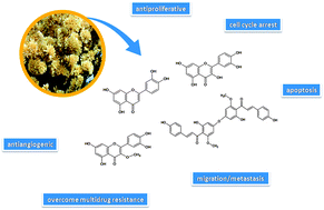
Cancer is the major public health problem worldwide; consequently, the search for new chemotherapeutic drugs is constant. Most of these agents are derived from natural sources, which are the major consistent basis for the search for modern anticancer medicines. In this context, numerous studies indicate flavonoids as a potential new class of secondary metabolites for anticancer therapy. In this review, special attention was addressed to flavonoids present in Achyrocline satureioides, a widely used medicinal plant with several and a well established range of biological properties. Two of these flavonoids are extensively studied for anticancer therapy, quercetin and luteolin, followed by 3-O-methylquercetin. Achyrobichalcone, recently isolated from A. satureioides by our group, can also represent a promising chemotherapeutic biomolecule due to its similarity with other cytotoxic bichalcones to cancer cell lines. The anticancer properties of these flavonoids, specially quercetin and luteolin, type of cell death, mechanisms and molecular targets involved were described. In general, these effects were observed due to the inhibition of cell proliferation, cell cycle arrest, apoptosis, inhibition of angiogenesis, prevention of migration/metastasis and overcoming multidrug resistance, alone or in combination with commonly used chemotherapeutic drugs. All these successful findings in preclinical studies suggest that these flavonoids are promising biomolecules for the development of new anticancer drugs in the future.
Natural products are privileged structures created by strong biological and ecological pressure that are able to interact with a wide variety of biological targets, consequently originating effective drugs in a large variety of therapeutic indications.4,5 Throughout human evolution, the importance of natural products for medicine and health has been enormous, from the earliest civilizations until today.6 The accumulated experience, knowledge and research over the years makes the secondary metabolites from natural sources, like plants, the most consistently successful approach for obtaining modern medicines.6,7
According to an extensive work published by Newman and Cragg, in 2012, 175 small molecules were approved worldwide over the time frame from around the 1940s to 2010 for cancer treatment. Of this total, the majority (74.9%) were inspired by compounds found in nature.8 Since the discovery of successful natural anticancer drugs such as paclitaxel (Taxol®) (from Taxus brevifolia), camptothecins (from Camptotheca acuminata), vinca alkaloids, such as vincristine and vinblastine (from Catharanthus roseus) and many others,4 the clinical interest for secondary metabolites for cancer therapy increased over the years and currently receives major attention from researchers.
Flavonoids are an emerging potential class of secondary metabolites from plants for anticancer chemotherapy.9 The existence of a large number of recently published papers focused on the chemotherapeutic and/or chemopreventive properties of flavonoids9–22 reinforces the great interest and importance of this issue today. Thus, this work aims to review the potential application of flavonoids for anticancer therapy focusing on the compounds present in Achyrocline satureioides (Lam.) DC. Asteraceae.
Two flavonoid derivatives reached close to approval and being launched in the market as new drugs for cancer treatment. Phenoxodiol, a synthetic analog of the plant isoflavone genistein, is one of these molecules, which is effective as anticancer agent and/or chemosensitizer for several traditional chemotherapeutic drugs in resistant tumors. The molecule is active against several human cancers, such as ovarian cancer,24–27 prostate cancer,28 melanoma,29,30 and squamous carcinoma,31 both in vitro and in vivo tests, reaching the step of clinical trials for some cancers.32 Phenoxodiol presented several promising and encouraging results, as the ability to increase the effectiveness of docetaxel in drug resistant ovarian carcinoma cells by at least 100 times,33 besides having an inhibitory effect against Topoisomerase 2 in the same range of etoposide, but stronger than genistein.34 Despite this, phenoxodiol failed in clinical trials for recurrent ovarian carcinoma after the “fast track” status for clinical use provided by FDA,9 the molecule (or its derivatives) still arouses great scientific35–38 and economical interest, especially from the pharmaceutical industry.39
Flavopiridol is another semisynthetic flavonoid with potential anticancer properties. The compound is a flavone derived from rohitukine, a natural anticancer agent isolated from an Indian tree.11 Due to its interesting biological properties both in vitro and in vivo, as cell cycle arrest in G1 and G2 phases, cytotoxicity against the majority of tumor cell lines in the nanomolar range, suppression of the tumor growth in animals and synergistic activity with most anticancer drugs and radiation, flavopiridol was the first cyclin-dependent kinase (CDK) inhibitor to be tested in human clinical trials.11,40,41 However, the molecule in Phase II clinical trials was ineffective against cancer and presented high toxicity to patients.40–42 Nevertheless, currently 60 anticancer clinical trials can be found with flavopiridol, alone or combined with other drugs, whereby 6 of them have shown to be active in the recruiting phase.43
Some works have reported the ethnopharmacological use of Achyrocline genus (A. satureioides and A. lehmanii Heiron) for anticancer therapy.50,85 A work of Santos and Elisabetsky, in 1999, investigated the ethnopharmacological use of plants to treat signs and sympoms related to cancer. From these analyses, A. satureioides was one of the species with the highest degree of importance.58 Another study reported that the plant was used by cancer patients as a complementary therapy to radioterapy.86
The anticancer properties of A. satureioides extracts could also be observed in biological assays. Methanolic extracts of its flowers inhibited growth of two human cancer cell lines: oropharyngeal epidermoid carcinoma cells (KB cells)54 and hepatoblastoma cells (HepG2 cells).63 The inhibition growth of KB cells was 66.8% after 72 hours of treatment with 50 μg mL−1 of the methanolic extract.54 The IC50% of HepG2 cells after 48 hours of treatment was 237 μg mL−1 (SD 56 μg mL−1), with inhibition growth in a concentration-dependent manner.63 The difference between these results can be related to the types of cell line, but also due to variations in methodology, as methods of evaluation of the cell death, treatment times or lack of standardization of the tested extracts. In another report, an aqueous extract of A. satureioides significantly inhibited cell division in the Allium cepa system, a vegetal test which measures the mitotic and replication indexes.87 Since the main flavonoids are present in the plant as aglycone form, only low concentrations are present in aqueous extracts. Thus, the observed effect reported by Fachinetto et al., in 2007, can be related to other constituents of the plant rather than the flavonoid aglycones.
Polydoro et al., in 2004, noted that A. satureioides freeze dried extracts (prepared from hydroethanol extractive solutions using 80% or 40% v/v ethanol) were cytotoxic to Sertoli cells, at the concentrations of 0.25 and 0.125 mg mL−1, increasing cell death over 30%. It was also observed that an enriched flavonoid fraction, which contained the highest total flavonoid aglycone, caused increase in the cell death and the extracts with higher quercetin contents (prepared with 80% hidroethanol solution) were more cytotoxic.67 Nevertheless, another study showed that an aqueous extract of A. satureioides (2% infusion) had low acute toxicity when administered intraperitoneally and no toxicity upon oral administration,88 that could be related to the very low flavonoid aglycone content, which presents reduced water solubility.
Most of the biological properties ascribed to A. satureioides extracts are related to the presence of flavonoids in its inflorescences.89 Often, quercetin (QCT; Fig. 1B), luteolin (LUT; Fig. 1C) and 3-O-methylquercetin (3OMQ; Fig. 1D) are the main flavonoids found in extracts, mainly in hydroalcoholic preparations.45,49,90,91,92 More recently, our group identified a new flavonoid in A. satureioides, named achyrobichalcone (ACB; Fig. 1E).91,93 Bearing in mind that a better understanding of the pharmacological activity of flavonoids is a key factor to improve the comprehension of the mode of action of many plant extracts,94 the next topics will address a compilation of the main studies found in literature concerning the anticancer properties of flavonoids from A. satureioides. Noting that much information is found in scientific databases about this issue, the intention of this review is to describe the most relevant and recent findings.
In general, the direct anticancer effects of flavonoids from A. satureioides were involved with cell proliferation, cell cycle arrest, apoptosis, angiogenesis, migration/metastasis and overcome multidrug resistance. These effects were observed for flavonoids alone or in combination with commonly used chemotherapeutic drugs.
Cancer | Cell culture system | Concentration used | Molecular mechanism | Biological effect | Reference |
---|---|---|---|---|---|
Bladder | 253J | 50 μM | Activation of BKCa channels | Antiproliferative | 102 |
T24 | 50–100 μM | Inhibition of ecto-5′-NT/CD73 activity | Antiproliferative | 103 | |
T24, EJ, J82 | 100 μM | Increase in G0/G1 phase, decrease in mutant p53 and survivin | Induction of apoptosis, cell cycle arrest | 104 | |
Neuro (glioma) | U87, T98G | 25–50 μM | Blocker of IL-6/STAT3 | Antiproliferative, inhibition of migration | 105 |
U87 | 50 μM | Inhibition of NFkB-induced by PLD1, decrease in MMP-2 | Antiproliferative, anti-invasive | 106 | |
A172 | 50 μM | Down-regulation of ERK, AKT and survivin | Induction of apoptosis | 107 | |
Colon | HT29 | 50–100 μM | Increase in G1 phase, activation of AMPK, up-regulation of p53 and p21 | Induction of apoptosis, cell cycle arrest | 108 |
HT29/MCF-breast | 25–200 μM | Increase in G1 phase, activation of AMPK-COX-2 | Induction of apoptosis, cell cycle arrest | 109 | |
HCT116 | 40 μM | Induction of NAG-1 | Induction of apoptosis | 110 | |
Liver | HepG2 | 40–160 μM | Increase in G0/G1 phase and p53, decrease in survivin and Bcl-2, activation of caspase-9 and -3 | Induction of apoptosis, cell cycle arrest | 111 |
Stomach | BGC823 | 30–120 μM | Increase in S phase, decrease in Bcl-2/Bax ratio, activation of caspase-3 | Induction of apoptosis, cell cycle arrest | 112 |
Blood (leukemia) | CCRF-CEM, HL60, K562 | 12.5–100 μM | Suppression of telomerase activity | Induction of apoptosis | 113 |
HL60 | 25–100 μM | Induction of FasL, activation of caspase-8 | Induction of apoptosis | 114 | |
Breast | MCF7 | 25–200 μM | Suppression of AMPK, decrease HIF-1α | Induction of apoptosis | 115 |
MCF7 | 10–175 μM | Increase in S phase, decrease in Bcl-2 and ΔΨm, activation of caspase-6, -8 and -9 | Induction of apoptosis, cell cycle arrest | 116 | |
Skin (melanoma) | B16-BL6 | 330 μM | Decrease in PKC and MMP-9 | Anti-invasive | 117 |
Prostate | PC3 | 50–200 μM | Increase in G0/G1 phase, decrease in Bcl-2 and ΔΨm, increase in Bax, activation of caspase-3, -8 and -9 | Induction of apoptosis, cell cycle arrest | 118 |
PC3 (and HUVECs) | 20–40 μM | Down-regulation of AKT, mTOR and P70S6K | Induction of apoptosis, antiangiogenic | 119 | |
Lung | A549 | — | Increase in G0/G1 phase, decrease in survivin and Bcl-2, activation of caspase-3 | Induction of apoptosis, cell cycle arrest | 120 |
A549, H1299 | 20–80 μM | Increase in G2/M phase and p53, decrease in survivin | Induction of apoptosis, cell cycle arrest | 121 | |
A549 | 14.5–58 μM | Activation of MEK–ERK | Induction of apoptosis | 122 |
In the bladder cancer cell lines T24, EJ and J82, QCT inhibited the cell growth by cell cycle arrest in G0/G1 and induced apoptosis by decrease in the expression of mutant p53 and surviving.104 Additionaly, Rockenbach et al., in 2013, observed the involvement of the ectonucleotidase pathway, by means of ecto-5′-NT/CD73, the enzyme responsible for AMP hydrolysis, in the antiproliferative effects of QCT on T24 cells.103 The large conductance Ca2+-activated K+ (BKCa) channel influenced the proliferation of 253J bladder cancer cells only in the presence of QCT, suggesting that this channel may represent a valuable target for antiproliferative effects of QCT in these cells.102
In U87 and T98G glioma cells, QCT exerted anticancer effects through abrogation of the IL-6/STAT3 signaling pathway, inhibiting IL-6 and consequently STAT3, a key player involved in cancer-related inflammation by activate cellular responses as cyclin D1, MMP2 and Bcl-2 responsible for cancer cell proliferation, invasion and survival, respectively.105 Other work demonstrated that QCT inhibited U87 cell proliferation by abolishing the phospholipase D (PLD1) expression, a regulator of cell proliferation and tumorigenesis.106 In A172 cells, QCT induced apoptosis through caspase-dependent mechanisms involving down-regulation of ERK, AKT and surviving.107
QCT promoted cell cycle arrest through the increase in G1 phase in HT29 colon cancer cells and upregulated apoptosis-related proteins, such as AMPK, p53 and p21. Additionaly, in vivo experiments showed that QCT resulted in a significant reduction in tumor volume over 6 weeks of treatment and apoptosis-related protein induction was significantly higher in the treated group compared to the control group.108 Other works observed that AMPK activation in HT29 colon cancer cells and MCF breast cancer cells seemed to be closely related to a decrease in COX-2 expression, suggesting that a possible mechanism involved in the anticancer activity of QCT is partly mediated through its anti-inflammatory action.109,123 The expression of NAG-1 (non-steroidal anti-inflammatory drug activated gene-1) was induced during treatment of HCT116 colon carcinoma cells with the QCT and apoptosis process was activated. Early growth response-1 (EGR-1) and p53 were required for QCT mediated activation of the NAG-1 promoter.110
The apoptotic effect and cell cycle arrest of QCT in HepG2 hepatoma cancer cells was promoted at G0/G1 phases, with suppression of survivin and Bcl-2, elevation of p53 and activation of caspase-9 and caspase-3.120,124 Similar results were found for the BGC-823 gastric cancer cell treated with QCT, where caspase-3 was activated and the Bcl-2/Bax ratio was decreased. Additionally, some morphological features of apoptosis were found, such as cell shrinkage or even the formation of an apoptotic body.112
Avci et al., in 2011, observed that QCT had antiproliferative and pro-apoptotic effects on acute lymphocytic leukemia (ALL) (CCRF-CEM), acute promyelocytic leukemia (HL60) and chronic myeloid leukemia (CML) (K562) cell lines by the suppression of the activity of telomerase, an enzyme that is one of the hallmarks of cancer because it is able to restore final sequences of DNA.113 QCT also activated the extrinsic apoptotic pathway in HL60 cells through interaction with death receptor Fasligand (FasL) in the plasma membrane, which is involved in ERK and JNK signaling pathways and consequent activation of caspase-8, induction of Bid cleavage and Bax conformation change.114
Despite its apoptotic effects, AMPK also can help the cancer cell in the adaptation and survival during hypoxic conditions. In MCF7 breast cancer cells under hypoxia, QCT was capable of inhibiting AMPK and decrease HIF-1α, which is a critical survival factor in hypoxia.115 Another study showed that QCT arrested the cell cycle in sub-G1 and S phases for MCF-7 cells. The arrest in S phase was mediated by decreasing the protein expression of CDK2, cyclins A and B while increasing the p53 and p57 proteins. Decreased levels of Bcl-2 protein and ΔΨm and increased activations of caspase-6, -8 and -9 also were observed.116 Similar results were found for metastatic breast cancer cells, as MDA-MB-231 (ref. 125 and 126) and MDA-MB-453 cells.127
Antiangiogenic activity of QCT was evaluated in vitro in PC3 prostate cancer cells and HUVEC endothelial cells, besides in vivo and ex vivo. QCT suppressed vascular endothelial growth factor (VEGF) that induced phosphorylation of VEGF receptor 2 and their downstream protein kinases AKT, mTOR and P70S6K in HUVECs, that are responsible by promoting proliferation, migration and tube formation of endothelial cells. QCT (20 mg kg−1) significantly reduced the volume and the weight of solid tumors in the prostate xenograft mouse model, inhibiting tumorigenesis by targeting angiogenesis. Apoptosis was induced in PC3 cells, which was correlated with the down-regulation of AKT, mTOR and P70S6K expressions.119 The actions of QCT suppressing tumor invasion, angiogenesis and metastasis in PC3 cells also were observed in other works128,129 and in B16-BL6 melanoma cells.117 QCT promoted cell cycle arrest, increasing G0/G1 phase, decreasing Bcl-2 and ΔΨm, increasing Bax and activating of caspase-3, -8 and -9 in PC3 cells.118 These findings are in agreement with the results of Senthilkumar et al., in 2010, that demonstrated the induction of both intrinsic and extrinsic pathways to mediate apoptosis in PC3 cells after treatment with QCT.130
QCT induced cytotoxicity and apoptosis in A549 and H1299 non-small lung carcinoma cells. Inhibition of cell growth was observed, with increase in G2/M phase and raised cyclin B1 and phospho-CDC2 proteins. Survivin proteins were decreased and levels of total p53, phospho-p53 and p21 proteins increased and translocated to the nuclei.121 Another work demonstrated that QCT reduced cell viability and DNA synthesis, with a rate of apoptosis equivalent, in A549 cells. QCT treatments led to increase in Bax and Bad, decrease in Bcl-2 and cleavage of caspase-3 and caspase-7. While Akt-1 was inhibited, ERK was phosphorylated following QCT treatment. The induction of MEK–ERK pathway in A549 cells led to activation of caspase-3 and apoptosis.122 However, QCT was not active against DMS114, a small lung carcinoma cell.131
Finally, some works pointed QCT as very potent inhibitor of topoisomerases (Top 1 and 2) having activity at concentrations similar to that of etoposide (50 μM).9
The resistance of death receptor to the fludarabine in leukemic cells isolated from chronic lymphocytic leukemia (CLL) patients was ameliorated by combined treatment with QCT.135 In another work, QCT cooperated with arsenic trioxide to induce apoptosis in U937, THP-1 and HL-60 leukemia cells, increasing the clinical efficacy of the antileukemic drug.136
QCT sensitized DB-1 melanoma (p53 wildtype) cells to temozolomide (TMZ) by a mechanism that involves the modulation of p53 family members. After treatment with TMZ, DB-1 cells demonstrated increased p53, however the cells were resistant to TMZ-induced apoptosis. QCT treatment in combination with TMZ abolished drug insensitivity and caused a more than additive induction of apoptosis compared to either treatment alone.137
The antiproliferative effect of QCT and cisplatin (CIS) alone or in combination was evaluated in SPC212 and SPC111 malignant mesothelioma cell lines. QCT significantly reduced the proliferation of cell lines, altered the cell cycle distribution and increased the level of caspase-9 and -3. Additionally, the combination of QCT and CIS was found to be more effective when compared to single-drug treatment.138 In another work, QCT exhibited synergistic effects with CIS against nasopharyngeal carcinoma cells. The dose reduction index found implied the possibility of reducing the CIS dosage required to treat with the addition of QCT, reducing the risk of CIS-associated toxicity.139 In vincristine (VCR) resistant A549 lung adenocarcinoma cells, QCT exerted increasing chemosensitivity when combined with CIS and VCR.140 It was also found that in lung cancer cells that QCT sensitizes, TRAIL-induced apoptosis and two independent pathways are involved: induction of DR5 through PKC and suppression of AKT mediated survivin expression.141
In the pancreatic cell line EPP85-181RDB, QCT seemed to sensitize resistant cells to daunorubicin (DAU). In parallel, the effect of both substances on the sensitive cell line was synergistic.142 QCT was an effective cytotoxic agent in gastric carcinoma cell lines. It had not only a synergistic effect with DAU on P-cells but also sensitized DOX-resistant cells. The analysis of p-glycoprotein (P-gp) activity showed that QCT may down-regulate this transporter.100
The combination treatment of 5-fluorouracil (5-FU) and QCT increased apoptosis in CO115 colorectal cells. Mitochondrial pathway and p53 were involved in cell death.143 In another work, QCT sensitized HCT116 colon cancer cells to CIS and etoposide (ETO) under hypoxic conditions by targeting AMPK.144
Cancer | Cell culture system | Concentration used | Molecular mechanism/cellular targets | Biological effect | Reference |
---|---|---|---|---|---|
Bladder | RT112 | 10–50 μM | Increase in sub-G1 phase, poly(ADP-ribose) polymerase cleavage, DNA fragmentation | Induction of apoptosis, cell cycle arrest | 145 |
Colon | COLO205, HCT116 | 40 μM | Inhibition of NFkB, activation of JNK | Induction of apoptosis | 146 |
HT29 | 40–60 μM | Increase in G1 and G2/M phases, poly(ADP-ribose) polymerase cleavage, decreased in p21CIP1/WAF1, survivin, Mcl-1, Bcl-xL, and Mdm-2, activation caspase-3, -7 and -9 | Induction of apoptosis, cell cycle arrest | 147 | |
Liver | HepG2 | 25–100 μM | Activation of AMPK, inhibition of NFkB | Induction of apoptosis | 148 |
HLF | 50 μM | Blocker of STAT3 | Antiproliferative, induction of apoptosis | 149 | |
Stomach | AGS | 20–80 μM | Increase in G2/M phase, Bax and p53, decrease of Bcl-2, activation of caspase-3, -6, -9 | Induction of apoptosis, cell cycle arrest | 150 |
Blood (leukemia) | H60 | 10–50 μM | DNA fragmentation | Induction of apoptosis | 151 |
Breast | MCF7, MDA-MB-231, SK-BR3 | 30 μM | Activation of ERK and p38 | Induction of apoptosis | 152 |
Skin (melanoma) | B16F10 | 5–50 μM | Inhibition of β3 integrin/FAK | Anti-invasive | 153 |
Prostate | PC3 (and HUVECs) | 20–40 μM | Suppression of VEGFR2, down-regulation of AKT, ERK, mTOR, P70S6K, MMP-2, MMP-9 | Induction of apoptosis, antiangiogenic | 154 |
PC3 | 10–40 μM | Induction of E-cadherin | Anti-invasive | 155 | |
Lung | A549, H460 | 5–80 μM | Production of superoxide, degradation of MKP-1, activation of JNK | Induction of apoptosis | 156 |
A549 | 20–80 μM | Increase in G2/M phase, increase in Bax, activation of JNK, inhibition of NFkB, activation of caspase-3, -9 | Induction of apoptosis, cell cycle arrest | 157 | |
A549 | 25–50 μM | Increase in G1 phase, decrease in ΔΨm, disruption of actin | Induction of apoptosis, cell cycle arrest, inhibition of migration | 158 |
The cytotoxic effects of LUT on two different cancer cell lines, including RT112 bladder carcinoma and K562 chronic myeloid leukemia, was described by Kilani-Jaziri et al., in 2012. The apoptosis was induced, with minor cell-cycle perturbations, but some increase in sub-G1 fraction was observed. The poly(ADP-ribose) polymerase was cleaved and a typical ladder of DNA fragments was observed in treated cells.145
In COLO205 and HCT116 colorectal cancer cells and cervical cancer HeLa cells, LUT inhibited TNFα, a cell mediator that activates both cell death and cell survival pathways, which render most cancer cells resistant to its cytotoxicity. With the inhibition of TNFα, the activation of NFkB was decreased and consequently the induction of NFkB-targeted anti-apoptotic genes was inhibited. In addition, the inhibition of NFkB led to augmentation of JNK activation.146 Another work showed that LUT induced G1 and G2/M cell cycle arrest, which was mediated by CDK2, CDK4 and CDC2 in HT29 colon cancer cells. In addition, LUT promoted apoptosis through down-regulation of several anti-apoptotic proteins.147 The same group described that LUT down-regulates the activation of the PI3K/AKT and ERK1/2 pathways via reduction in insulin-like growth factor-I receptor (IGF-IR) signaling in HT29 cells.159
LUT strongly induced cell death in HepG2 hepatoma cells and dramatically reduced the tumor volume in a tumor xenograft model. Both effects were accompanied by AMPK activation and a strong inhibitory effect on NFkB. LUT treatment causes reactive oxygen species (ROS) production and these intracellular ROS in turn mediate AMPK-NFkB signaling.148 Selvendiran et al., in 2006, observed the pro-apoptotic effect of LUT on HLF hepatoma cells in vitro and in vivo. Apoptosis was found by caspase-8 activation and enhanced expression in Fas/CD95. Decrease of STAT3, a known negative regulator of Fas/CD95 transcription, lead to down-regulation on these target gene products, such as cyclin D1, survivin, Bcl-xL and vascular endothelial growth factor. An overexpression in STAT3 led to resistance to LUT, suggesting that STAT3 was a critical target of LUT. In nude mice with xenografted tumors using HAK-1B hepatoma cells, LUT significantly inhibited the growth of the tumors.149
Antiproliferative and chemosensitizing effects of LUT on AGS gastric cancer cells treated with LUT and/or other chemotherapeutic agents were evaluated by Wu et al., 2008. LUT induced cell cycle arrest by an increase of the G2/M phase, besides reducing protein levels of CDC2, cyclin B1 and CDC25C and increasing the cyclin-dependent kinase inhibitor p21 (cip1/Waf1). The pro-apoptotic proteins, including caspase-3,- 6, -9, Bax and p53 were increased and the levels of anti-apoptotic protein Bcl-2 was reduced, thus shifting the Bax/Bcl ratio in favor of apoptosis. The combined treatment of CIS and LUT induced more effective cell growth inhibition, compared to CIS treatment alone.150
In MCF7, MDA-MB-231 and SK-BR3 breast cancer cells, LUT induced nuclear translocation of AIF (apoptosis-inducing factor), which was mediated by activation of ERK and p38. LUT induces caspase dependent and independent apoptosis involving AIF nuclear translocation mediated by activation of ERK and p38.152 Another work with MDA-MB-231 cells showed that LUT arrested the cell cycle at the G2/M and S stages and activated the apoptosis by decreasing AKT, PLK1, cyclin B1, cyclin A, CDC2, CDK2 and Bcl-xL and increasing p21 and Bax expression. LUT-supplementation significantly reduced tumor burden in nude mice inoculated with MDA-MB-231 cells.160
LUT suppressed the hypoxia-induced changes in B16F10 melanoma cells, as increased cellular adhesion and invasion. Hypoxia significantly decreased the expression of E-cadherin while increasing the expression of N-cadherin in the cells, which was reversed by LUT. LUT up-regulated the E-cadherin at least partly via inhibiting the β3 integrin/FAK signal pathway. In experimental metastasis model mice, treatment with LUT (10–20 mg kg−1) reduced metastatic colonization in the lungs by 50%.153
The antiangiogenic activity of LUT was evaluated by Pratheeshkumar et al., in 2012, in HUVECs cells and PC3 prostate cancer cells, using in vitro, ex vivo and in vivo models. In vitro, LUT at non-toxic concentrations significantly inhibited microvessel sprouting and proliferation, migration, invasion and tube formation of endothelial cells, key events in the process of angiogenesis, which was also confirmed by ex vivo studies. LUT suppressed VEGF induced phosphorylation of VEGF receptor 2 and their downstream protein kinases AKT, ERK, mTOR, P70S6K, MMP-2 and MMP-9 in HUVECs. Proinflammatory cytokines such as IL-1b, IL-6, IL-8 and TNF-α were significantly reduced after treatment of PC3 cells with LUT. The apoptosis also was activated in PC3 cells. LUT (10 mg kg−1) significantly reduced the volume and the weight of solid tumors in the prostate xenograft mouse model.154 In another work, LUT inhibits invasion of PC3 prostate cancer cells through the induced expression of E-cadherin through MDM2. LUT inhibits MDM2 through down-regulation of AKT, suggesting that LUT regulates E-cadherin through the AKT/MDM2 pathway. In vivo experiments showed that LUT inhibited spontaneous lung metastasis of PC3 cells implanted onto the nude mice.155 LUT also acted as an antiproliferative and anti-invasion agent in LNCaP prostate cancer cells.161
In A549 and H460 non-small cell lung carcinoma, LUT induced apoptotic and non-apoptotic death mediated by the induction of superoxide radicals. JNK was potently activated after superoxide accumulation and suppression of superoxide completely blocked LUT-induced JNK activation. The induction of JNK suppressed MKP-1 and promoted cell death.156 Kim et al., in 2006, observed that caspase activation culminated in apoptosis in Lewis lung carcinoma cells treated with LUT.162 In another work, LUT blocked the NFkB pathway, sensitizing H23, H2009, H460 and A549 lung carcinoma cells to apoptosis.163
The antiproliferative effects of LUT in multi-drug resistance (MDR) breast cancer cells (ADR/RES and MCF-7/MitoR) that expressed high levels of P-glycoprotein and ABCG2 were evaluated by Rao et al., in 2012. LUT induces apoptosis in P-glycoprotein- and ABCG2-expressing MDR cancer cells without affecting the transport functions of these drug transporters. LUT induced apoptosis by ROS generation, DNA damage, activation of ATR → CHK2 → p53 signaling pathway, inhibition of NFkB signaling pathway, activation of p38 pathway and depletion of anti-apoptotic proteins.164
Tang et al., 2011, showed that LUT acted as a potent inhibitor of nuclear factor erythroid 2-related factor 2 (Nrf2). This transcription factor is redox-sensitive and regulates the expression of a battery of genes generally involved with cell defense, enhancing cancer cell survival and resistance to anticancer drugs. In A549 non-small lung cancer cells, which possess constitutively active Nrf2, LUT promoted a dramatic reduction in Nrf2, leading to decreased Nrf2 binding to antioxidant response elements (AREs), down-regulation of ARE-driven genes and depletion of reduced glutathione. Additionally, LUT significantly sensitized A549 cells to the anticancer drugs oxaliplatin (OXA), bleomycin (BLEO) and DOX.166
Antiproliferative effect of 3OMQ was observed in mouse skin epidermal JB6P1 cells, with induction of cell cycle arrest at the G2/M phase. It also suppressed neoplastic cell transformation, with inhibitory efficacy greater than QCT. The activation of AP-1 was suppressed after the cell treatment with 3OMQ. The inhibition of ERK kinase activity and attenuated phosphorylation of ERKs were also observed.167
3OMQ caused significant growth inhibition of lapatinib (LAP) sensitive (SK–Br-3) and resistant (SK–Br-3–Lap R) breast cancer cells. 3OMQ had no effect on AKT or ERK signaling in resistant cells. However, it caused a pronounced G2/M arrest mainly through the CHK1–CDC25c–cyclin B1/CDK1 pathway in both cell types. Differently, LAP produced an accumulation of cells in G1 phase mediated through cyclin D1, but only in LAP sensitive cells. 3OMQ induced apoptosis by increasing levels of cleaved caspase-3, -7 and poly(ADP-ribose) polymerase (PARP) in both cell lines.168
Rubio et al., in 2006, evaluated the cytotoxicity of 3OMQ against several human cancer cell lines (HL60-leukemia, A431-epidermoid carcinoma, HeLa-cervical cancer, SKOV3-ovarian carcinoma and HOS-hosteosarcoma). The study also compared the cytotoxicity of 3OMQ and QCT in these cells. 3OMQ acted at a lower range and values of IC50% (15–50 μM) when compared to QCT (30–100 μM). Both were not active for HOS cells. The methylation of the hydroxyl group at position C3 of QCT yields 3OMQ, a molecule with a higher antiproliferative activity. The authors presuppose that methylated derivatives can pass through biological membranes more easily than QCT, consequently having higher intracellular concentration and lower IC50% values.169
3OMQ isolated from Inula viscosa showed antiproliferative activity against the MCF7 breast cancer cells and Hep2 hepatoma cells (IC50% values of 11.23 μg mL−1 and 26.12 μg mL−1, respectively). The toxicity was limited against Vero cells, with IC50% values above 150 μg mL−1. 3OMQ induced apoptosis in MCF7 cells and DNA fragmentation. The molecule increased the number of apoptotic cells with fragmented DNA, compared with untreated MCF7 cells. Cytomorphological alterations corresponding to typical morphology of apoptosis were detected in MCF7 cells treated with 3OMQ, that included cell shrinkage, membrane blebbing and formation of apoptotic bodies.170
MAPK | Mitogen-activated protein kinase |
PKC | Protein kinase C |
PI3K or AKT | Phosphatidylinositol 3-kinase |
NFkB | Nuclear factor kappa B |
CDKs(1–9) | (1–9)Cyclin-dependent kinases |
ERK | Extracellular signal-regulated kinase |
VEGF | Vascular endothelial growth factor |
IGF-IR | Insulin-like growth factor-I receptor |
P-gp | P-glycoprotein |
Cip1/Waf1 | Cyclin-dependent kinase inhibitor p21 |
AIF | Apoptosis-inducing factor |
Nrf2 | Nuclear factor erythroid 2-related factor 2 |
PARP | Poly(ADP-ribose) polymerase |
Bax | Bcl-2-associated X protein |
PUMA | p53 Upregulated modulator of apoptosis or BBC3, Bcl-2-binding component 3 |
NOXA | Phorbol-12-myristate-13-acetate-induced protein 1 |
Bak | Bcl-2 homologous antagonist/killer |
Bid | BH3 interacting-domain death agonist |
Bcl-2 | Derived from B-cell lymphoma 2 |
Bcl-xL | Derived from B-cell lymphoma-extra large |
Mcl-1 | Induced myeloid leukemia cell differentiation protein |
IAP | Inhibitor of apoptosis |
p53 | Protein 53 or tumor protein 53 |
TRAIL | TNF-related apoptosis-inducing ligand |
survivin or BIRC5 | baculoviral inhibitor of apoptosis repeat-containing 5 |
IL | Interleucine |
STAT3 | Signal transducer and activator of transcription 3 |
MMP | Matrix metalloproteinase |
COX-2 | Cyclooxygenase-2 |
PLD1 | Phospholipase D1 |
p21 | Cyclin-dependent kinase inhibitor 1 or CDK-interacting protein 1 |
NAG-1 | Nonsteroidal anti-inflammatory drug-activated gene |
EGR-1 | Early growth response protein 1 |
HIF-1α | Hypoxia-inducible factor 1-α |
FasL or CD95L | Fas ligand |
p57 | Cyclin-dependent kinase inhibitor 1C |
ΔΨm | Mitochondrial transmembrane potential |
mTOR | mammalian target of rapamycin |
P70S6K | serine/threonine kinase |
TNFα | Tumor necrosis factor |
JNK | c-Jun NH2-terminal kinase |
PLK1 | Serine/threonine-protein kinase |
ABCG2 | ATP-binding cassette sub-family G member 2 |
MKP-1 | Mitogen-activated protein kinase phosphatases |
ARE | Antioxidant responsive element |
ROS | Reactive oxygen species |
QCT | Quercetin |
LUT | Luteolin |
3OMQ | 3-O-methylquercetin |
ACB | Achyrobichalcone |
DOX | Doxorubicin |
TMZ | Temozolomide |
CIS | Cisplatin |
VCR | Vincristine |
DAU | Daunorubicin |
5-FU | 5-Fluorouracil |
ETO | Etoposide |
MDR | Multi-drug resistance |
OXA | Oxaliplatin |
BLEO | Bleomycin |
LAP | Lapatinib. |
This journal is © The Royal Society of Chemistry 2014 |