Naked-eye detection of inorganic fluoride in aqueous media using a new azo-azomethine colorimetric receptor enhanced by electron withdrawing groups†
Hamid Khanmohammadi* and
Khatereh Rezaeian
Department of Chemistry, Faculty of Science, Arak University, Arak 38156 8 8349, Iran. E-mail: h-khanmohammadi@araku.ac.ir; Fax: +98-861-4173406; Tel: +98-861-2777401
First published on 8th October 2013
Abstract
A new azo-azomethine receptor, containing active phenolic sites, has been designed and synthesized for quantitative detection and colorimetric sensing of inorganic fluoride ion in aqueous media. The introduction of four electron withdrawing groups into the backbone of the receptor makes the two phenolic groups efficient hydrogen bonding sites. Hence, the receptor is capable of competing with water molecules to detect trace amounts of inorganic fluoride ions in aqueous solutions. Accordingly, the receptor showed a remarkable colour change from light yellow to orange upon the addition of aqueous solution of NaF, enabling naked-eye F− sensing without any spectroscopic instrumentation. The recognition details of F− sensing were also evaluated using UV-Vis spectroscopy and 1H NMR titration techniques. The 1H NMR titration revealed that the colorimetric response was considered to be the direct consequence of hydrogen-bond formation between the phenolic groups of the receptor and fluoride ions followed by deprotonation. Importantly, the current sensor can detect inorganic fluoride in water even at 0.058 ppm, which is lower than the World Health Organization (WHO) permissible level. To the best of our knowledge, there have been very few examples of detection limits lower than the WHO permissible level for detecting fluoride ion in drinking water (below 1 ppm). The designed sensor has also shown highly promising results for the qualitative and quantitative detection of fluoride in real samples like sea water, toothpaste and mouthwash.
Introduction
Anions have been recognized as playing key roles in a wide range of chemical and biological processes.1 Thus, the design and also construction of molecular sensors for the recognition and sensing of physiologically important anions have been in the forefront of chemistry research.2 In particular, the design of sensors for the colorimetric detection of anions is increasingly appreciated since naked eye detection is low cost and does not require any spectroscopic instrumentation.3a–c Among the biologically important anions, fluoride has attracted the interest of chemists owing to its established role in dental care and the treatment of osteoporosis.3d–jTo date a number of colorimetric receptors which are capable of detecting the fluoride ion with high affinity, have been reported.4 The common recognition moieties of the reported receptors involve amide, urea and thiourea, amidourea, phenol, pyrrole, imidazolium and indole.5 Generally, the formation of hydrogen bonds between active sites, N–H and/or O–H, of these subunits with F− plays a pivotal role in anion sensing. However, most receptors are restricted to the detection of tetrabutylammonium fluoride (TBAF) in organic solvents and there is a paucity of reports regarding the colorimetric detection of fluoride ion in aqueous media.6 This obstacle is attributed to the fact that water is a highly competitive solvent and participates in the detection process of F− by mediating interactions between the binding partners. In this regard, F− becomes solvated strongly even with a trace amount of water.7 The mentioned blockages limit the discrimination and detection of inorganic fluoride anion in water.
On the other hand, highly selective fluoride ion receptors are strongly favoured for practical applications. However, it is quite difficult to design colorimetric chemosensors for the recognition of F− in water. Indeed, only a few examples of sensors capable of detecting fluoride ion in water have been reported.6 Furthermore, most of the determination processes are limited to either recognition of TBAF or use of test papers which need several minutes to complete detection and are far away from real-life applications.8
Therefore, the development of sensors for the direct detection of inorganic fluoride in aqueous media accompanied by a visually striking colour change, naked eye sensing, is an important target for real-life applications. Kruger et al. developed a 1,8-naphthalimide-based colorimetric anion sensor containing the aminothiourea group for the recognition of NaF in aqueous solutions.9 Subsequently, Das et al. have designed a receptor for the selective and quantitative extraction of fluoride anions from aqueous solution of sodium fluoride.10 Nevertheless, the evaluation and discrimination of inorganic fluoride in aqueous media still remains as important a challenge to chemical analysis.
Until now, a number of azo sensors for the detection of fluoride have been reported. However, most of these sensors have been designed for use in noncompetitive organic solvents.3a,4i–k Recently, Mahapatra and co-workers reported new chromogenic azo-azomethine receptors for the recognition of fluoride.4i Although the synthesized receptors have been shown to possess good selectivity and sensitivity for fluoride ion in CH3CN, the sensory system was not able to compete with water. Thus, F− sensing in aqueous media was restricted to the use of test strips which requires several minutes for the detection process. In pursuit of these, we report here a new member of the azo-azomethine sensors, receptor 1, (Scheme 1) for the rapid detection and colorimetric sensing of inorganic fluoride ion in aqueous media. To the best of our knowledge, this is the first azo-azomethine receptor used for the qualitative and quantitative detection of fluoride in water and real samples. The prepared molecular sensor, 1, contains azo phenol moieties serving as sensing units to form hydrogen bonds via the disposal of OH groups in the presence of anions. In particular, the presence of four electron withdrawing substituents in the molecular structure of 1 enhances the acidity of its active sites and hence allows the formation of stronger hydrogen bonds between the OH groups of the sensor and the anionic guest. Therefore, receptor 1 can compete with water for the naked-eye detection of inorganic fluoride in aqueous media.
 | ||
| Scheme 1 Synthesis of receptor 1. | ||
Results and discussion
The synthesis of receptor 1 was achieved by the straightforward condensation reaction of α,α′-bis(o-aminophenylthio)-1,2-xylene with 1-(3-formyl-4-hydroxyphenylazo)-2,4-dichlorobenzene as depicted in Scheme 1. The prepared receptor was characterized using standard spectroscopic techniques (ESI†).As an initial test, we checked the colour changes of receptor 1 in DMSO upon the addition of 10 equiv. of tetrabutylammonium (TBA) salts of F−, Cl−, Br−, I−, H2PO4−, ClO4−, N3−, NO2−, NO3−, AcO− and HSO4−. As shown in Fig. 1(a), a noticeable colour change was observed from light yellow to red in the presence of F−, AcO− and H2PO4−. In contrast, other anions caused no obvious changes in colour. To further study the colorimetric behaviour of receptor 1, UV-Vis absorption experiments were also performed upon the addition of 10 equiv. of tetrabutylammonium salts of anions to 1 (2 × 10⁻⁵mol L−1 in DMSO), Fig. 1(b). Free receptor 1 exhibited one absorption band at 369 nm and a weak tail at ∼490 nm. A distinct change was observed in the spectral pattern in the presence of F−, AcO− and H2PO4−; the extent of the changes was much more for F−. Moreover, the addition of anion-based salts to the DMSO solution of 1 gave a bathochromic shift from 369 nm to ca. 500 nm with F−, AcO− and H2PO4−. While other anions induced negligible spectral responses.
 | ||
| Fig. 1 (a) Colour changes of receptor 1 (2 × 10⁻⁵ mol L−1 in DMSO) after addition of 10 equiv. of different anions. From left to right: none, H2PO4−, ClO4−, Br−, Cl−, AcO−, F−, N3−, HSO4−, NO2−, NO3− and I− (TBA+ salts). (b) UV-Vis absorption spectra of receptor 1 (2 × 10⁻⁵mol L−1 in DMSO) in the presence of 10 equiv. of various anions. | ||
In order to further assess the binding characteristics of receptor 1, we carried out UV-Vis spectrophotometric titrations by adding a standard solution of TBAF to the dry DMSO solution of 1 (2 × 10⁻⁵mol L−1). As shown in Fig. 2, upon the addition of fluoride ion, the band at 369 nm, the π → π* transition of the chromophores, decreased gradually and simultaneously a new strong absorption band, attributed to the charge transfer (CT), formed at 503 nm indicating interaction between fluoride ion and receptor. An analogous investigation was performed for the addition of the TBA salt of H2PO4− and AcO− to a solution of 1 in dry DMSO (Fig. S11 and S23, ESI†). The Benesi–Hildebrand method11 confirmed 1![[thin space (1/6-em)]](https://www.rsc.org/images/entities/char_2009.gif) :1 stoichiometry with binding constants of 2.36 × 10⁴, 1.87 × 10⁴ and 9.54 × 10³ M−1for F−, AcO− and H2PO4−, respectively. The 1:1 stoichiometry was also determined by Job's plot (Fig. S10, S13 and S24, ESI†).
:1 stoichiometry with binding constants of 2.36 × 10⁴, 1.87 × 10⁴ and 9.54 × 10³ M−1for F−, AcO− and H2PO4−, respectively. The 1:1 stoichiometry was also determined by Job's plot (Fig. S10, S13 and S24, ESI†).
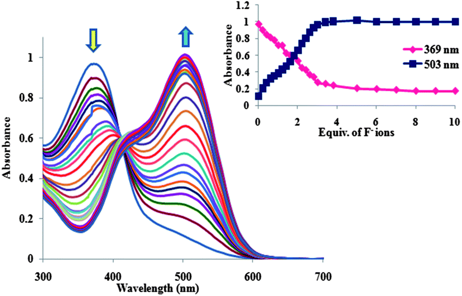 | ||
| Fig. 2 UV-Vis absorption spectra of receptor 1 (2 × 10⁻⁵mol L−1) in dry DMSO upon the addition of TBAF (0–10 equiv.). Inset showing the binding isotherm at selected wavelengths in DMSO. | ||
Based on UV-Vis measurements, the detection limits of receptor 1 towards F−, AcO− and H2PO4− in DMSO were found to be 2.06 × 10⁻⁶, 3.39 × 10⁻⁶ and 2.22 × 10⁻⁶ mol L−1, respectively. The surprising results of our present study were that upon the addition of competitive protic solvents such as methanol or water to the solution of 1 + 10 equiv. F− in DMSO, the colour and also absorption spectrum were not reversed12 (Fig. S14, ESI†). However, progressive addition of a protic solvent to the solution of 1 + 10 equiv. H2PO4− in DMSO caused the colour of the solution to turn from red to yellow. Correspondingly, the absorption band recovered from 497 nm to 369 nm with a clean isosbestic point at 407 nm (Fig. S15, ESI†). Similar behaviour was also observed for AcO− (Fig. S25, ESI†). This phenomenon is attributed to the fact that protic solvents would compete with H2PO4−and AcO− anions for the binding sites of the host and disturb the interaction between the host and guest.4f
From the above findings we concluded that receptor 1 is capable of detecting fluoride ions in aqueous solution. Hence, to investigate the applicability of 1, further UV-Vis spectrophotometric titrations were performed to detect sodium fluoride in water. Fig. 3 depicts the UV-Vis titration of 1 (2 × 10⁻⁵mol L−1) in 9:1 DMSO–water, with a standard solution of NaF in water. Upon addition of increasing concentrations of NaF, the colour changed dramatically from light yellow to orange (Fig. S18, ESI†), this was associated with a gradual decrease in the characteristic absorption band at 369 nm and the simultaneous growth of a new intense band at 467 nm. Clearly, the distinct isosbestic point at 397 nm indicates the interaction between 1 and F− ion. These findings corroborate that the addition of inorganic fluoride to an aqueous solution of 1 produces absorption spectral changes similar to those seen when TBAF is added to a receptor solution in dry DMSO. Furthermore, titration of 10% aqueous solution of 1 with TBAF afforded similar spectral changes with a binding constant of 2.25 × 10⁴ M−1 close to that found in organic solution (Fig. S16, ESI†). In fact, the presence of four electron withdrawing chlorine substituents improved the ability of the phenolic OH protons of 1 to form strong hydrogen bonds7 with F−, present either as inorganic fluoride or TBAF in competitive aqueous media. The Benesi–Hildebrand plot11 of 1/[A − A0] vs. 1/[F−] for the titration of receptor 1 with NaF provided a straight line (Fig. 4), corroborating the formation of a 1:1 H-bonded complex with a binding constant of 1.50 × 10⁴ M−1.
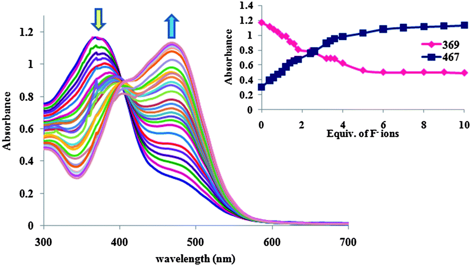 | ||
| Fig. 3 UV-Vis titration of the receptor (2 × 10⁻⁵ mol L−1) in 9:1, DMSO–water with increasing concentrations of NaF (0–10 equiv.) in water. Inset showing the binding isotherm at selected wavelengths in 9:1, DMSO–water. | ||
 | ||
| Fig. 4 Benesi–Hildebrand plot of receptor 1 binding with F− anion associated with absorbance change at 369 nm in 9:1, DMSO–water. | ||
Interestingly, a comparison of the binding constant of 1 towards F− in aqueous media with that found in organic solution revealed that the binding affinity remained strong even in water. It is worth noting that the current system can detect inorganic fluoride even at 0.058 ppm level, which is in accordance with the system reported by Das and co-workers.10 Remarkably, this limit of detection is much lower than the World Health Organization (WHO) permissible level for the detection of fluoride in drinking water (below 1 ppm).13
It has been reported that excess of fluoride ions not only in potable water but also in toothpaste and osteoporosis drugs is responsible for causing dental problems and skeletal fluorosis.14 Hence, for practical purposes and also to demonstrate the potential application of the present receptor for the rapid detection of fluoride ion in real samples, we extended our work to the qualitative detection of fluoride in toothpaste. Finally, we were successful in the naked-eye sensing of fluoride ions. The results were also verified using UV-Vis spectroscopy (Fig. S19, ESI†).
Recently, we were inspired by Trivedi and co-workers,15 who reported the quantitative detection of fluoride ion in real-life applications. Therefore, receptor 1 was tested for the detection of fluoride ion in sea water, collected from the Persian Gulf (latitude 27°49′94′′, longitude 52°62′19′′) and in commercial mouthwash. To our surprise, the addition of one drop of sea water to receptor 1, resulted in a noticeable colour change from light yellow to orange, suggesting that it is possible to use 1 for the naked-eye detection of fluoride ion in sea water. Moreover, F− was detected quantitatively by applying a calibration curve obtained from a plot of absorbance vs. various concentrations of fluoride anions (Fig. 5). The value obtained from the standard plot for sea water, as an unknown sample, was 0.92 ppm, which is in good accordance with the standard value.13b Our control studies showed that there was no interference from phosphate in the colour change resulting from the reaction of 1 with fluoride in sea water (Fig. S21, ESI†). Additionally, the mentioned sensor system allowed the rapid recognition and quantitative detection of fluoride in mouthwash (Fig. S20, ESI†).
 | ||
| Fig. 5 Calibration curve for the determination of the concentration of fluoride anion in sea water. | ||
To investigate the effect of pH on the host–guest binding affinity, we probed the changes in intensity of the absorption band at 467 nm attributed to the formation of the receptor 1–F− complex , over a broad pH range 2–12. As indicated in Fig. 6, there is a drastic drop in the intensity of the absorption band in highly acidic conditions, this is probably due to the protonation of fluoride ion, to form weakly ionized hydrofluoric acid, thereby lowering the affinity of fluoride anion to bind to the receptor active sites.16 As the pH rises, the intensity of the characteristic band increases, this is probably due to an increase in the accessibility of the deprotonated fluoride to form strong hydrogen bonds with the receptor. These pH responses of 1 are not surprising since similar findings have been reported for other colorimetric fluoride sensors.8b,9,10
 | ||
| Fig. 6 Absorbance changes of receptor 1 corresponding to various pHs of aqueous solution containing 4 × 10⁻⁵ mol L−1NaF. | ||
To further study the nature of the interaction between receptor 1 and fluoride, 1H NMR titration experiments were conducted. Fig. 7 shows the 1H NMR spectra of 1 with different amounts of TBAF in DMSO-d6. It is clear that the OH protons of the azo phenol moieties at 13.87 ppm thoroughly disappeared when only 0.5 equiv. of F− was added into a solution of 1. This indicates the formation of strong hydrogen bonds between fluoride anions and active OH groups, as previously reported.17 With increasing further equiv. of F−, the signals of Ha and Hb showed progressive upfield shifts, whereas the signal of Hc exhibited a progressive reverse shift toward downfield. Simultaneously, an upfield shift of the imine proton (Hd) from 9.00 to 8.76 ppm was also observed. The evidence above can be attributed to the two possible effects which are responsible for observed chemical shifts upon OH–fluoride interaction:4e,18 (1) through-bond effects, which increase the electron density on the phenyl ring and promote an upfield shift of the C–H protons and (2) through-space effects, which induce polarization of the C–H bonds, where the partial positive charge creates a downfield shift.
 | ||
| Fig. 7 1H NMR spectra of receptor 1 in DMSO-d6 (2 × 10⁻²mol L−1) in the absence and in the presence of different amounts of TBAF. | ||
Interestingly, a typical HF2− signal was observed in 1H NMR titration experiments at higher concentration of F−, indicating that deprotonation of receptor 1 occurred (Fig. S26, ESI†).4i Consequently, the 1H NMR titration experiments corroborated that F− recognition occurs through initial hydrogen bond formation between anion and sensor, followed by deprotonation. Furthermore, the formation of an initial 1–F− H-bonding host–guest complex is supported by the pKa value, determined from spectrophotometric pH titration (Fig. S22, ESI†).
Conclusion
To sum up, we have designed and synthesized a new azo-azomethine based anion-receptor for the facile detection of fluoride present in aqueous media without any spectroscopic instrumentation. Selective binding of the receptor with fluoride ion through hydrogen-bonding interactions gave rise to a dramatic colour change from light yellow to orange with a concomitant bathochromic shift. Our achieved detection limit of F− ion was 0.058 ppm, which is lower than the WHO recommended level (below 1 ppm) for safe drinking. Notably, we demonstrated that the synthesized sensor can also be considered as a tool for the detection and quantification of fluoride ion in real samples.Experimental
General
All chemicals were purchased from Sigma-Aldrich and used without further purification. Electronic spectral measurements were carried out using an Optizen 3220 UV spectrophotometer in the range of 200–900 nm. FT-IR spectra were recorded with pressed KBr discs, using an Unicom Galaxy Series FT-IR 5000 spectrophotometer in the region of 400–4000 cm−1. Melting points were determined with an Electrothermal 9200 apparatus. NMR spectra were recorded with Bruker AV 300 MHz and Bruker Avance III 400 MHz spectrometers. C. H. N. analyses were performed using a Vario EL III elemental analyzer. Mass spectra were recorded with a FINNIGAN-MATT 8430 mass spectrometer operating at an ionization potential of 70 eV.Synthesis of α,α′-bis(o-aminophenylthio)-1,2-xylene
A mixture of 2-aminothiophenol (12.52 g, 0.10 mol) and sodium hydroxide (4.00 g, 0.10 mol) in deionized water (60 mL) was heated to reflux for 30 min. The mixture was allowed to cool to 60 °C and α,α′-dibromo-o-xylene (13.20 g, 0.05 mol) was added dropwise over a period of 30 min. The mixture was then heated to reflux for 2 h. After cooling to room temperature, the reaction mixture was extracted with chloroform. The organic layer was dried over anhydrous MgSO4 and then evaporated to dryness. The obtained product was further washed thoroughly with diethyl ether and dried under vacuum at room temperature. Yield: 70%,. m. p. 103–106 °C (lit.106 °C).19 1H NMR (DMSO-d6, 400 MHz, ppm): δ 4.02 (s, 4H), 5.31 (br, 4H), 6.46 (t, 2H, J = 7.60 Hz),6.73 (d, 2H, J = 8.00 Hz), 7.05 (m, 8H). IR (KBr, cm−1); 3352 and 3449 (NH), 1607 and 1477 (C![[double bond, length as m-dash]](https://www.rsc.org/images/entities/char_e001.gif) CAr).MS (EI, 70 eV): m/z (%) = 352 (M+, 63),228 (100), 194 (94), 124 (69), 80 (63).
CAr).MS (EI, 70 eV): m/z (%) = 352 (M+, 63),228 (100), 194 (94), 124 (69), 80 (63).
Synthesis of 1-(3-formyl-4-hydroxyphenylazo)-2,4-dichlorobenzene
A mixture of 2,4-dichloroaniline (1.62 g, 10 mmol) in hydrochloric acid (9 mL) and water (4 mL) was heated to 70 °C to to form a solution. The clear solution was poured into an ice-water mixture and was diazotized at 0–5 °C with sodium nitrite (0.7 g, 10 mmol in water, 10 mL). The cold diazo solution was added to the solution of salicylaldehyde (1.22 g, 10 mmol) in water (20 mL) containing sodium hydroxide (0.40 g, 10 mmol) and sodium carbonate (4.24 g, 40 mmol) over a period of 30 min at 0 °C. The reaction mixture was stirred for 1 h at 0 °C and then allowed to warm slowly to room temperature. The product was collected by vacuum filtration and washed with NaCl solution (100 mL, 10%). The obtained solid was dried under vacuum at 80 °C overnight. Yield: 86%, m. p. 120–123 °C. 1H NMR (DMSO-d6, 300 MHz, ppm): δ 7.29 (d, 1H, J = 8.84 Hz), 8.09 (dd, 1H, J = 8.84 Hz, J = 2.16 Hz), 7.55 (d, 1H, J = 8.76 Hz), 7.68 (d, 1H, J = 8.76 Hz), 7.89 (s, 1H), 8.19 (d, 1H, J = 2.16 Hz), 10.37 (s, 1H), 11.88 (br, 1H). IR (KBr, cm−1); 1668 (CHO), 1620 (CC), 1578 (phenol ring), 1485 (NN), 1287 (C–O), 1246, 1148, 866 and 768. Anal.calcd.forC13H8Cl2N2O2: C, 52.91; N, 9.49; H, 2.73%. Found: C, 52.62; N, 9.38; H, 2.65%. λmax (nm) (ε (M−1 cm−1)): 260 (29950), 356 (49260) and 465 (11853) in DMSO.
Synthesis of 1,2-bis(4-((2,4-dichlorophenyl)diazenyl)-2-((2-(methylthio)phenylimino)methyl)phenol)methylenbenzene (1)
A solution of α,α′-bis(o-aminophenylthio)-1,2-xylene (0.35 g, 1 mmol) in absolute EtOH (10 mL) was added to a stirring solution of azo-coupled precursor (0.59 g, 2 mmol) in absolute EtOH (50 mL) during a period of 20 min at 60 °C. The solution was heated in a water bath overnight at 80 °C with stirring. The mixture was filtered whilst hot and the obtained precipitate was washed with hot ethanol (three times) and then with diethyl ether. The resulting product was dried in air. Yield: 96%, m. p. 172–175 °C. 1H NMR (DMSO-d6, 300 MHz, ppm): δ 4.30 (s, 4H), 7.04 (d, 2H, J = 8.80 Hz), 7.20 (m, 2H), 7.30 (m, 6H), 7.46 (m, 6H), 7.60 (d, 2H, J = 8.71 Hz), 7.80 (s, 2H), 7.91 (d, 2H, J = 8.80 Hz), 8.15 (s, 2H), 8.99 (s, 2H), 13.86 (br, 2H). IR (KBr, cm−1); 1614 (CN), 1578 (phenol ring), 1470 (NN), 1288 (C–O), 1097, 831 and 756. Anal. calcd.for C46H32Cl4N6O2S2: C, 60.93; N, 9.27; H, 3.56; S, 7.07%. Found: C, 61.73; N, 9.26; H, 3.16; S, 7.00%.λmax (nm) (ε (M−1 cm−1)): 264 (41200) and 369 (50850) in DMSO.MS (EI, 70 eV): [M]+ = 908 molecular ion peak was not observed. m/z (%) = 399 (25), 226 (94),198 (44), 161 (61), 145 (100), 124 (89), 80 (69).
Acknowledgements
We are grateful to the Arak University for financial support of this work.Notes and references
- (a) H. Sun, X. Dong, S. Liu, Q. Zhao, X. Mou, H. Y. Yang and W. Huang, J. Phys. Chem. C, 2011, 115, 19947–19954 CrossRef CAS; (b) C. Suksai and T. Tuntulani, Chem. Soc. Rev., 2003, 32, 192–202 RSC; (c) P. A. Gale, S. E. Garcia-Garrido and J. Garric, Chem. Soc. Rev., 2008, 37, 151–190 RSC; (d) K. J. C. M. Keaveney and D. A. Leigh, Angew. Chem., Int. Ed., 2004, 43, 1222–1224 CrossRef PubMed; (e) S. Li, C. Zhang, S. Huang, F. Hu, J. Yin and S. H. Liu, RSC Adv., 2012, 2, 4215–4219 RSC; (f) C. Caltagirone and P. A. Gale, Chem. Soc. Rev., 2009, 38, 520–563 RSC; (g) X. Yong, M. Su, W. Wan, W. You, X. Lu, J. Qu and R. Liu, New J. Chem., 2013, 37, 1591–1594 RSC; (h) P. A. Gale, Chem. Commun., 2011, 47, 82–86 RSC; (i) K. Bowman-James, Acc. Chem. Res., 2005, 38, 671–678 CrossRef CAS PubMed.
- (a) X. Cao, W. Lin, Q. Yu and J. Wang, Org. Lett., 2011, 13, 6098–6101 CrossRef CAS PubMed; (b) F. P. Schmidtchen and M. Berger, Chem. Rev., 1997, 97, 1609–1646 CrossRef CAS PubMed.
- (a) E. J. Cho, B. J. Ryu, Y. J. Lee and K. C. Nam, Org. Lett., 2005, 7, 2607–2609 CrossRef CAS PubMed; (b) R. M. Duke, E. B. Veale, F. M. Pfeffer, P. E. Kruger and T. Gunnlaugsson, Chem. Soc. Rev., 2010, 39, 3936–3953 RSC; (c) F. M. Pfeffer, P. E. Kruger and T. Gunnlaugsson, Org. Biomol. Chem., 2007, 5, 1894–1902 RSC; (d) X. Yong, M. Su, W. Wang, Y. Yan, J. Qu and R. Liu, Org. Biomol. Chem., 2013, 11, 2254–2257 RSC; (e) L. M. P. Lima, A. Lecointre, J. F. Morfin, A. de Blas, D. Visvikis, L. J. Charbonnière, C. Platas-Iglesias and R. Tripier, Inorg. Chem., 2011, 50, 12508–12521 CrossRef CAS PubMed; (f) S. Guha and S. Saha, J. Am. Chem. Soc., 2010, 132, 17674–17677 CrossRef CAS PubMed; (g) S. Ayoob and A. K. Gubta, Crit. Rev. Environ. Sci. Technol., 2006, 36, 433–487 CrossRef CAS; (h) J. Aaseth, M. Shimshi, J. L. Gabrilove and G. S. Birketvedt, J. Trace Elem. Exp. Med., 2004, 17, 83–92 CrossRef CAS; (i) M. Kleerekoper, Endocrinol. Metab. Clin. North Am., 1998, 27, 441–452 CrossRef CAS; (j) K. L. Kirk, Biochemistry of the Halogens and Inorganic Halides, Plenum Press, New York, 1991, p. 58 Search PubMed.
- (a) D. A. Jose, D. K. Kumar, B. Ganguly and A. Das, Org. Lett., 2004, 6, 3445–3448 CrossRef CAS PubMed; (b) V. Kumar, M. P. Kaushik, A. K. Srivastava, A. Pratap, V. Thiruvenkatam and T. N. Guru Row, Anal. Chim. Acta, 2010, 663, 77–84 CrossRef CAS PubMed; (c) P. Bose and P. Ghosh, Chem. Commun., 2010, 46, 2962–2964 RSC; (d) P. Dydio, D. Lichosyt and J. Jurczak, Chem. Soc. Rev., 2011, 40, 2971–2985 RSC; (e) X. Peng, Y. Wu, J. Fan, M. Tian and K. Han, J. Org. Chem., 2005, 70, 10524–10531 CrossRef CAS PubMed; (f) L. Wang, X. He, Y. Guo, J. Xu and S. Shao, Org. Biomol. Chem., 2011, 9, 752–757 RSC; (g) H. M. Chawla, R. Shrivastava and S. N. Sahu, New J. Chem., 2008, 32, 1999–2005 RSC; (h) J. V. Ros-Lis, R. Martinez-Manez, F. Sancenon, J. Soto, K. Rurack and H. Weißhoff, Eur. J. Org. Chem., 2007, 2449–2458 CrossRef CAS; (i) A. K. Mahapatra, S. K. Manna and P. Sahoo, Talanta, 2011, 85, 2673–2680 CrossRef CAS PubMed; (j) V. Reena, S. Suganya and S. Velmathi, J. Fluorine Chem., 2013, 153, 89–95 CrossRef CAS PubMed; (k) R. Gu, S. Depraetere, J. Kotek, J. Budka, E. Wagner-Wysiecka, J. F. Biernat and W. Dehaen, Org. Biomol. Chem., 2005, 3, 2921–2923 RSC.
- (a) V. Amendola, M. Bonizzoni, D. Esteban-Gomez, L. Fabbrizzi, M. Licchelli, F. Sancenon and A. Taglietti, Coord. Chem. Rev., 2006, 250, 1451–1470 CrossRef CAS PubMed; (b) S. O. Kang, R. A. Begum and K. Bowman-James, Angew. Chem., Int. Ed., 2006, 45, 7882–7894 CrossRef CAS PubMed; (c) S. Zhang and L. Echegoyen, J. Am. Chem. Soc., 2005, 127, 2006–2011 CrossRef CAS PubMed; (d) C. Li, M. Numata, M. Takeuchi and S. Shinkai, Angew. Chem., Int. Ed., 2005, 44, 6371–6374 CrossRef CAS PubMed; (e) R. Yang, W. X. Liu, H. Shen, H. H. Huang and Y. B. Jiang, J. Phys. Chem. B, 2008, 112, 5105–5110 CrossRef CAS PubMed; (f) D. H. Lee, H. Y. Lee, K. H. Lee and J. I. Hong, Chem. Commun., 2001, 1188–1189 RSC; (g) C. J. Woods, S. Camiolo, M. E. Light, S. J. Coles, M. B. Hursthouse, M. A. King, P. A. Gale and J. W. Essex, J. Am. Chem. Soc., 2002, 124, 8644–8652 CrossRef CAS PubMed; (h) Q. S. Lu, L. Dong, J. Zhang, J. Li, L. Jiang, Y. Huang, S. Qin, C. W. Hu and X. Q. Yu, Org. Lett., 2009, 11, 669–672 CrossRef CAS PubMed; (i) G. W. Bates, P. A. Gale and M. E. Light, Chem. Commun., 2007, 2121–2123 RSC.
- (a) P. Sokkalingam and C.-H. Lee, J. Org. Chem., 2011, 76, 3820–3828 CrossRef CAS PubMed; (b) R. Sakai, E. B. Barasa, N. Sakai, S. Sato, T. Satoh and T. Kakuchi, Macromolecules, 2012, 45, 8221–8227 CrossRef CAS.
- (a) S. Kubik, Chem. Soc. Rev., 2010, 39, 3648–3663 RSC; (b) A. Bencini, C. Coluccini, A. Garau, C. Giorgi, V. Lippolis, L. Messori, D. Pasini and S. Puccioni, Chem. Commun., 2012, 48, 10428–10430 RSC; (c) F. Huang, C. Cheng and G. Feng, J. Org. Chem., 2012, 77, 11405–11408 CrossRef CAS PubMed.
- (a) A. K. Mahapatra, G. Hazra and P. Sahoo, Beilstein J. Org. Chem., 2010, 6 CrossRef CAS PubMed; (b) Z. H. Lin, S. J. Ou, C. Y. Duan, B. G. Zhang and Z. P. Bai, Chem. Commun., 2006, 624–626 RSC; (c) Y. M. Zhang, Q. Lin, T. B. Wei, D. D. Wang, H. Yao and Y. L. Wang, Sens. Actuators, B, 2009, 137, 447–455 CrossRef CAS PubMed; (d) Z. H. Lin, Y. G. Zhao, C. Y. Duan, B. G. Zhang and Z. P. Bai, Dalton Trans., 2006, 3678–3684 RSC.
- T. Gunnlaugsson, P. E. Kruger, P. Jensen, J. Tierney, H. D. P. Aliand and G. M. Hussey, J. Org. Chem., 2005, 70, 10875–10878 CrossRef CAS PubMed.
- P. Das, A. K. Mandal, M. K. Kesharwani, E. Suresh, B. Ganguly and A. Das, Chem. Commun., 2011, 47, 7398–7400 RSC.
- H. A. Benesi and J. H. Hildebrand, J. Am. Chem. Soc., 1949, 71, 2703–2707 CrossRef CAS.
- (a) M. Vázquez, L. Fabbrizzi, A. Taglietti, R. M. Pedrido, A. M. González-Noya and M. R. Bermejo, Angew. Chem., Int. Ed., 2004, 43, 1962–1965 CrossRef PubMed; (b) V. Thiagarajan, P. Ramamurthy, D. Thirumalai and V. T. Ramakrishnan, Org. Lett., 2005, 7, 657–660 CrossRef CAS PubMed; (c) B. Liu and H. Tian, Chem. Lett., 2005, 34, 686–687 CrossRef CAS; (d) H. Miyaji and J. L. Sessler, Angew. Chem., Int. Ed., 2001, 40, 154–157 CrossRef CAS.
- (a) J. Fawell, K. Bailey, J. Chilton, E. Dahi, L. Fewtrell and Y. Magara, Fluoride in Drinking-water, IWA Publishing, London, 2001, p. 32 Search PubMed; (b) WHO Technical Report Series-846, Fluoride and Oral Health, Geneva, 1994.
- (a) G. Saikia, A. K. Dwivedi and P. K. Iyer, Anal. Methods, 2012, 4, 3180–3186 RSC; (b) WHO, Guidelines for Drinking-Water Quality, World HealthOrganization, Geneva, 2004 Search PubMed.
- M. Prasad, A. N. Shetty and D. R. Trivedi, RSC Adv., 2012, 2, 3133–10504 RSC.
- I. I. Abbas, H. H. Hammud and H. Shamsaldeen, Eur. J. Chem., 2012, 3, 156–162 CrossRef CAS.
- (a) P. Thiampanya, N. Muangsin and B. Pulpoka, Org. Lett., 2012, 14, 4050–4053 CrossRef CAS PubMed; (b) J. Yoo, M. S. Kim, S. J. Hong, J. L. Sessler and C. H. Lee, J. Org. Chem., 2009, 74, 1065–1069 CrossRef CAS PubMed; (c) A. S. F. Farinha, A. C. Tomé and J. A. S. Cavaleiro, Tetrahedron, 2010, 66, 7595–7599 CrossRef CAS PubMed.
- M. Boiocchi, L. Del Boca, D. E. Gómez, L. Fabbrizzi, M. Licchelli and E. Monzani, J. Am. Chem. Soc., 2004, 126, 16507–16514 CrossRef CAS PubMed.
- Y. Gok, S. Karabocek, N. Karabocek and Y. Atalay, New J. Chem., 1995, 19, 1275–1283 CAS.
Footnote |
| † Electronic supplementary information (ESI) available: Characterization data of all compounds, UV-Vis spectrophotometric titarations, stoichiometric determination by Benesi–Hildebrand method, Job's plot, UV-Vis spectral changes upon addition of protic solvent, figures and photographs showing the sensing behavior, procedure details of interference study and details of qualitative and quantitative detection of fluoride by prepared sensor. See DOI: 10.1039/c3ra42709a |
| This journal is © The Royal Society of Chemistry 2014 |