DOI:
10.1039/C3RA42462F
(Paper)
RSC Adv., 2014,
4, 1015-1024
Unraveling the mode of binding of the anticancer drug topotecan with dsDNA†
Received
17th May 2013
, Accepted 8th October 2013
First published on 9th October 2013
Abstract
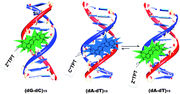
The binding interactions between antitumor drugs and DNA are of burgeoning interest due to increasing demand in medicinal science. In the present work, we have tried to examine the mode of binding of topotecan (TPT) with DNA. TPT, an eminent anti-cancer drug from the Camptothecin family, is found to interact with DNA Topoisomerase-I and inhibits the DNA replication process. Steady state, time resolved fluorescence, circular dichroism and thermal melting studies have been utilized to explore the mode of binding of TPT with synthetic polynucleotides ((dA-dT)15, (dG-dC)15) and natural DNA (CT-DNA). The mode of binding of TPT with the DNA double helix has been substantiated to be principally groove binding. It is found that even though the ground state cationic form (C) of the drug binds to dsDNA irrespective of DNA sequences, the emission mainly appears from Z*, and it is attributed to the intermolecular excited state proton transfer (ESPT) reaction between the drug and surrounding water molecules. However, in the case of (dA-dT)15, the emission profile indicates the existence of a small population of excited state cationic form (C*) of the drug in the minor groove of DNA. The different photophysical behavior of TPT in the case of (dA-dT)15 compared to others is attributed to the narrower and deeper minor groove of (dA-dT)15 than that of the others. The exact molecular picture of the binding interaction between the drug and DNAs has been explored from molecular modeling studies.
Introduction
Chemotherapeutic agents have shared the spotlight over the decades due to their increasing demand in medicinal science. Although the anti-cancer activity of chemotherapeutic drugs is well known, the exact mode of action in many cases is still under debate. Therefore, chemotherapeutic drugs are of burgeoning interest to researchers.1–4 Among many others, topotecan (TPT), a pentacyclic water soluble alkaloid member of the Camptothecin family, is our point of interest due to its proven inhibitory activity against animal as well as human tumors. CPT was first isolated by Wall et al. in 1966 from a Chinese tree, Camptotheca acuminate.5 Water solubility is the main advantage of TPT compared to other drugs of the Camptothecin family, and it mainly exists in two forms in water, namely, the lactone form, responsible for TPT's anticancer activity, and the carboxylate form, which does not show any anti-tumor action.6,7 Note that, whereas most of the well known anti-cancer drugs are found to inhibit DNA Topoisomerase-II, TPT selectively interacts with Topoisomerase I (Topo-I) and inhibits DNA replication, and leads to necrosis or cell death.4,8–10 This overall process is believed to be governed by the formation of a ternary complex between DNA–TPT and Topo-I,11 though it was shown that TPT can interact solely with DNA in the absence of Topo-I.12
More recently, Nunzio et al. reported elaborate pH dependent studies and solvent effects on the structural dynamics of TPT, and it was found that TPT exists in different protolytic forms depending on the pH as well as on the solvent.13,14 It was observed that under physiological conditions (pH 7), the drug exists in equilibrium between the enol (E), cationic (C) and zwitterionic (Z) forms in the ground state (Scheme 1).13,14 Interestingly, TPT exhibits a single fluorescence peak (∼530 nm) in aqueous solution responsible for the emission from Z*, which is believed to result from the excited-state proton transfer (ESPT) from TPT to water.15 At physiological pH, TPT hydrolyses to a toxic carboxylate form,16 and this appears to be a strong challenge for researchers. Therefore, ongoing efforts have been invested to carry the active lactone form to the desired location. Results show that the stability of the lactone moiety could be improved by encapsulating the drug either in liposome17,18 or in cyclodextrin,19 which might solve the problem of carriage. Next, the mode of interaction with DNA and its mechanism is also a prime point of interest, which can improve the therapeutic importance of TPT. The mode of interaction for lactone TPT and DNA devoid of Topo-I was determined by Yang et al. using HPLC followed by NMR techniques.20 The results conclude that the lactone form predominantly exists in the presence of dsDNA and binds to DNA through intercalation, though the relative shifts (∼0.15–0.2 ppm) reported from NMR spectra do not impressively match with those of a regular intercalation process.21–23 Again the possibility of intercalation of the lactone form of TPT was questioned when Streltsov et al. showed by linear dichroism study that the angle of orientation for bound TPT (quinoline part) with DNA base pairs is found to be less than 54°.24 This value is clearly less than that of a classical intercalator (62–76°) and close to that of minor groove binders (<55°), which suggests TPT acts as a minor groove binder.24 Not only the mode of binding, but the sequence specificity of TPT for DNA is also under strong debate. Yang et al. reported that the lactone form of TPT is highly selective towards poly(dG-dC) compared to poly(dA-dT) for intercalation.20 Interestingly, Yao et al. reported that sequence specificity of TPT is towards dT of dsDNA instead of the GC rich region,12 which contradicts previous reports. The ongoing quest into TPT's mode of binding along with sequence selectivity has attracted our attention, and we have carried out thorough investigations to eliminate the contradictions surrounding the TPT and DNA interaction. Absorption, steady state emission, circular dichroism (CD), and thermal melting studies have been employed to investigate the binding mode, specificity and affinity of TPT with DNA. The dynamic aspects of the interactions are also highlighted through time-resolved fluorescence as well as rotational relaxation measurements.
 |
| Scheme 1 Different prototropic forms of TPT in aqueous solution. | |
TPT being an important anticancer drug, the detailed mechanism of interaction is helpful to enhance the therapeutic efficiency of the drug, as stronger binding to the DNA backbone along with increased time in the bound state enhances the therapeutic efficiency of drugs.25,26 Moreover, specific binding of TPT to DNA makes it possible to use this drug as a fluorescent marker in histochemical studies to exclusively locate DNA or chromosomes in cells.27–29 TPT's sequence specific binding in DNA may further be useful to trace particular short sequence repeats, which are of great importance in DNA polymorphism.30,31 We hope this new understanding will pave the way to reveal specific details of the interaction of a renowned anti-carcinogen TPT with DNA, which may help to better design, target and trace several cellular phenomena in vitro as well as in cells.
Experimental section
1. Materials, methods and instrumentation
Topotecan (high purity, ≥99%) and Calf Thymus DNA (CT-DNA) were purchased from Sigma-Aldrich and used without any further purification. (dA-dT)15 and (dG-dC)15 were procured from Integrated DNA Technologies (IDT) and purified using the SDS-PAGE gel purification technique. The concentrations of (dA-dT)15 and (dG-dC)15 were calculated using the cumulative molar extinction coefficient of all bases generated by SciTools on the IDT website (332
200 M−1 cm−1 and 251
400 M−1 cm−1, respectively). CT-DNA concentration was calculated using the molar extinction coefficient 6600 M−1 cm−1 per base.32 Phosphate buffer (PB) of pH 5.1 was used for all measurements, and each of the constituents of the buffer was also procured from Sigma-Aldrich. The working concentration of topotecan was calculated using the molar extinction coefficient at 380 nm (ε380 = 20
000 M−1 cm−1)24 and kept at 20 μM for all the studies.
Measurements of solution pH were performed by a pH-1500 instrument (Eutech Instruments, USA) and cross verified by a silicon micro sensor pocket sized pH meter (ISFETCOM. Co. Ltd., Japan). Absorption spectra of free and DNA bound TPT were recorded in a Evolution-300 UV-Visible spectrophotometer (Thermo Fisher Scientific, USA). Steady state fluorescence measurements were executed in a Fluoromax 400 instrument (Horiba Jobin Yvon, USA). Fluorescence lifetime and time resolved anisotropy measurements were performed using a time-correlated single photon counting (TCSPC) set-up from IBH Horiba Jobin Yvon (USA), the detailed description of which is mentioned elsewhere.33–36 The lifetime and anisotropy data were collected by excitation at 375 nano LED (FWHM = 90 ps) and analyzed using IBH DAS6 software. The quality of lifetime and anisotropy fits were judged on the basis of χ2 values and from the visual inspection of the residuals. The value of χ2 ≈ 1 was considered as the best fit for the plots. Experimental error in our TCSPC measurements was ±5%.
Circular dichroism (CD) spectra were recorded on a J-815 CD instrument (JASCO, USA). Each CD profile is an average of 3 scans of the same sample collected at a scan speed of 100 nm min−1, with a proper baseline correction of the blank buffer. During CD measurement, DNA concentration was fixed and the concentrations of TPT were increased steadily. Melting studies were performed using a Varian Cary 300 Bio UV-Vis Spectrophotometer. All the steady state and time-resolved experiments were performed at 300 K.
2. Molecular docking
The crystal structure of (dA-dT)n [d(ATATATATAT)], obtained from RSCB Protein Data Bank (PDB ID 3EY0 (ref. 37)), was used for the docking study. We did not obtain a similar crystal structure of the (dG-dC)n sequence, and therefore we have chosen a crystal structure [d(CCGCCGGCGG), PDB ID 1QC1 (ref. 38)] of close resemblance to our sequence. 1DCV (ref. 39) [d(CCGCTAGCGG)] has been utilized for the docking study in place of CT-DNA. The energy optimization of the TPT conformer was performed using the HF/6-31G level of the Gaussian 98 suite, and the resultant energy minimized geometry was saved in Autodock 4.2 compatible format. We have used the Lamarckian Generic Algorithm to elucidate the mode of binding of TPT with DNA in Autodock 4.2 software.40–42 At the beginning of the study, all water molecules were removed and Gasteiger charges were computed followed by the addition of hydrogen as required by the Lamarckian Generic Algorithm.41 As the approximate location of binding was not known, we fixed a grid box with dimensions 120 × 120 × 120 along the x, y, and z axes, with a grid spacing of 0.56 Å to cover all the DNA atoms, and a blind docking was performed to locate the possible position of the probe. TPT was docked in DNA with a population size of 150 GA and 100 GA runs. The lowest possible energy structure was found using rank by energy presentation. After selecting the possible locations of TPT, the size of the grid box was decreased to 60 × 60 × 60 in respective directions with a grid spacing of 0.375 Å to generate the most stable docked form through finite docking studies. The respective structure was used for visualization through Python molecular viewer. The final structure for presentation has been generated using Chimera.43
Results and discussion
1. Absorption study
All the experiments with TPT were carried out at pH 5.1, as TPT exclusively exists in the active lactone form at this pH (the pKa of the carboxylic group is ∼6.5).14 Although the drug can exist in different prototropic forms (Scheme 1), it exists mainly in the cationic form in aqueous solution at pH 5.1, as the pKa values of dimethylamino and 10-hydroxyl groups are 9.5 and 6.99, respectively.14 Notably, the DNA structure is not perturbed at this pH, as the pKa values for deprotonation of adenine and cytosine nucleobases are 3.5 and 4.2.44 TPT in phosphate buffer at pH 5.1 exhibits dual absorption bands at ∼368 nm and at ∼381 nm (Fig. 1), which are attributed to the π–π* type absorption of the quinoline moiety. The very negligible absorbance at ∼410 nm indicates that the ground state zwitterionic (Z) form does not exist at this pH (Fig. 1). With the gradual addition of (dG-dC)15 and (dA-dT)15, a decrease in absorption of both the peaks is observed (Fig. 1). However, the peak at ∼368 nm almost vanishes at the maximum concentration of DNA (100 μM), whereas the peak at ∼381 nm is retained with a decreased absorption. Such changes in absorption spectral features of TPT clearly indicate the strong interaction between DNA and TPT. Considering favorable electrostatic stabilization between the cationic form of the drug and the negatively charged phosphate backbone of DNA, we believe the cationic form of the drug preferentially interacts with DNA compared to other forms (enol or zwitterionic form) of the drug. Similar observations have been reported in the case of sanguinarine, where the cationic form of the drug also strongly interacts with DNA compared to the enol form.45 The existence of an isosbestic point at ∼387 nm indicates the presence of an equilibrium between the free drug and the DNA-bound drug. To check the interaction behavior of the drug with natural DNA, we have also probed the interaction behavior between TPT and CT-DNA, and it shows close resemblance to the synthetic polynucleotide absorption changes (see Fig. S1a in ESI†). The association constants for the TPT–DNA (insets of Fig. 1) interaction have been calculated from the half reciprocal plot46 using the changes observed at 381 nm, which are found to be 1.35 × 10⁴ (±10%) M−1, 1.23 × 10⁵ (±10%) M−1 and 4.8 × 10³ (±10%) M−1 for (dG-dC)15, (dA-dT)15 and CT-DNA, respectively. The association constants along with the free energy changes (−5.71 kcal mol−1, −7.03 kcal mol−1 and −5.08 kcal mol−1 for (dG-dC)15, (dA-dT)15 and CT-DNA, respectively) infer that the ground state binding between the drug and DNA is a thermodynamically favorable process.
 |
| Fig. 1 Absorption spectra of TPT (20 μM) in the presence of (a) (dG-dC)15 and (b) (dA-dT)15. 1 → 10 represents DNA concentrations 0, 1, 2, 5, 10, 15, 20, 40, 80, and 100 μM, respectively. Inset shows the half reciprocal plot used to calculate binding constants. | |
2. Fluorescence study
The DNA induced modification of the steady-state emission profile of the drug is depicted in Fig. 2. The emission profile of TPT in aqueous buffer medium at pH 5.1 is characterized by a single unstructured band at 530 nm upon excitation at 380 nm. It is already established that the excited state zwitterionic (Z*) form of TPT is the main emitting species in water (pH 6.25), while the fluorescence from C* is not detectable in water (pH 6.25).13 Therefore, the emission peak at 530 nm of TPT in aqueous solution originates from the Z* form of TPT. It is interesting to note that, although the cationic form of TPT (C-TPT) is the major ground state species at this pH, the resultant fluorescence is from Z*. This observation is attributed to the excited state proton transfer (ESPT) reaction from the 10-hydroxyl group of C-TPT to water, which leads to the formation of Z* in the excited state.15 A similar kind of ESPT process was already reported in the case of 10-hydroxy camptothecin by Solntsev et al.47 As displayed in Fig. 2a, upon gradual addition of (dG-dC)15, the emission intensity at 530 nm progressively decreases, and it exhibits almost 50% quenching at the maximum concentration of DNA (∼100 μM) without any shift in emission position. On the other hand, addition of (dA-dT)15 produces relatively lower intensity quenching (∼35%) with a slight blue shift of the emission position (∼5 nm). Astonishingly, in the case of (dA-dT)15 a small, emerging peak appeared at ∼420 nm, and at maximum DNA concentration (∼100 μM) it appears as a prominent peak (Fig. 2b). Decreasing intensity at 530 nm for both the synthetic dsDNAs infers the relative lowering of the Z* population of TPT through the interactions with the abovementioned DNA. The appearance of the emission peak at 420 nm (Fig. 2b) infers that some population of TPT remains in the C* state instead of converting to Z* in the excited state after binding to (dA-dT)15. It is interesting to note that the population of C* is not present in the case of (dG-dC)15 (Fig. 2a) and CT-DNA (Fig. S1b in ESI†), as the 420 nm peak does not appear in those systems. Although the exact reason for such selective interaction is difficult to conclude at this point, after confirmation of the binding modes of TPT, we hope that we will be able to predict more precisely why (dA-dT)15 selectively preserves the excited state cationic (C*) form of TPT.
 |
| Fig. 2 Fluorescence emission spectra of TPT (20 μM) in the presence of (a) (dG-dC)15 and (b) (dA-dT)15. 1 → 10 represents DNA concentrations 0, 1, 2, 5, 10, 15, 20, 40, 80 and 100 μM, respectively. Inset shows the double logarithmic plot for the determination of the binding constant. | |
After exploring the trends in emission, we have tried to elucidate the possible mechanism of quenching. Usually, there are two distinct possibilities of quenching, namely, photo-induced electron transfer (PET) or photo-induced energy transfer. The possibility of energy transfer can easily be ruled out as it requires overlap between the emissions of the donor (Z* TPT) and the absorption of the acceptor (nucleobases), which is not possible in the present case. Therefore, the most plausible mechanism for fluorescence quenching is PET between TPT and nucleobases, as it is already reported that PET between drugs and nucleobases leads to fluorescence quenching.48–51 Note that guanine is the better electron donor among all nucleobases,49 and hence, the poly(dG-dC) sequence exhibits a greater quenching effect compared to poly(dA-dT). When nucleobase(s) of dsDNA participate in quenching it demands a close proximity to the drug. There are three distinct possibilities for a drug to interact with a dsDNA in close proximity, namely intercalation, groove binding and electrostatic interaction. Although the first two processes can lead to quenching of fluorescence intensity, electrostatic interaction alone cannot lead to fluorescence quenching to such an extent. The changes in emission intensity have been utilized to calculate the Stern–Volmer (S–V) quenching constant. The S–V plot shows a straight line fit for (dG-dC)15 (KSV = 9.6 × 10³ M−1), whereas a curved plot towards the x-axis is obtained for the (dA-dT)15 sequence (Fig. S2†). A straight line S–V plot is strongly indicative of the fact that TPT molecules are bound to (dG-dC)15 DNA in a single mechanistic manner, whereas curvature towards the x-axis in the S–V plot is suggestive of the concomitant existence of two different distributions of TPT molecules in the presence of (dA-dT)15. The quenching results are corroborative with our fluorescence spectral features, where it has been observed that the zwitterionic form (Z*) of TPT is the sole emitting species in the case of (dG-dC)15, but in the presence of (dA-dT)15 both C* and Z* are the emitting species, although the latter species are in the majority.
The binding constant is calculated for the quantitative representation of drug–DNA binding affinity using the following equation:52–54
|
log[(F0 − F)/F] = log K + n log[Q]
| (1) |
where
F0 and
F are the fluorescence intensities of the drug (at 530 nm) in the absence and the presence of polynucleotides, respectively. A graph of log[(
F0 −
F)/
F]
versus log[Q] yields
K and
n (see insets in
Fig. 2), which represent the binding constant and number of binding sites, respectively. A linear plot for the (dG-dC)
15 system generates
K and
n values of 3.3 × 10
3 (±10%) M
−1 and 0.9, respectively. For (dA-dT)
15, we have obtained a curved plot. Hence, the binding constant is calculated using experimental points up to which the plot is linear, and is calculated to be 0.8 × 10
3 (±10%) M
−1. The binding constant for TPT binding with CT-DNA is found to be 0.54 × 10
3 (±10%) M
−1. The corresponding free energy changes are −4.83 kcal mol
−1, −4.0 kcal mol
−1 and −3.77 kcal mol
−1 for (dG-dC)
15, (dA-dT)
15 and CT-DNA, respectively. The binding affinity of the drug obtained from the fluorescence study is slightly less than that from the absorption study, presumably because in emission we have monitored the intensity of the zwitterionic form, which probably has a lower binding affinity compared to the cationic form of the drug in the ground state. We have also carried out similar experiments with (dG-dC)
7 and found that the binding constant with TPT is 1.03 × 10
3 (±10%) M
−1 (Fig. S3 in ESI
†), which closely resembles the association constant of TPT with (dG-dC)
15. These findings clearly demonstrate that the length of flanking bases has a negligible role on the binding affinity of TPT with dsDNA.
Up to this point, we are certain that the drug is interacting with dsDNA, however, we cannot precisely predict the mode of binding. To elucidate the mechanism of interaction between the drug and dsDNA, we have carried out several studies, such as intercalator displacement assay, KI titration and salt concentration dependent fluorescence, circular dichroism, time-resolved experiments and molecular modeling studies, which are discussed below.
3. Ethidium bromide (EtBr) displacement assay and KI titration
Among different modes of interaction (namely, intercalation, groove binding, and electrostatic), we first consider the possibility of intercalation through an EtBr displacement assay experiment (Fig. S4†). It is already evident from fluorescence experiments that TPT emits at ∼530 nm in the presence of (dG-dC)15, whereas a dual emission at 530 nm and 421 nm appears in the case of (dA-dT)15. With increasing EtBr concentration (up to 40 μM) in TPT–DNA systems, we observe a dominant emission peak at ∼600 nm, which is the signature peak of EtBr intercalating into DNA.55,56 Interestingly, no emission enhancement is observed at 530 nm in the presence of EtBr. If TPT acts as an intercalator, then one would expect an instant replacement of the TPT molecule from DNA by one of the most efficient intercalators EtBr, and as an outcome the emission of TPT would retract back to its initial intensity as obtained in aqueous medium. As no such changes in the TPT spectra have been observed through the EtBr displacement assay, one can definitely conclude that TPT is not binding to either (dG-dC)15 or (dA-dT)15 through an intercalative binding mode.
Monitoring the quenching of fluorescence of free TPT and DNA-bound TPT also paves a way for a simple but efficient technique for exploring the mode of binding of the drug with DNA. I− being an excellent quencher can be used to clarify the type of interaction between drug and DNA. I− can quench either electrostatically bound or groove bound drugs, as these positions are fully and partially accessible by the quencher, whereas for intercalated drugs hardly any quenching can be observed.52,54 When KI is employed as an external quencher, we found that its quenching efficiency toward TPT is reduced in the presence of DNA compared to the TPT–KI interaction in the absence of DNA (Fig. S5†). The observation of a quenching effect of TPT even in the presence of either (dA-dT)15 or (dG-dC)15 supports an electrostatic or groove binding mode of binding of TPT into DNA double strands, since electrostatic or groove binding leads to ample exposure of the fluorophore to the bulk aqueous buffer phase. The quenching effect is greater in the case of (dG-dC)15 compared to (dA-dT)15, and it is attributed to the wider minor groove of (dG-dC)15 than (dA-dT)15.57,58 TPT bound to the wider groove of (dG-dC)15 can be relatively more exposed to KI compared to (dA-dT)15, where the drug is located in the narrow minor groove. In a nutshell, the fluorescence studies clearly infer that TPT is not involved in an intercalative mode of binding to the dsDNA. This conclusion is further confirmed by the thermal melting study of DNA in the presence of TPT (Fig. S6 in ESI†). DNA intercalation, being the strongest mode of interaction, shows large changes in melting temperature.22,59 However, we observed no prominent change in melting temperature either for (dA-dT)15 or (dG-dC)15. Therefore, the thermal melting study corroborates well our conjecture from fluorescence studies, where we have also found that TPT does not interact with dsDNA through an intercalative mode of binding.
4. Salt concentration dependence assay
Salt concentration dependent assays are a good tool to verify the role of electrostatic interactions between a drug and DNA. To elucidate the type of interaction between TPT and DNA, we have carried out the same experiment of TPT–DNA titration in the presence of a high concentration of NaCl (Fig. S7†). Interestingly, when the TPT was titrated by DNA in PBS having an NaCl concentration of 100 mM, we have observed that the quenching effect of TPT is lower compared to the changes observed in non-NaCl buffer medium (PB). This observation suggests that electrostatic interactions also play a role in the TPT–DNA interaction. Here it is pertinent to mention that NaCl (a source of Na+ ion) can effectively screen the negative charge of the phosphate backbone of DNA and weakens the possible electrostatic attraction between the positively charged drug and DNA. Moreover, Na+ and other monovalent alkali metals are very well known for their groove binding and groove narrowing effects.60–62 Eventually, the minor groove becomes narrower when Na+ binds to the outer edge of the groove having a direct contact with the phosphates.60–62 The lower extent of change for (dA-dT)15 is likely to be due to the narrower groove size (3–4 Å) of poly(dA-dT) compared to that of poly(dG-dC) (5–7 Å).57,58,60 In the presence of 100 mM NaCl, the groove width of poly(dA-dT) becomes narrower. Therefore, it is very likely that the molecule cannot fully access the minor groove in these conditions, and as a result the quenching effect is lower in the presence of NaCl. The peak of the cationic (C*) form of TPT at 420 nm, which becomes visible after addition of (dA-dT)15, could not be observed in phosphate buffer containing 100 mM NaCl (Fig. S7b†). This may be attributed to the fact that the drug molecule cannot fully access the minor groove at high salt concentrations (as Na+ ion reduces the groove size). Therefore, the drug is more exposed in water, and as a result the cationic form (C) of TPT cannot exist in this exposed condition, as it subsequently converts to the Z* form of drug in an aqueous environment. In the case of natural DNA, a similar effect is observed in the presence of 100 mM NaCl.
5. Circular dichroism
Circular dichroism (CD) is a very sensitive technique for elucidating the modification of the secondary structure of biopolymers as a result of interaction with small molecules.60,63–65 Therefore, we have exploited the CD technique in order to confirm the probable mode of binding of TPT with (dA-dT)15, (dG-dC)15, and natural CT-DNA using CD spectroscopy (Fig. 3). In this study polynucleotide concentration is kept constant throughout the measurements and changes of CD spectra were monitored with increasing concentrations of drug. The CD spectra of both the synthetic DNAs shows a positive peak at ∼275 nm, and a negative peak in the 245–250 nm region, which is a signature of the right handed B-form63,64 (Fig. 3). These bands originate from stacking interactions between the bases and the helical structure of the polynucleotide, respectively.63–65 With the progressive addition of TPT, the intensity of the positive peak of both the DNAs is found to be almost unperturbed and no induced CD is observed in the visible region. The unaffected base stacking interaction and the absence of induced CD indicates that intercalation is not the binding mode of TPT into the DNA helix, as intercalation leads to significant disruption of the base stacking interaction.66 On the other hand, the reduced helicity of both the DNAs (reflected by the decrease of ellipticity at around ∼245–250 nm, Fig. 3) dictates that TPT interacts with dsDNA through a groove binding mode, as it is known that groove binding can lead to the perturbation of ellipticity of the DNA helix.51 Similar observations are also found for natural CT-DNA (Fig. S8 in ESI†). Therefore, CD results are well supportive of the fluorescence and thermal melting results, where we have also observed that TPT interacts with DNA mainly through a groove binding mode, although in the previous section we have found that electrostatic interactions also have a slight role in the interaction process between TPT and DNA.
 |
| Fig. 3 CD spectral profiles of (a) (dG-dC)15 and (b) (dA-dT)15 in the presence of varying concentrations of TPT. | |
6. Time resolved study
6a. Lifetime measurements. To obtain clear insight into the drug–DNA interaction, the time-resolved decays of TPT have been recorded in the absence and in the presence of various concentrations of DNA, exciting at 375 nm. The decays are shown in Fig. S9, ESI† and the results are summarized in Table 1. It is clear from the fitting results that the drug exhibits a bi-exponential decay feature at pH 5.1. The long and major component (5.79 ns, 93%) reflects the lifetime of the Z* form of TPT. However, the origin of the fast component at 0.79 ns, which has a contribution of almost 7% to the decay profile, is not clear to us. Notably, we have already mentioned that, although we are selectively exciting the C form of the drug, Z* becomes the major emitting species (λem = 530 nm) due to an ESPT process. We believe that a small population of C* is also contributing to the decay profile collected at 530 nm, due to the existence of protolytic equilibrium between C* and Z*. The data compiled in Table 1 reveals that the drug lifetime is almost unperturbed in the case of (dG-dC)15, even at the highest DNA concentration. However, we have observed a quenching effect of almost 60% in the steady state emission profiles for the (dG-dC)15 sequence. Therefore, it is logical to conclude that TPT forms a ground state dark complex when it binds to the minor groove of DNA, and this dark complex does not emit in the excited state. This conclusion remains the same even for CT-DNA, as we did not notice any change in lifetime for the CT-DNA and TPT interaction.
Table 1 Time-resolved fluorescence decay parameters of TPT in the absence and in the presence of dsDNAs collected at 530 nm (a) and 420 nm (b)
(a) |
Sample |
τ1 |
τ2 |
a1 |
a2 |
τava |
χ2 |
τav = (τ1a1 + τ2a2). τav = (τ1a1 + τ2a2 + τ3a3). |
TPT in PB pH 5.1 |
0.79 |
5.79 |
0.07 |
0.93 |
5.43 |
1.01 |
(dG-dC)15 10 μM |
0.69 |
5.80 |
0.07 |
0.93 |
5.42 |
1.07 |
(dG-dC)15 100 μM |
1.26 |
5.80 |
0.05 |
0.95 |
5.59 |
1.11 |
(dA-dT)15 10 μM |
1.61 |
6.01 |
0.12 |
0.88 |
5.46 |
1.04 |
(dA-dT)15 100 μM |
2.28 |
6.60 |
0.26 |
0.74 |
5.49 |
1.14 |
CT-DNA 10 μM |
1.7 |
5.80 |
0.05 |
0.95 |
5.6 |
1.21 |
CT-DNA 100 μM |
1.09 |
5.80 |
0.07 |
0.93 |
5.45 |
0.97 |
(b) |
Sample |
τ1 |
τ2 |
τ3 |
a1 |
a2 |
a3 |
τavb |
χ2 |
(dA-dT)15 10 μM |
0.06 |
0.42 |
1.7 |
0.61 |
0.32 |
0.07 |
0.30 |
1.3 |
(dA-dT)15 40 μM |
0.08 |
0.50 |
2.1 |
0.5 |
0.42 |
0.08 |
0.42 |
1.2 |
(dA-dT)15 100 μM |
0.08 |
0.47 |
1.9 |
0.45 |
0.44 |
0.10 |
0.45 |
1.3 |
In the case of (dA-dT)15, the lifetime of the short component progressively increases with the gradual increase of polynucleotide concentration (Table 1, Fig. S9b in ESI†). At the highest DNA concentration, the lifetime of the short component is increased to 2.3 ns, together with an increased relative contribution of 26%. As the short component is already assigned to the cationic form of TPT, the increased lifetime along with the increased relative contribution of the short component infers that the cationic form of TPT is being stabilized in the presence of DNA due to electrostatic interactions between the negatively charged phosphate backbone and the positive charge of the drug. The increased lifetime of the long component, which is assigned to the lifetime of the Z* form of TPT, is attributed to stability gained by the Z* form of TPT when it binds to the minor groove of DNA. The decreased relative contribution of the Z* form of TPT is due to the slight shifting of equilibrium between Z* and C* towards the latter side. In the case of (dA-dT)15, we have also measured the lifetime of the cationic form (C*) of TPT at 420 nm (Table 1), as this peak appears in the emission profile of the abovementioned synthetic DNA. It is clear from the analysis that the average lifetime of the C* form of TPT increases from 300 ps to 450 ps, inferring stability is gained by the C* form of TPT in the presence of (dA-dT)15. This corroborates well with the steady state observation, where we have observed prominent enhancement of the 420 nm emission peak in the presence of (dA-dT)15. In a nutshell, the lifetime results infer that (dA-dT)15 offers a distributed binding of different forms of TPT, whereas (dG-dC)15 binds specifically one form of the drug. The molecular picture of the interaction between different forms of the drug and dsDNA will be clear from the molecular modeling study discussed in a later part of the manuscript.
6b. Anisotropy measurements. Time resolved anisotropy offers a vivid glance into the system induced restrictions over the tumbling motion of emitting species in the excited state.67 The anisotropy decays of TPT in buffer and in the presence of DNA are shown in Fig. 4. The rotational relaxation of TPT in buffer solution collected at 530 nm exhibits a single exponential decay with a rotational diffusion time of 220 ps. Upon addition of (dA-dT)15, the rotational motion of TPT is retarded. At the maximum (dA-dT)15 concentration, TPT shows a dominant 4.3 ns component, which may be attributed to DNA bound TPT (Fig. 4). This significant alteration in rotational time of the drug is only possible when the drug resides in a highly restricted milieu. These results are in accordance with steady state and time resolved lifetime results, where we have inferred that the Z* form of TPT undergoes a groove binding interaction with (dA-dT)15. Other protolytic forms of TPT, namely the C* form, which is generated in the presence of (dA-dT)15, also exhibits restriction in rotational motion (Table 2), as it also binds in the minor groove region of (dA-dT)15. No such changes have been observed in the case of (dG-dC)15, as it is already evident from steady state and time resolved studies that TPT forms a dark complex when it binds to (dG-dC)15. For CT-DNA, TPT also does not exhibit any change of rotational motion, likewise (dG-dC)15. The anisotropy results for (dG-dC)15 and CT-DNA substantiate well with the steady state and time resolved lifetime results. Therefore, it is logical to conclude from the above observation that in the natural variety of DNA, TPT prefers to bind to GC rich regions rather than AT rich regions.
 |
| Fig. 4 Time-resolved fluorescence anisotropy decay profile of TPT in buffer (pH 5.1) and in the presence of (dA-dT)15. λex = 375 nm, λcollection = 530 nm. | |
Table 2 Anisotropy decay parameters of TPT in the absence and in the presence of dsDNAs (λex = 375 nm)
Sample |
τ1r |
τ2r |
a1r |
a2r |
χ2 |
TPT in PB pH 5.1, (λem = 530 nm) |
0.24 |
— |
1 |
— |
1.02 |
(dG-dC)15 100 μM, (λem = 530 nm) |
0.23 |
— |
1 |
— |
1.02 |
(dA-dT)15 100 μM, (λem = 530 nm) |
4.28 |
0.32 |
0.46 |
0.54 |
1.07 |
CT-DNA 100 μM, (λem = 530 nm) |
0.24 |
— |
1 |
— |
1.04 |
(dA-dT)15 100 μM, (λem = 420 nm) |
1.70 |
0.08 |
0.60 |
0.40 |
1.2 |
The change in rotational diffusion time determined for free and trapped TPT has been exploited to evaluate the hydrodynamic radius of the free and DNA bound drug using the following Stokes–Einstein relationship:68
|
 | (2) |
where
Dr,
η,
V and
T are the rotational diffusion coefficient, viscosity of the medium, hydrodynamic molecular volume of the complex and the temperature, respectively. The hydrodynamic diameters calculated from anisotropy data are found to be ∼12 Å for the free drug and ∼33.5 Å for TPT bound to (dA-dT)
15 (100 μM). An almost ∼20 Å increase in the hydrodynamic diameter of the drug–DNA complex is conclusive for groove binding interactions of TPT. As TPT form dark complexes with (dG-dC)
15 and CT-DNA, we are not able to calculate the change in hydrodynamic radius when TPT binds to either of the abovementioned DNA.
7. Molecular modeling
Though crystal structures of the drug–DNA complex can provide detailed insight into the interaction, the binding location and orientation of the drug molecule can be qualitatively predicted through docking studies. For the docking studies, we have used the cationic (C) form of topotecan, which is speculated to be interacting with dsDNA from our experimental results. The zwitterionic form (Z) of TPT is considered for the docking study, as it is formed in the excited state. The crystal structures of both poly(dA-dT) and poly(dG-dC) clearly indicate that the minor groove of the former is narrower and deeper than the latter, which is consistent with literature reports.57,58 According to docking studies, C-TPT binds in the minor groove of both of the synthetic polynucleotides, with variable orientation due to the cumulative effect of hydrogen bonding, electrostatic attraction and van der Waals forces. The most stable structures on the basis of binding energy are depicted in Fig. 5. In the case of the GC-duplex, C-TPT binds with binding energy −8.09 kcal mol−1 (Fig. 5a). For C-TPT, almost 100 docking structures with very small energy differences have been found. However, in most of the cases, TPT is orientated in the GC-minor groove in such a way that its 10-hydroxy and 9-dimethylamino methylene groups are projecting outwards from the groove, and therefore, it can access the water environment, which is essential for the excited state conversion to the zwitterionic form (Z*) due to the ESPT process. Interestingly, in the case of poly(dA-dT), two orientations of DNA bound C-TPT are observed in the docking cluster with a small difference in binding energy. Among the two different orientations, the –OH group of TPT is projected inside the groove (Fig. 5b) in one orientation (−11.67 kcal mol−1); whereas in other orientation (−11.42 kcal mol−1), the –OH group is away from the groove (Fig. 5c). In the first case, the –OH group of TPT cannot access the water, and as a result the water assisted ESPT process, which leads to C to Z* conversion, does not take place. On the other hand, in the second orientation, the –OH group away from the groove can access water, hence C to Z* conversion takes place. Therefore, in the case of poly(dA-dT), an emission peak appeared at 420 nm along with that at 530 nm, although the intensity in the former case is significantly lower than that of the latter. This may be one of the reasons for observing emission from both the C* and Z* forms of TPT in the case of poly(dA-dT), whereas for poly(dG-dC) sole emission from Z* is observed. Another plausible reason for not detecting C* emission for poly(dG-dC) is the larger minor groove width of poly(dG-dC) compared to poly(dA-dT). Due to a larger minor groove width, water accessibility to the minor groove of poly(dG-dC) is greater than to the minor groove of poly(dA-dT). Natural DNA (1DCV39) bound TPT (Fig. S10, ESI†) also exhibits similar binding orientation to poly(dG-dC), and therefore exhibits emission only at 530 nm, as does poly(dG-dC).
 |
| Fig. 5 Docking structures of C-TPT when binding with (a) poly(dG-dC) and (b and c) poly(dA-dT). | |
Conclusion
The present work deals with the age old quest into TPT's mode of binding to DNA and its sequence specificity. TPT at pH 5.1 has been studied with various synthetic dsDNAs ((dG-dC)15, (dA-dT)15) as well as natural DNA (calf thymus DNA) to find the exact mode and mechanism of binding. From the EtBr displacement assay and thermal melting experiments, the possibility of intercalation of the TPT lactone in dsDNA has been ruled out. However, KI titration and circular dichroism experiments confirm that TPT interacts with dsDNA through a minor groove binding mode, although salt concentration experiments indicate that electrostatic interactions also play some role in this binding process. Interestingly, it has been found that, irrespective of DNA sequence, TPT emission becomes quenched when it binds to DNA, and it has been attributed to photoinduced electron transfer between nucleobases and the drug. Moreover, TPT emission arises mainly from the Z* form of the drug irrespective of DNA sequences, and it has been ascribed to an excited state ESPT process by which the excited cation (C*) converts to the Z* form. Astonishingly, a slight population of the excited state cationic form (C*) of TPT is observed in the case of (dA-dT)15 and exhibits an emission peak at around ∼420 nm, although the emission from Z* dominates over the C* emission. The observed difference in photophysical behaviors of the drug in different DNA sequences is attributed to the narrower and deeper minor groove width of (dA-dT)15 compared to those of (dG-dC)15 and CT-DNA. The groove width of (dA-dT)15 allows for tighter binding of the drug, and this is supported by time-resolved anisotropy measurements, where we found the drug tumbling motion drastically slowed when it binds to the (dA-dT)15, irrespective of the form(s) of drug binding to DNA. Finally, molecular modeling studies have been performed to obtain insight into the molecular picture of the interaction between the drug and dsDNA. In the case of (dG-dC)15, it has been observed that the orientation of the C-TPT form in the minor groove is such that 10-OH and 9-dimethylamino methylene groups are projecting outward from the groove, and therefore it can access the water environment, which is essential for the excited state conversion to the Z* form by an ESPT process. In the case of natural DNA, the orientation of C-TPT is found to be similar to that of (dG-dC)15. However, it is found that in the case of the (dA-dT)15 bound C-TPT conformer, there are some conformers where the –OH group is projecting inside the minor groove, and it cannot access water. As a result, the ESPT process, which leads to the conversion of C to Z* in the excited state, does not take place. Hence, we have observed a slight emission from C* along with Z* in the case of (dA-dT)15.
Acknowledgements
This work is partly supported by SERC, the Department of Science and Technology (DST), and Council of Scientific and Industrial Research (CSIR), Government of India. HJ thanks DST for the fellowship. AS is thankful to CSIR for providing a CSIR-SRF fellowship. The authors thank the Director, IISER-Pune for providing excellent experimental and computation facilities, and the reviewers for their constructive comments and suggestions.
References
- B. D. Bax, P. F. Chan, D. S. Eggleston, A. Fosberry, D. R. Gentry, F. Gorrec, I. Giordano, M. M. Hann, A. Hennessy, M. Hibbs, J. Huang, E. Jones, J. Jones, K. K. Brown, C. J. Lewis, E. W. May, M. R. Saunders, O. Singh, C. E. Spitzfaden, C. Shen, A. Shillings, A. J. Theobald, A. Wohlkonig, N. D. Pearson and M. N. Gwynn, Nature, 2010, 466, 935–940 CrossRef PubMed.
- C.-C. Wu, T.-K. Li, L. Farh, L.-Y. Lin, T.-S. Lin, Y.-J. Yu, T.-J. Yen, C.-W. Chiang and N.-L. Chan, Science, 2011, 333, 459–462 CrossRef CAS PubMed.
- K. Garber, Nature, 2012, 489, S4–S6 CrossRef CAS PubMed.
- L. F. Liu, P. Duann, C.-T. Lin, P. D'Arpa and J. Wu, Ann. N. Y. Acad. Sci., 1996, 803, 44–49 CrossRef CAS PubMed.
- M. E. Wall, M. C. Wani, C. E. Cook, K. H. Palmer, A. T. McPhail and G. A. Sim, J. Am. Chem. Soc., 1966, 88, 3888–3890 CrossRef CAS.
- L. B. Grochow, E. K. Rowinsky, R. Johnson, S. Ludeman, S. H. Kaufmann, F. L. McCabe, B. R. Smith, L. Hurowitz, A. DeLisa and R. C. Donehower, Drug Metab. Dispos., 1992, 20, 706–713 CAS.
- L. P. Rivory and J. Robert, Pharmacol. Ther., 1995, 68, 269–296 CrossRef CAS PubMed.
- Y.-H. Hsiang, M. G. Lihou and L. F. Liu, Cancer Res., 1989, 49, 5077–5082 CAS.
- Y. H. Hsiang, R. Hertzberg, S. Hecht and L. F. Liu, J. Biol. Chem., 1985, 260, 14873–14878 CAS.
- M. L. Rothenberg, Ann. Oncol., 1997, 8, 837–855 CrossRef CAS PubMed.
- B. L. Staker, K. Hjerrild, M. D. Feese, C. A. Behnke, A. B. Burgin and L. Stewart, Proc. Natl. Acad. Sci. U. S. A., 2002, 99, 15387–15392 CrossRef CAS PubMed.
- S. Yao, D. Murali, P. Seetharamulu, K. Haridas, P. N. V. Petluru, D. G. Reddy and F. H. Hausheer, Cancer Res., 1998, 58, 3782–3786 CAS.
- M. R. di Nunzio, Y. Wang and A. Douhal, J. Phys. Chem. B, 2012, 116, 7522–7530 CrossRef CAS PubMed.
- M. R. di Nunzio, Y. Wang and A. Douhal, J. Phys. Chem. B, 2012, 116, 8182–8190 CrossRef CAS PubMed.
- K. Gavvala, A. Sengupta, R. K. Koninti and P. Hazra, ChemPhysChem, 2013, 14, 3375–3383 CrossRef CAS PubMed.
- J. Fassberg and V. J. Stella, J. Pharm. Sci., 1992, 81, 676–684 CrossRef CAS PubMed.
- P. Tardi, E. Choice, D. Masin, T. Redelmeier, M. Bally and T. D. Madden, Cancer Res., 2000, 60, 3389–3393 CAS.
- J. Du, W.-L. Lu, X. Ying, Y. Liu, P. Du, W. Tian, Y. Men, J. Guo, Y. Zhang, R.-J. Li, J. Zhou, J.-N. Lou, J.-C. Wang, X. Zhang and Q. Zhang, Mol. Pharmaceutics, 2009, 6, 905–917 CrossRef CAS PubMed.
- C. Foulon, J. Tedou, T. Queruau Lamerie, C. Vaccher, J. P. Bonte and J. F. Goossens, Tetrahedron: Asymmetry, 2009, 20, 2482–2489 CrossRef CAS.
- D. Yang, J. T. Strode, H. P. Spielmann, A. H. J. Wang and T. G. Burke, J. Am. Chem. Soc., 1998, 120, 2979–2980 CrossRef CAS.
- J. Feigon, W. A. Denny, W. Leupin and D. R. Kearns, J. Med. Chem., 1984, 27, 450–465 CrossRef CAS PubMed.
- F. A. Tanious, J. M. Veal, H. Buczak, L. S. Ratmeyer and W. D. Wilson, Biochemistry, 1992, 31, 3103–3112 CrossRef CAS PubMed.
- W. D. Wilson, F. A. Tanious, D. Ding, A. Kumar, D. W. Boykin, P. Colson, C. Houssier and C. Bailly, J. Am. Chem. Soc., 1998, 120, 10310–10321 CrossRef CAS.
- S. Streltsov, A. Sukhanova, A. Mikheikin, S. Grokhovsky, A. Zhuze, I. Kudelina, K. Mochalov, V. Oleinikov, J.-C. Jardillier and I. Nabiev, J. Phys. Chem. B, 2001, 105, 9643–9652 CrossRef CAS.
- F. Zunino, F. Animati and G. Capranico, Curr. Pharm. Des., 1995, 1, 83–94 CAS.
- J. B. Gibbs, Science, 2000, 287, 1969–1973 CrossRef CAS PubMed.
- G. Manolov, Y. Manolova, A. Levan and G. Klein, Hereditas, 1971, 68, 235–244 CrossRef CAS PubMed.
- B. Weisblum and P. L. De Haseth, Proc. Natl. Acad. Sci. U. S. A., 1972, 69, 629–632 CrossRef CAS.
- E. Schröck, S. d. Manoir, T. Veldman, B. Schoell, J. Wienberg, M. A. Ferguson-Smith, Y. Ning, D. H. Ledbetter, I. Bar-Am, D. Soenksen, Y. Garini and T. Ried, Science, 1996, 273, 494–497 Search PubMed.
- W. Powell, G. C. Machray and J. Provan, Trends Plant Sci., 1996, 1, 215–222 CrossRef.
- A. van Belkum, S. Scherer, L. van Alphen and H. Verbrugh, Microbiol. Mol. Biol. Rev., 1998, 62, 275–293 CAS.
- M. E. Reichmann, S. A. Rice, C. A. Thomas and P. Doty, J. Am. Chem. Soc., 1954, 76, 3047–3053 CrossRef CAS.
- A. Sengupta and P. Hazra, Chem. Phys. Lett., 2010, 501, 33–38 CrossRef CAS.
- A. Sengupta, W. D. Sasikala, A. Mukherjee and P. Hazra, ChemPhysChem, 2012, 13, 2142–2153 CrossRef CAS PubMed.
- K. Gavvala, W. D. Sasikala, A. Sengupta, S. A. Dalvi, A. Mukherjee and P. Hazra, Phys. Chem. Chem. Phys., 2013, 15, 330–340 RSC.
- K. Gavvala, A. Sengupta and P. Hazra, ChemPhysChem, 2013, 14, 532–542 CrossRef CAS PubMed.
- T. Moreno, J. Pous, J. A. Subirana and J. L. Campos, Acta Crystallogr., Sect. D: Biol. Crystallogr., 2010, 66, 251–257 CrossRef CAS PubMed.
- Y. Timsit and D. Moras, EMBO J., 1994, 13, 2737–2746 CAS.
- B. F. Eichman, J. M. Vargason, B. H. M. Mooers and P. S. Ho, Proc. Natl. Acad. Sci. U. S. A., 2000, 97, 3971–3976 CrossRef CAS.
- P. A. Holt, J. B. Chaires and J. O. Trent, J. Chem. Inf. Model., 2008, 48, 1602–1615 CrossRef CAS PubMed.
- G. M. Morris, D. S. Goodsell, R. S. Halliday, R. Huey, W. E. Hart, R. K. Belew and A. J. Olson, J. Comput. Chem., 1998, 19, 1639–1662 CrossRef CAS.
- G. M. Morris, R. Huey, W. Lindstrom, M. F. Sanner, R. K. Belew, D. S. Goodsell and A. J. Olson, J. Comput. Chem., 2009, 30, 2785–2791 CrossRef CAS PubMed.
- E. F. Pettersen, T. D. Goddard, C. C. Huang, G. S. Couch, D. M. Greenblatt, E. C. Meng and T. E. Ferrin, J. Comput. Chem., 2004, 25, 1605–1612 CrossRef CAS PubMed.
- R. M. Izatt, J. J. Christensen and J. H. Rytting, Chem. Rev., 1971, 71, 439–481 CrossRef CAS PubMed.
- A. Sen and M. Maiti, Biochem. Pharmacol., 1994, 48, 2097–2102 CrossRef CAS PubMed.
- C. V. Kumar and E. H. Asuncion, J. Am. Chem. Soc., 1993, 115, 8547–8553 CrossRef CAS.
- K. M. Solntsev, E. N. Sullivan, L. M. Tolbert, S. Ashkenazi, P. Leiderman and D. Huppert, J. Am. Chem. Soc., 2004, 126, 12701–12708 CrossRef CAS PubMed.
- C. Eggeling, J. R. Fries, L. Brand, R. Günther and C. A. M. Seidel, Proc. Natl. Acad. Sci. U. S. A., 1998, 95, 1556–1561 CrossRef CAS.
- D. Řeha, M. Kabeláč, F. Ryjáček, J. Šponer, J. E. Šponer, M. Elstner, S. Suhai and P. Hobza, J. Am. Chem. Soc., 2002, 124, 3366–3376 CrossRef.
- T. Heinlein, J.-P. Knemeyer, O. Piestert and M. Sauer, J. Phys. Chem. B, 2003, 107, 7957–7964 CrossRef CAS.
- Y. Sun, F. Ji, R. Liu, J. Lin, Q. Xu and C. Gao, J. Lumin., 2012, 132, 507–512 CrossRef CAS.
- D. Sahoo, P. Bhattacharya and S. Chakravorti, J. Phys. Chem. B, 2010, 114, 2044–2050 CrossRef CAS PubMed.
- Y. Shi, C. Guo, Y. Sun, Z. Liu, F. Xu, Y. Zhang, Z. Wen and Z. Li, Biomacromolecules, 2011, 12, 797–803 CrossRef CAS PubMed.
- B. K. Paul and N. Guchhait, J. Phys. Chem. B, 2011, 115, 11938–11949 CrossRef CAS PubMed.
- F. M. Pohl, T. M. Jovin, W. Baehr and J. J. Holbrook, Proc. Natl. Acad. Sci. U. S. A., 1972, 69, 3805–3809 CrossRef CAS.
- Q. Guo, M. Lu, L. A. Marky and N. R. Kallenbach, Biochemistry, 1992, 31, 2451–2455 CrossRef CAS PubMed.
- U. Heinemann, C. Alings and M. Bansal, EMBO J., 1992, 11, 1931–1939 CAS.
- H. C. M. Nelson, J. T. Finch, B. F. Luisi and A. Klug, Nature, 1987, 330, 221–226 CrossRef CAS PubMed.
- I. D. Vladescu, M. J. McCauley, I. Rouzina and M. C. Williams, Phys. Rev. Lett., 2005, 95, 158102 CrossRef PubMed.
- F. Zsila, Z. Bikadi and M. Simonyi, Org. Biomol. Chem., 2004, 2, 2902–2910 CAS.
- M. Egli, Chem. Biol., 2002, 9, 277–286 CrossRef CAS PubMed.
- N. V. Hud and M. Polak, Curr. Opin. Struct. Biol., 2001, 11, 293–301 CrossRef CAS PubMed.
- A. Rodger and B. Nordén, Circular Dichroism & Linear Dichroism, Oxford University Press Inc., New York, United States, 1997 Search PubMed.
- V. A. Bloomfield, D. M. Crothers and I. Tinoco, Nucleic Acids: Structures, Properties and Functions, University Science Books, Sausalito, 2000 Search PubMed.
- J. Kypr, I. Kejnovská, D. Renčiuk and M. Vorlíčková, Nucleic Acids Res., 2009, 37, 1713–1725 CrossRef CAS PubMed.
- D. Sarkar, P. Das, S. Basak and N. Chattopadhyay, J. Phys. Chem. B, 2008, 112, 9243–9249 CrossRef CAS PubMed.
- J. R. Lakowicz, Principles of Fluorescence Spectroscopy, Springer, 3rd edn, 2006 Search PubMed.
- G. R. Fleming, Chemical Applications of Ultrafast Spectroscopy, Oxford University Press, New York, 1986 Search PubMed.
Footnote |
† Electronic supplementary information (ESI) available. See DOI: 10.1039/c3ra42462f |
|
This journal is © The Royal Society of Chemistry 2014 |
Click here to see how this site uses Cookies. View our privacy policy here.