The influence of Pluronics nanovehicles on dark cytotoxicity, photocytotoxicity and localization of four model photosensitizers in cancer cells†
Jan
Sobczyński
*a,
Solveig
Kristensen
a and
Kristian
Berg
ab
aSchool of Pharmacy, Department of Pharmacy, University of Oslo, P.O. Box 1068, Blindern, 0316 Oslo, Norway
bDepartment of Radiation Biology, Institute for Cancer Research, Norwegian Radium Hospital, Oslo University Hospital, Oslo, Norway. E-mail: jan.sobczynski@farmasi.uio.no
First published on 24th September 2013
Abstract
Many photosensitizers (PSs) for use in photodynamic therapy (PDT) are characterized by poor solubility and a tendency to aggregate in aqueous environments. Nanovehicles of Pluronics block copolymers may be used for drug delivery of antineoplastic agents and may also exert a separate effect in enhancing drug efficiency. The objective of this study was to determine the effects of selected Pluronics (F127, P123, L44 and F68) on the dark cytotoxicity, photocytotoxicity and localization of four model photosensitizers, tetraphenyl porphyrins 4-substituted on the phenyl groups with trimethylamine (TAPP), hydroxyl (THPP), sulfonate (TSPP) and carboxyl (TCPP) in cancer cells. The selected PSs showed a 3 log range in sensitivity to cellular photoinactivation. Pluronics were found to efficiently deaggregate the PSs and improve PS solubility as analyzed by fluorescence spectroscopy and dynamic light scattering. The Pluronics had moderate to profound effects on intracellular localization of the PSs and cellular sensitivity to photoinactivation. Confocal microscopy was used to determine the localization of PSs in colon adenocarcinoma cell line (WiDr), guided by co-staining with nuclear (Hoechst 33342) and endolysosomal (LysoTracker Green DND® 26 and Dextran Alexa Fluor® 488) markers. Of the most significant effects P123 and F127 strongly attenuated the uptake and photocytotoxicity of THPP and redirected the cellular uptake to endocytosis. P123 stimulated translocation of TAPP from endocytic vesicles to a cytosolic and nuclear localization followed by an enhanced phototoxicity. In the absence of Pluronics TCPP was found to localize partly in endocytic vesicles and partly in the cytosol and nucleus, while P123 and F127 lowered the fraction in endocytic vesicles followed by a reduced sensitivity to photoinactivation. F68 had only moderate effects on intracellular localization of the evaluated PSs with the exception of a higher endocytic accumulation of TCPP and lowered photocytotoxicity of TCPP and THPP. In conclusion, Pluronics are attractive solubilizers of porphyrin-based PSs which have in many cases substantial effects on intracellular localization and efficacy of the PSs.
1. Introduction
Photodynamic therapy (PDT) of cancer is based upon the activation of nontoxic photosensitizers (PS) by light of appropriate wavelengths, and subsequent interaction with molecular oxygen.1 This process yields reactive oxygen species (ROS) that exert cytotoxic effects. PSs which are drug candidates for PDT are often characterized by low solubility, extensive aggregation in aqueous environments and limited bioavailability.2 Development of a nanoparticulate drug delivery system can be used to overcome these problems and subsequently increase biological effects of the PS.2Agents used in this study for delivery of PSs are nonionic, triblock copolymers, registered under the trademark of Pluronic®. The Pluronic triblock copolymer consists of hydrophilic ethylene oxide (EO) and hydrophobic propylene oxide (PO) blocks arranged in the A–B–A manner (Fig. 1). US Food and Drug Administration (FDA) approves several Pluronics for oral or intravenous administration.3 They are widely employed as solubilizers, emulsifiers or coating agents4 due to their biocompatibility and high drug loading. Certain Pluronics increase the efficiency of chemotherapeutic agents against multiple drug resistant cancer cells, by inhibiting glycoprotein P (Pgp) mediated drug efflux5,6 when used below the critical micelle concentration (cmc). Above cmc, Pluronics self-assemble into small micelles (10–100 nm) in aqueous environments. These nanoparticles exhibit prolonged in vivo circulation time prior to dissociation.4 Pluronic drug loading, particle size, and interactions with cells depend on the length of hydrophilic polyethyleneoxide (PEO) and hydrophobic polypropyleneoxide (PPO) blocks (Fig. 1) and physicochemical properties of the solubilized drug.
 | ||
| Fig. 1 Left side: structural formula of the porphyrin photosensitizers possessing different ligands in the p-position of the phenyl groups: trimethylaminium (TAPP, R = N(CH3)3+); hydroxyl (THPP, R = OH); sulfonic (TSPP, R = SO3H); carboxylic (TCPP, R = COOH). Chemical names are given in the Materials and methods section. Right side: the Pluronic block copolymers L44, F68, F127 and P123. Bottom: average number of ethylene oxide groups (NEO), average number of propylene oxide groups (NPO), the HLB values and the cmc values.6 | ||
It has previously been demonstrated that a proper selection of Pluronics enables development of a tailor-made drug delivery system. Pluronics hinder PS aggregation through encapsulation of the PS or by reducing formation of aggregating PS zwitterions. The aim of this study was to determine how the Pluronic composition influences cytotoxicity, photocytotoxicity and cellular localization of four model PSs in cancer cells. The model PSs selected for this study are structurally related symmetrical tetraphenyl-porphyrins, possessing different substituents and hence various physico-chemical properties (Fig. 1). The results indicate that it is possible to develop a tailor-made formulation through thorough selection of a block copolymer of appropriate hydrophobicity for each PS. However, the use of Pluronic nanocarriers causes conflicting effects and a high degree of solubilization may impede PS uptake by cancer cells and subsequently decrease PS efficiency.
2. Materials and methods
2.1. Materials
5,10,15,20-Tetrakis(4-trimethylaniliniumphenyl)porphine tetrachloride (TAPP, Mw = 989.02 g mol−1); 5,10,15,20-tetrakis(4-hydroxyphenyl)porphine (THPP, Mw = 678.74 g mol−1); 5,10,15,20-tetrakis(4-sulphonatophenyl)porphine dihydrochloride (TSPP, Mw = 1007.92 g mol−1); 5,10,15,20-tetrakis(4-carboxyphenyl)porphine (TCPP, Mw = 790.8 g mol−1; purity >97%) were purchased from Frontier Scientific (Logan, UT, USA). The compounds were stored desiccated at +4 °C and used as received. LysoTracker Green DND® 26 (LT) and Dextran Alexa Fluor® 488 (DA) were purchased from Invitrogen (Paisley, UK). 3-(4,5-Dimethylthiazol-2-yl)-2,5-diphenyltetrazolium bromide (MTT), bisBenzimide H 33342 trihydrochloride (Hoechst), sodium hydroxide (NaOH), Dulbecco's phosphate buffered saline modified with calcium chloride and magnesium chloride (PBS), dimethylsulfoxide (DMSO), Pluronic P123 (Mw = 5750 g mol−1), Pluronic F127 (Mw = 12![[thin space (1/6-em)]](https://www.rsc.org/images/entities/char_2009.gif) 600 g mol−1), Roswell Park Memorial Institute medium (RPMI), McCoy's medium modified, penicillin and streptomycin were purchased from Sigma Aldrich (St. Louis, MO, USA). Pluronic F68 (Mw = 8400 g mol−1) and Pluronic L44 (Mw = 2200 g mol−1) were purchased from Merck (Darmstadt, Germany). Fetal calf serum (FCS) was purchased from Life Technologies (Paisley, UK). Methanol (HiPerSolv Chromanorm; HPLC purity) was from VWR (Oslo, Norway). Eagle's minimum essential medium (MEM) was purchased from PAA Laboratories GmbH (Pasching, Austria). Dulbecco's modified Eagle's medium (DMEM) was purchased from Lonza Group Ltd (Basel, Switzerland). Cell culture flasks were purchased from Nunc (Roskilde, Denmark). 35 mm p-D-lysine coated, glass bottom microwell dishes (No. 1.0) were purchased from MatTek Corporation (Ashland, MA, USA). Millex®GP 0.22 μm syringe filters were purchased from Millipore (Corrigtwohill, Co. Cork, Ireland). Disposable 70 μl microcuvettes were purchased from Plastibrand (Brand GMBH, Wertheim, Germany).
600 g mol−1), Roswell Park Memorial Institute medium (RPMI), McCoy's medium modified, penicillin and streptomycin were purchased from Sigma Aldrich (St. Louis, MO, USA). Pluronic F68 (Mw = 8400 g mol−1) and Pluronic L44 (Mw = 2200 g mol−1) were purchased from Merck (Darmstadt, Germany). Fetal calf serum (FCS) was purchased from Life Technologies (Paisley, UK). Methanol (HiPerSolv Chromanorm; HPLC purity) was from VWR (Oslo, Norway). Eagle's minimum essential medium (MEM) was purchased from PAA Laboratories GmbH (Pasching, Austria). Dulbecco's modified Eagle's medium (DMEM) was purchased from Lonza Group Ltd (Basel, Switzerland). Cell culture flasks were purchased from Nunc (Roskilde, Denmark). 35 mm p-D-lysine coated, glass bottom microwell dishes (No. 1.0) were purchased from MatTek Corporation (Ashland, MA, USA). Millex®GP 0.22 μm syringe filters were purchased from Millipore (Corrigtwohill, Co. Cork, Ireland). Disposable 70 μl microcuvettes were purchased from Plastibrand (Brand GMBH, Wertheim, Germany).
2.2. Cell cultures
The cell lines used in this study were purchased from ATCC (Manassas, VA, USA). The cells were grown in 75 cm2/175 cm2 flasks (Nunc, Roskilde, Denmark) at 37 °C in a humidified atmosphere, containing 5% CO2. Human colorectal adenocarcinoma (WiDr), human fibrosarcoma (HT-1080), human breast carcinoma (Zr-75-1 and MDA-MB-231) and human prostate carcinoma (DU-145) cell lines were cultivated in RPMI medium. Human ovarian carcinoma (SK-OV-3), human breast carcinoma (SK-BR-3) and human uterine sarcoma (MES-SA) cell lines were cultivated in McCoy's medium modified. Human vulvar leiomyosarcoma (SK-LMS-1) and human epidermoid carcinoma (A-431) cell lines were cultivated in DMEM. Human breast carcinoma cell line (BT-20) was cultivated in MEM. Cell media were enriched with sodium bicarbonate, L-glutamate (2 mM), 10% FCS, 100 units per ml penicillin and 100 μg ml−1 streptomycin.2.3. Preparation of formulations
Stock solution of TAPP (5 mg ml−1) was prepared in PBS. Stock solutions of TSPP and TCPP (5 mg ml−1) were prepared in PBS, enriched with sodium hydroxide (NaOH, final concentration = 12.5 mM). THPP stock solution (1 mg ml−1) was prepared in methanol and 0.1 M NaOH in PBS (1:1; v/v). Porphyrin stock solutions were stored at 4 °C for up to 30 days protected from light exposure. A stock solution of P123 (25 mM) was prepared in PBS and stored at 4 °C prior to use within 7 days. F68 and F127 were dissolved in cold cell culture medium and used within one day. Pluronics solutions in medium (F127 and F68) or in PBS (P123) and PS solutions were further diluted in cell culture medium to the appropriate concentrations (< or > cmc) for cell experiments (0.5 μM–1 mM Pluronic) and preheated at 37 °C before application to cell cultures. Samples containing Pluronics and PS were obtained by direct dissolution.
2.4. Cell viability analysis
Cells were seeded out in 96-well plates (2500 cells per well for HT-1080; 3000 cells per well in the case of SK-BR-3 and MES-SA; 4000 cells per well in the case of WiDr, SK-LMS-1 and DU-145; 7000 cells per well for SK-OV-3; 10000 cells per well for Zr-75-1; 15000 cells per well for BT-20). After 24 hours the medium was replaced with the medium containing a Pluronic, PS, or PS–Pluronic formulation and 10% FCS. After 18 hour incubation, the cells were washed twice with FCS-free medium and incubated with FCS containing medium for 48 hours. In photocytotoxicity experiments the cells were exposed to irradiation after 4 hours of washing. The light source (LumiSource®, PCI Biotech, Oslo, Norway) emits blue light with a peak at 435 nm and a fluence rate of 13.5 mW cm−2. Cell viability was assessed by use of the MTT test 48 hours after irradiation. The cells were incubated for 4 hours with a medium containing MTT (0.25 mg ml−1). The medium was then removed and precipitated formazan crystals were dissolved in DMSO. The plates were shaken on a Titramax 101 shaker (Heidolph Instruments, Kelheim, Germany) at 450 rpm for 5 min. The absorbance was measured at 570 nm using the PowerWave XS2 plate reader and the Gen5 software (BioTek, Winooski, VT, USA). Cell viability was assessed by including untreated controls and blank wells.
LD50 (50% reduction in cell viability) was calculated for every PS and Pluronic combination, except for formulations where cell survival was <80% in the absence of light. The PS efficiency (E), i.e. the enhancement effect of Pluronics on the photocytotoxicity of the PS, was calculated by using the following equation:
| E = LDPS50/LDPLUR50, |
PS efficiency was plotted against Pluronic concentration.
2.5. Microscopy
WiDr cells (200000 in 35 mm p-D-lysine coated, glass bottom microwell dishes) were seeded out and after 24 hours treated with medium containing 10% FCS and 1 μM of THPP or TAPP or 70 μM of TSPP or TCPP in the presence or absence of 100 μM P123 or F127 or F68 for 18 hours. 1 nM Hoechst, 4 nM LT and 10 μM DA solutions were prepared in RPMI with 10% FCS. The cells were subsequently washed twice with FCS-free medium, incubated for 4 hours in RPMI containing 10% FCS (including incubation with LT for 1 hour and with Hoechst for 15 min) before microscopy. After the incubation period, the cells were washed twice with FCS-free medium and fresh FCS-containing medium was applied prior to microscopy. Cells treated with TAPP were co-incubated with DA for 18 hours. Microscopy was performed using the LSM-710 confocal microscopy system (Zeiss, Obercoechen, Germany) at 63× magnification with an oil immersion objective. Colocalization coefficients were calculated by use of ZEN 2009 imaging software (version 5.5.0.375; Zeiss, Obercoechen, Germany).
2.6. Spectroscopy
Fluorescence emission spectra were recorded using a Perkin Elmer LS50B spectrofluorometer (Perkin Elmer, Waltham, MA, USA). Porphyrin stock solutions (1 mM) were prepared in methanol (THPP and TCPP) or PBS (TSPP and TAPP). Stock solutions of Pluronics (0.2–1 mM) were prepared in PBS or in PBS enriched with 10% FCS. Pluronic samples were prepared by diluting an aliquot of stock solution in PBS or in PBS containing 10% FCS. Samples (n = 5 for samples containing THPP and samples containing FCS; n = 3 otherwise) were prepared by adding 0.5 μl of porphyrin stock solution to 5 ml of the Pluronic solutions. Thus, the methanol content was kept at 0.01% (v/v). In experiments with porphyrin solutions in PBS, excitation wavelengths were: 423 nm, 406 nm, 407.5 nm and 406 nm for THPP, TSPP, TCPP and TAPP respectively. In experiments with porphyrin solutions containing FCS, the excitation wavelengths were 423 nm, 413 nm, 414 nm and 407 nm for THPP, TSPP, TCPP and TAPP, respectively. In experiments with porphyrin solutions containing Pluronics, the excitation wavelengths were: 423 nm, 411.5 nm, 413 nm and 406 nm for THPP, TSPP, TCPP and TAPP, respectively. Excitation/emission slit 10/10 nm (THPP and TAPP) or 6/6 nm (TCPP and TSPP) were used. Measurements were performed at ambient temperature using quartz cuvettes (1 cm path length). Optical density of the FCS solution in PBS was determined at wavelengths used for PS excitation. The optical density was fixed at ≤0.015.2.7. Particle size measurements
2 mM stock solutions of Pluronics were prepared in PBS. Aliquots of a stock solution of porphyrins (see section 2.3) were diluted to 5 ml with 2 mM Pluronic in PBS in plastic 5 ml vials, wrapped in aluminum foils and the samples were shaken overnight using a vial reconstitution shaker. All samples were filtered (Millex®GP 0.22 μm) prior to measurements and 70 μl was transferred to a plastic microcuvette. Particle size was determined by use of Zetasizer (Malvern Nanosizer ZS, Malvern Instruments, UK). Detection limit of the instrument is defined as 0.3 nm–10 μm by the producer. Measurements were performed at 25 ± 0.1 °C and the samples were equilibrated for 120 seconds before each measurement (≥5 runs). Automatic attenuation was used. The refractive index of polystyrene particles (1.59) was selected. PBS was selected as theoretical dispersant. The quality of data was determined by assessing count rate and attenuation level.3. Results
3.1. Particle size measurements of Pluronics and their formulation with PS
Particle diameter was assessed by means of dynamic light scattering in order to study the ability of Pluronics to remove PS aggregation at a concentration relevant for PDT when dissolved in PBS. The PSs aggregate in PBS at a concentration relevant for the cell study (Fig. 2). As the shape and the refractive index of PS aggregates are not known the specific aggregate sizes are considered inaccurate. THPP particle size determined in this study is lower, as compared to ones reported previously.7 TSPP shows 3 groups of particles (∼1 nm, ∼20 nm and >1 μm), as in accordance with Micali and coworkers (2000).8 Upon addition of Pluronics THPP and TSPP became deaggregated and solubilized, giving rise mainly to nanoparticles similar to pure Pluronics in PBS (PDI ≤ 0.24, Fig. 2). P123 hindered TCPP aggregation, while F127 and F68 did not. The size of TCPP aggregates increased in the presence of Pluronics F127 and F68 (Fig. 2D). Micellar clusters (∼100 nm) were also observed in THPP formulations with F68 (Fig. 2B). | ||
| Fig. 2 Particle size distribution by intensity of Pluronics in PBS: 0.2 mM P123 (- - -), 2.0 mM F68 (===), or 0.2 mM F127 (•••) (A). Size distribution by intensity of PS aggregates (—) in PBS: 1 μM THPP (B), 70 μM TSPP (C) or 30 μM TCPP (D) and after addition of Pluronics. | ||
3.2. Fluorescence spectra of PS
The fluorescence spectroscopy analyses, used to study interactions between PSs and Pluronics, were performed at a submicromolar range due to the inner filter effect that would have otherwise influenced the results at the concentrations used in the cell studies. An increase in fluorescence with increasing concentration of Pluronics up to plateau was observed for all evaluated PSs (Fig. 3A, C, E and G). THPP was only sparingly soluble in PBS, resulting in a very low fluorescence. Pluronics solubilized THPP as seen by an increase in fluorescence (Fig. 3G and S1†). THPP was most efficiently solubilized by P123, followed by F127, F68 and L44. Fluorescence of TAPP, TSPP and TCPP exhibited a moderate increase upon Pluronics addition (Fig. 3A, C and E). The emission maximum (λmax) of all the PSs in PBS was red-shifted upon addition of Pluronics with the exception of TAPP in the presence of P123 (Fig. 3B, D, F and S1†). Although all the evaluated Pluronics were found to increase the fluorescence of the selected PSs at nontoxic concentration, this effect was the weakest for L44, which was excluded from further studies. | ||
| Fig. 3 Left side: relative fluorescence (Rel. fluor.) at the maximum wavelength (λmax) of 0.05 μM TAPP (A), TSPP (C), TCPP (E) and THPP (G) in PBS (open symbols) or 10% FCS (closed symbols) at fluorescence maximum as a function of Pluronic concentration as indicated in the figures. In (C) the symbol representing relative fluorescence of TSPP in PBS (280.1 ± 3.9 a.u.) overlaps with the one representing fluorescence in 10% FCS (287 ± 4.1 a.u.). The bars are SD from 3 (A, C, E) or 5 (G) parallels. Right side: fluorescence emission spectra of 0.05 μM TAPP (B), TSPP (D) and TCPP (F) in PBS (—), PBS enriched with 0.2 mM P123 (===), in 10% FCS (- - -) and in 10% FCS enriched with 0.2 mM P123 (•••). | ||
Addition of FCS caused a red-shift of λmax for all evaluated PSs, as compared to the spectra in PBS (Fig. 3B, D and F). Addition of FCS to THPP solutions in PBS resulted in more than a ten-fold increase in THPP fluorescence (Fig. 3G). A further addition of Pluronics to THPP in 10% FCS (Fig. S2†) had only moderate influence on THPP fluorescence (L44) or caused an increase of fluorescence only at high Pluronic concentrations (>100 μM; Fig. 3G). The FCS-induced red-shift of λmax for PSs in PBS was not seen for PS solubilized by Pluronics (Fig. 3B, D, F and S2†). Addition of FCS caused a decrease in fluorescence for TAPP and TCPP dissolved in PBS and solutions of TCPP, TSPP and TAPP containing Pluronics (Fig. 3A, C and E). The presence of FCS reduced the sample transmittance (optical density, determined at the excitation wavelengths of the PSs was ≤0.133). TSPP fluorescence in PBS was apparently not influenced by addition of FCS.
3.3. Dark toxicity of Pluronics and their formulation with PS
Cell viability of WiDr cells was assessed after 18 hours of treatment with Pluronics in the presence and absence of PSs in subdued light (Fig. S3†). 0.1 μM or 1 μM THPP, 1 μM TAPP, 30 μM TCPP or 70 μM TSPP were selected concentrations for the photocytotoxicity experiments. Cell survival decreased with increasing Pluronic concentration for P123 above 20 μM and for L44 above 200 μM, while F68 and F127 were nontoxic in the evaluated concentration range (0.5 μM–1 mM; Fig. S3†). Samples of PS in 10% FCS decreased cell viability and this effect was most prominent for TCPP (Table S1, Fig. 4 and S3†). PS solubilized by Pluronics had overall minor effects on cell viability with the exception of TCPP and particularly in combination with F127 above 100 μM (Table S1 and Fig. S3†). | ||
| Fig. 4 Cell survival of WiDr cells after 18 hours incubation with PS and 10% FCS (A: TAPP, B: THPP, C: TSPP, D: TCPP) and subsequently exposed to light. The data points are presented as the mean ± SD (n = 6) of one experiment, and are representative of two separate experiments. Photocytotoxicity of PS (expressed as LD50 (μM); after 60 s of light exposure) after 18 hours incubation. The bars represent SE of two separate experiments (E). Cell survival of SK-OV-3 cells after 18 hours incubation with 0.1–1.1 μM THPP and subsequently exposed light (F). The results are presented as mean ± SD (n = 6) of one experiment, and are representative of two separate experiments. | ||
Pluronic dark toxicity (18 hours treatment) was also examined in ten other cancer cell lines (Fig. S4 and Table S2†). Cell viability is similar for all evaluated cell lines and exceeds 75% at concentrations ≤1 mM, ≤0.2 mM, ≤60 μM and 20 μM for F68, L44, F127 and P123, respectively. Above these values however, cell viability is characterized by a cell line-dependent decrease. In general dark toxicity of Pluronics in the 10 cell lines followed the similar trend as in the WiDr cells. Nevertheless, certain cell lines were only moderately affected by Pluronic addition (Zr-75-1, SK-OV-3 and SK-BR-3) and some cell lines were susceptible towards particular Pluronics (L44 in A-431, P123 in HT-1080 and F127 in BT-20, Table S2†).
3.4. Photocytotoxicity of PS in the absence of Pluronics
WiDr cells were subjected to 18 hours treatment with the four selected PS and exposure to light. Cell survival decreased with increasing PS concentration and light doses (Fig. 4). Briefly, THPP was found most phototoxic, followed by TAPP, TCPP and TSPP. Based on these experiments the PS doses were selected for further studies in the presence of Pluronics. At their selected therapeutic doses THPP (1 μM) and TSPP (70 μM) exhibited no dark toxicity, while TCPP (≤30 μM) and TAPP (≤4 μM) reduced cell survival by ≤20% in the absence of light (Fig. 4).3.5. Photocytotoxicity of PSs in Pluronic formulations
Photocytotoxicity of PSs in the presence of Pluronics after 18 hours incubation of WiDr cells followed by exposure to light is presented in Fig. 5 and Fig. S5–S8.† The photocytotoxicity is presented as efficacy relative to doses of light required to kill 50% of the cells (see the Materials and methods section) or as survival curves in those cases where 50% cell killing was not obtained. Examples of cell survival curves used as bases for the efficacy assessments are presented as ESI (Fig. S5–8†). The efficacy assessments are only presented when the cytotoxicity (dark toxicity) was less than 20% and not significant. The results show that the Pluronic P123 increased the efficacy of the photosensitizers TAPP and TSPP, while F127 and F68 had little effect on the sensitizing effect of these PSs. For both TCPP and THPP all the Pluronics (P123, F127 and F68) reduced the photocytotoxicity of these PSs although F68 caused a slightly increased efficacy at lower concentrations (<100 μM). | ||
| Fig. 5 Photocytotoxicity (closed symbols) and cytotoxicity (open symbols) of photosensitizers towards WiDr cells as a function of type and concentration of Pluronics described as the doses of light required to kill 50% of the cells normalized to cells treated in the absence of Pluronics (PS efficiency). Results are based on two experiments (each with 6 replicates), bars represent SE of two separate experiments. Cell survival is used to describe photocytotoxicity when PDT does not reach 50% kill (D, E) and for cytotoxicity in the absence of light. Results are presented as mean ± SD (n = 6) of one experiment and are representative of two separate experiments. In (D) photocytotoxicity is presented on the cell survival scale where indicated (arrows). Cell survival is shown for cells treated with 0.1 μM THPP (D) or 1 μM THPP (E) and subsequently exposed to light for 150 s (D) or 30 s (E). The figures are based on interpolations from experiments as presented in Fig. S5–8.† Cells were incubated with 10% FCS. | ||
The effect of P123 on photocytotoxicity of all the PSs was further evaluated in the SK-OV-3 ovarian carcinoma cells (Fig. 6 and Fig. S9†). The results were in accordance with the response of the WiDr cells in that P123 enhanced the efficacy of both TAPP and TSPP and reduced the efficacy of THPP. In addition, P123 was found to attenuate the efficacy of TCPP which was not evaluated in the WiDr cells due to too high dark toxicity.
 | ||
| Fig. 6 Photocytotoxicity (closed symbols) and cytotoxicity (open symbols) of photosensitizers towards WiDr cells as a function of concentration of P123 described as the dose of light required to kill 50% of the cells normalized to cells treated in the absence of Pluronics (PS efficiency). Results are based on two experiments (each with 6 replicates), bars represent SE of two experiments (A). Cell survival is used to describe photocytotoxicity when PDT does not reach 50% kill (B) and for cytotoxicity in the absence of light. Results are presented as mean ± SD (n = 6) of one experiment and are representative of two separate experiments. Cell survival is shown for cells treated with 0.1 μM THPP and subsequently exposed to 150 s of light. Cells were incubated with 10% FCS. | ||
3.6. Localization of PS in WiDr cells
Intracellular PS localization was assessed by means of confocal microscopy. The colocalization of PS and fluorescing organelle-specific dyes was further analyzed in order to determine the effect of Pluronics on intracellular localization and accumulation of fluorescing PSs. In this respect Hoechst 33342 was used to identify the nucleus, LysoTracker Green DND® 26 (LT) as a marker for acidic vesicles such as late endosomes and lysosomes and Dextran Alexa Fluor® 488 (DA) as a marker for endocytosis.Fig. 7 and 8 show the intracellular fluorescence from the four selected PSs in the presence and the absence of Pluronics. The fluorescence micrographs within each figure are directly comparable since the sensitivity ranges and exposure times are identical for all the PSs analyzed that also exert similar absorptivity. In Fig. S10† the fluorescence micrographs from Fig. 7 and 8 are fused together for a direct comparison of all treatment combinations. TSPP and TAPP were both found to localize in granules identified as endocytic vesicles by colocalization with DA (TAPP, Fig. 9) or LT (TSPP, Fig. S11†). The cellular accumulation of fluorescing TCPP was found to be low and distributed all over the cell in addition to a fraction located in endocytic LT-positive vesicles (Fig. 10C and S12†). In contrast to the other PSs, THPP was found to express strong extranuclear fluorescence covering most of the cytoplasm (Fig. 8). The fluorescence micrographs may be utilized for semi-quantitative estimation of cellular accumulation of photoactive PSs.9 In this respect, THPP is much more efficiently accumulated in the WiDr cells than the other evaluated PSs. The cells were treated with a 70-fold higher concentration of TSPP and TCPP (70 μM) than THPP, but showed lower levels of fluorescence. Further, the cells were treated with only 1 μM TAPP, while the intracellular local fluorescence intensity was comparable to that of TSPP (Fig. 8).
 | ||
| Fig. 7 Confocal microscopy images of WiDr cells incubated for 18 hours with 10% FCS and 1 μM TAPP or 70 μM TSPP (red fluorescence, second and fourth column) alone or in preparations with 100 μM P123 or F127 or F68. The fluorescence in the images is directly comparable to each other in that the exposure times and the set sensitivity ranges are identical. | ||
 | ||
| Fig. 8 Confocal microscopy images of WiDr cells incubated for 18 hours with 10% FCS and 1 μM THPP or 70 μM TCPP (red fluorescence, second and fourth column) alone or in preparations with 100 μM P123 or F127 or F68. The fluorescence in the images is directly comparable to each other in that the exposure times and the set sensitivity ranges are identical. | ||
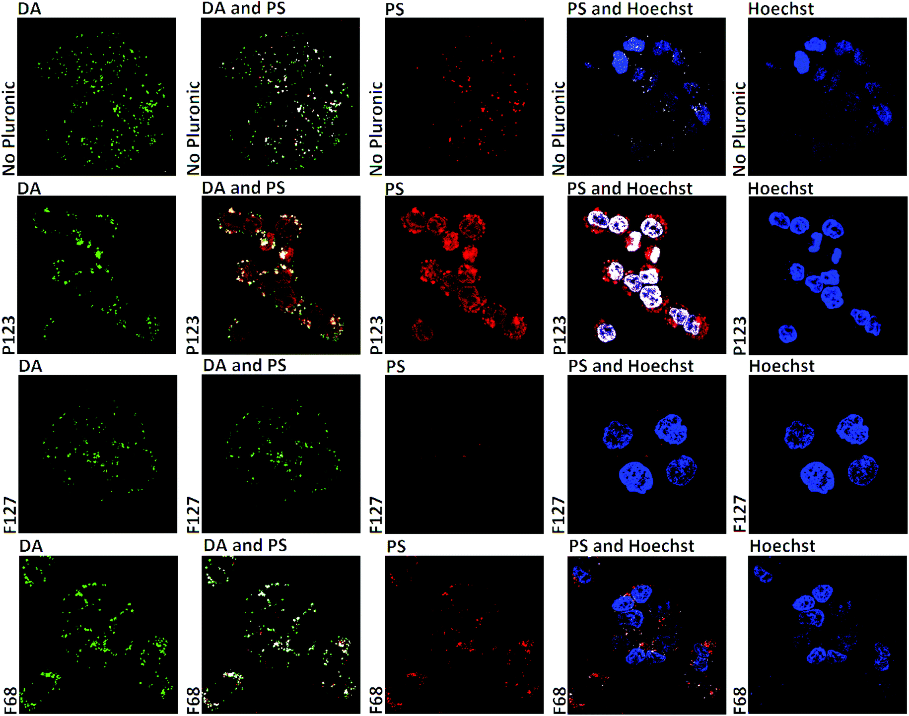 | ||
| Fig. 9 Confocal microscopy images of WiDr cells incubated for 18 hours with 10% FCS and 1 μM TAPP (red fluorescence), DA alone or in preparations with 100 μM P123 or F127 or F68. Cells were exposed to Hoechst (15 min) before imaging. Images showing the fluorescence from TAPP and florescence dyes are presented both separately and combined. White color indicates colocalized fluorochromes. The color contrast and brightness in the images were adjusted to optimize the visualization. | ||
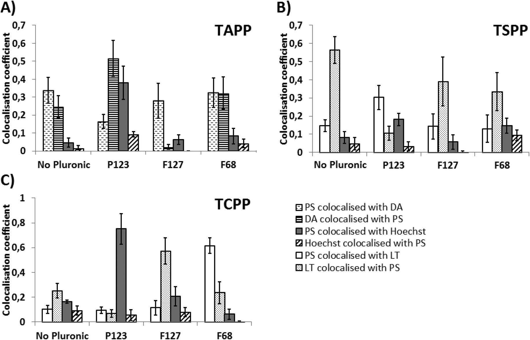 | ||
| Fig. 10 Colocalisation coefficients extracted from pictures of WiDr cells incubated for 18 hours with samples of PS (A: 1 μM TAPP, B: 70 μM TSPP, C: 70 μM TCPP) in medium (alone or in combination with 100 μM P123, F127 or F68). The figure is based on analysis of at least 50 cells per treatment. | ||
With the exception of TSPP all the Pluronics had substantial influence on the intracellular localization and accumulation of fluorescing PS in the WiDr cells: (1) P123 caused a partial translocation of TAPP from the endocytic vesicles to the nucleus (Fig. 9), while the uptake of THPP was strongly inhibited and resulted in some accumulation into endocytic vesicles (Fig. S13†). P123 increased TCPP accumulation in the nucleus (Fig. 10C and S12†). (2) F127 strongly inhibited the accumulation of both TAPP and THPP in the WiDr cells, while F127 had little effect on TCPP accumulation although a weak nuclear localization was found in some cells (Fig. 8, 10C and S12†). F68 showed no visible effect on accumulation or localization of TAPP or THPP, while the localization of TCPP was shifted to endocytic vesicles (Fig. 10C and S12†).
4. Discussion
4.1. Pluronic–porphyrin interactions
Pluronics exhibit distinct interactions with the evaluated PSs. The solubility of PS by Pluronics in PBS or 10% FCS is indicated by the increase in fluorescence intensity (Fig. 3, S1 and S2†) and removing of PS aggregates (Fig. 2), while the red-shifted fluorescence maximum suggests specific PS–Pluronic interactions (Fig. 3D and F). P123 seems to be the best solubilizer of THPP, followed by F127, F68 and L44 (Fig. 3G and S1†). This may be attributed to the high percentage of PPO blocks in the Pluronic molecule (Fig. 1), leading to a high lipophilicity and low cmc which eases formation of Pluronic micelles encapsulating the hydrophobic PS.4Serum can solubilize lipophilic porphyrins or bind hydrophilic compounds inducing their aggregation.10,11 Hydrophilic PSs may be bound to different serum components, as compared to hydrophobic ones. The association of hydrophilic PSs with serum is dictated by their charge.11 The positively charged TAPP and moderately hydrophilic TCPP are likely attracted by proteins. TSPP is bound by albumin; however its affinity, expressed as binding constant, is low,12 which may explain its unchanged fluorescence in the presence of 10% FCS (Fig. 3). Alakhov and coworkers have shown a change in particle size upon enriching Pluronic micelles with serum.4 Particle size measurements could not be performed in the presence of FCS, due to low resolution. It is therefore, difficult to speculate on structures formed by Pluronics in the presence of PS and FCS. Our results suggest, that serum constituents are better solubilizers of THPP than the evaluated Pluronics (Fig. 3G and S1†). However, as 10% FCS is used in this study, the amount of lipoproteins likely exceeds the amount of Pluronic micelles. Moreover, FCS seems less suitable as a solubilizer for PSs, as compared to copolymeric nanocarriers. Pluronics are efficient solubilizers of PSs as long as the appropriate Pluronics are selected for each PS.
It has previously been demonstrated, that the selected PS aggregate at micromolar concentration.13 Results suggest that both unimers and micelles can solubilize THPP, as shown by the continuous increase in THPP fluorescence below and above Pluronic cmc values (Fig. 3G and S1†). Hydrophilic PSs are bound to Pluronic unimers,14 as shown by moderate fluorescence increase at low Pluronics concentration, followed by a plateau (Fig. 3A–F). They may also exhibit slight interactions with the hydrophilic micellar coronas.15 Structures formed by block copolymers differ with respect to the mechanism of their cellular uptake16 and biological effects.17 As a consequence THPP, that shows greater changes in the solubility with increasing Pluronic concentration, is expected to exhibit substantial changes in cell localization and uptake.
4.2. Pluronic toxicity
Pluronics have been shown to exhibit dark toxicity which depends on both cell line and the type of Pluronic,18 which was confirmed by the use of cell lines representing sarcomas and carcinomas. The cellular distribution of Pluronics is strongly affected by the total molecular hydrophobicity,5 concentration,16,19 cell type and incubation time.20,21 We assume, that the hydrophilic Pluronic F68 will be poorly internalized and present mostly in endocytic vesicles, similar to Pluronic F108 (hydrophilic–lipophilic balance, HLB = 27). A Pluronic of intermediate polarity, F127, will be efficiently internalized, spread throughout the cytoplasm and reach intracellular compartments, including nuclei similar to L35 (HLB = 19). The hydrophobic Pluronic P123 will be retained in the plasma membrane and slowly transported into the cells, resulting in endocytic localization, similar to L121 (HLB = 1).5 Only unimers influence membrane fluidity,17 thus the low cmc value prohibits extensive interactions with the cells.Hydrophobic P123 and L44 were more toxic, compared to F127 and F68, as toxicity is expected to depend on the degree of Pluronic internalization in cells and their ability to integrate with phospholipids in plasma membranes and disrupt its structure.19 P123 micelles are efficiently internalized by endocytosis, as compared to unimers,17 which explains the reduction in cell survival at a concentration above cmc (Fig. 1, S3 and S4†). F68 was found to be the least toxic copolymer in our experimental settings. This is explained by its poor internalization by cells and inability to decrease membrane viscosity.5 Certain cell lines showed an increased susceptibility towards specific Pluronics (Table S2†). Although Pluronic biocomptability has previously been determined, the results of the present study show that the development of nanoparticulate PS formulation should include thorough toxicity tests using a model relevant for the targeted cancer tissue.
4.3. Intracellular localization of PSs in the absence of Pluronics
It has been shown that the mechanism of cell entry is dependent on various PS properties, i.e. lipophilicity, total charge and charge distribution within the PS.22 The hydrophobic THPP binds to LDL that targets the LDL receptor,23 but THPP penetrates the plasma membrane by passive diffusion.24,25 The charged PSs were assumed to be internalized by endocytosis. TAPP was found to be the 2nd most efficient PS after THPP in sensitizing cells to photoinactivation (Fig. 4E) and apparently more efficiently entering the cells than TSPP and TCPP (Fig. S10†). The relatively high cellular uptake of TAPP as indicated from the fluorescence micrographs (Fig. S10†) may be attributed to strong columbic attractions between the cationic PS and negatively charged cell membrane resulting in efficient endocytic uptake of TAPP as documented by colocalization with DA (Fig. 9). Cells treated with TSPP showed granular fluorescence (Fig. 7), documented to be endocytic vesicles by its colocalization with LT (Fig. S11†). In contrast, TCPP localized partly in granules (Fig. 8), found to colocalize with LT (Fig. S12†), and partly diffusely distributed all over the cytoplasm and nucleoplasm. The cellular uptake of TCPP has previously been shown to be exerted by endocytosis, partly through clathrin-related and not caveolae-mediated processes, in SW480 colon carcinoma cells.26 The intravesicular pH in the late endosomes and lysosomes is around 5.5 which is close to the pKa of the carboxyl-groups of TCPP.13 One may therefore, speculate that when TCPP enters the acidified endocytic vesicles the carboxyl-groups on TCPP become sufficiently protonated to allow translocation into the cytosol in which TCPP again becomes ionized and distributes all over the cytoplasm and nucleoplasm.4.4. Intracellular localization of PS and PS efficiency in the presence of Pluronics
Polymeric micelles have been shown to influence the intracellular localization of fluorescent probes.27 P123 exhibited an efficient solubilizing effect on THPP even at concentrations <cmc (Fig. 3G and S1†) presumably owing to the lipophilic properties of P123 unimers, the highly hydrophobic micellar core and low cmc value (Fig. 1). Solubilization with P123 reduced the cellular uptake of the hydrophobic PS curcumin through passive diffusion.18 Accordingly, THPP solubilized with P123 was found to accumulate in endocytic vesicles (Fig. S13†), indicating endocytosis as the main mechanism for cellular uptake of P123-solublized THPP, as in accordance with similar studies.28 The cellular uptake of THPP is substantially reduced by P123 (Fig. 8), which is reflected in a strongly reduced cell sensitivity to light (Fig. 5D–E) and is in accordance with the reduced photototoxic effect upon encapsulation of hydrophobic dyes within a hydrophobic nanocarrier as previously reported.29,30P123 increased the photodynamic effect of TSPP (Fig. 5B). Pluronics may decrease the number of lysosomes and decrease their acidity.4 This may be reflected in the co-localisation coefficients (Fig. 10B). Cells treated with TSPP show high co-localization between LT and PS (Fig. S11†), as all the lysosomes and late endosomes are acidic. On the other hand, in cells treated with TSPP and P123 the fraction of the PS localized in acidic vesicles seems to be reduced, while a larger fraction of the acidic vesicles contains the PS (Fig. 10B). This may be caused by a Pluronic-induced reduction of the pH gradient between the vesicles and the cytosol,4 while TSPP localization in the various endocytic compartments remains unchanged. It is not clear what impact the intravesicular pH could have on TSPP-based photosensitivity. The pKa of the sulfonate groups are too low to be protonated in the lysosomes, while the overall charge of the two imino groups13 may increase upon acidification in the lysosomes.31 Thus, increased ionization of the core imino groups may cause intermolecular charge repulsion and reduced aggregation and thereby increased photodynamic efficacy. TSPP has previously been shown to be partly aggregated inside lysosomes32 and P123-induced intralysosomal pH increase may therefore cause enhanced treatment effect. It is also possible that a membrane destabilizing effect of P123 can reduce the light dose required to rupture the endocytic vesicles and thereby increase the photocytotoxic efficacy of TSPP. We have recently found that the amphiphilic PS AlPcS2a may alter the fluid phase endocytosis by inhibiting trafficking from early endosomes to late endosomes and lysosomes.33 The influence of drugs and excipients on intracellular trafficking has not been much studied and warrant further investigations, also with respect to the impact of Pluronic–PS complexes.
Upon incubation with P123, partial translocation of TAPP from the endocytic vesicles to other compartments including the nucleus was observed (Fig. 9). This is depicted by an increase in colocalisation of PS with Hoechst (Fig. 10A). Similar drug relocalization pattern was observed with the hydrophobic and amphiphilic Pluronics L61, P85 and P105 (HLB = 3, 16 and 15, respectively20,34,35). Pluronic nanocarriers, including P123, have been proven efficient as vectors for gene and oligonucleotide transfection36 substantiating the destabilizing effect of several Pluronics on endocytic vesicles. The increase of membrane fluidity and heightened relocalization of TAPP into the nucleus may be the reason for the enhanced PS efficiency in the presence of P123.
Although P123 increases TCPP colocalization with the nucleus, photodynamic activity of this PS is not increased by P123 (Fig. S7C†). However, we found that TCPP can easily be translocated to most cellular compartments also in the absence of P123, so P123 does not provide any new functionality to the mechanism of action of this PS.
F68 has been reported to be distributed to endosomes/lysosomes.21 As a hydrophilic Pluronic, F68 is expected to be internalized by endocytosis. Hydrophilic Pluronics have been found to solidify membranes and subsequently enhance the Pgp ATPase activity.5 It is therefore expected, that F68 would limit lysosomal escape of PSs into the cytoplasm. In this study it was demonstrated, that cells incubated with TCPP and F68 show higher colocalization of PS with LT, as compared to cells incubated with TCPP alone (Fig. 10 and S12†). TCPP diffusion through lysosomal membrane is apparently hindered in the presence of F68. As a result photodynamic activity of TCPP decreases with increasing F68 concentration (Fig. 5C). Similar reduction of photodynamic efficiency was observed for TSPP (Fig. 5B), while photodynamic efficiency of TAPP was not changed. F68 is not able to solubilize THPP as efficiently as F127 or P123 (Fig. 3G and S1†), probably due to its short PPO block (Fig. 1). Therefore, small concentrations of F68 enhance THPP photoreactivity (Fig. 5) probably due to reduced aggregation and subsequent increased uptake. At higher concentrations, as the solubilizing capacity of F68 increases, PS efficiency is reduced.
P123 and F127 increase the survival of cells exposed to THPP (Fig. 5D and E). Even though the total hydrophobicity of F127 is lower than P123, the length of the central PPO block is similar (Fig. 1). As the latter property is crucial for the strong hydrophobic drug–polymer affinity,15 inhibition of THPP uptake and efficiency seems similar for the two Pluronics. Uptake of TAPP depends on columbic attraction between the PS and the cellular membrane. Solubilization of TAPP by F127 may reduce this interaction and the cellular uptake (Fig. 7). However, the efficiency of TAPP is not changed much in the presence of F127 (Fig. 5) and may reflect the higher efficacy of PSs when located extralysosomally.37 Woodburn and coworkers found mitochondria as the optimal target for photodynamic inactivation of cancer cells while lysosomes were accordingly less optimal.37 As a result the smaller amount of internalized PS present in cells treated with TAPP in the presence of F127 appears to be located in intracellular compartments more sensitive to PDT.
5. Conclusions
The influence of Pluronics nanoparticles on dark cytotoxicity, photocytotoxicity, and localization of four model photosensitizers in cancer cells was investigated. Pluronics efficiently solubilized and deaggregated porphyrin PSs at nontoxic concentrations. While this effect was less pronounced for hydrophilic PSs, THPP fluorescence was significantly increased in the presence of Pluronics. Photodynamic efficiency of THPP was inversely proportional to the solubilization capability and lipophilic Pluronics had negative impact on THPP photocytotoxicity. On the other hand, the hydrophilic F68 attenuated THPP photodynamic activity at high concentration. In case of TSPP and TAPP, Pluronic P123 exerted positive effect on cell photokilling, due to enhancement of PS relocalization from late endosones and lysosomes. Other Pluronics did not affect the cell survival following PDT treatment with TAPP or TSPP. Cell inactivation by TCPP was diminished by addition of all evaluated Pluronics. Pluronics have been shown to be efficient solubilizers of the selected PSs. However, the impact of formulation on photocytotoxicity should be thoroughly evaluated.Acknowledgements
This work was kindly supported by the Norwegian Research Council. Authors would like to thank Tore Geir Iversen, PhD (Department of Biochemistry, Norwegian Radium Hospital) for providing a Zetasizer instrument and Roman Generalov (Department of Radiation Biology, Norwegian Radium Hospital) for technical assistance with confocal microscopy.References
- P. Agostinis, K. Berg, K. A. Cengel, T. H. Foster, A. W. Girotti, S. O. Gollnick, S. M. Hahn, M. R. Hamblin, A. Juzeniene, D. Kessel, M. Korbelik, J. Moan, P. Mróz, D. Nowis, J. Piette, B. C. Wilson and J. Gołąb, Photodynamic Therapy of Cancer: An Update, CA-Cancer J. Clin., 2011, 61, 250–281 CrossRef PubMed.
- D. Bechet, P. Couleaud, C. Frochot, M.-L. Viriot, F. Guillemin and M. Barberi-Heyob, Nanoparticles as vehicles for delivery of photodynamic therapy agents, Trends Biotechnol., 2008, 26, 612–621 CrossRef CAS PubMed.
- FDA, Inactive Ingredient search for approved drug products, accessed 06.05.2013 (http://www.accessdata.fda.gov/scripts/cder/iig/index.cfm).
- V. Alakhov, E. Klinski, S. Li, G. Pietrzynski, A. Venne, E. V. Batrakova, T. Bronitch and A. V. Kabanov, Block copolymer-based formulation of doxorubicin. From cell screen to clinical trials, Colloids Surf. B: Biointerfaces, 1999, 16, 113–134 CrossRef CAS.
- E. V. Batrakova, S. Li, V. Y. Alakhov, D. W. Miller and A. V. Kabanov, Optimal structure requirements for Pluronic block copolymers in modifying P-glycoprotein drug efflux transporter activity in bovine brain microvessel endothelial cells, J. Pharmacol. Exp. Ther., 2003, 304, 845–854 CrossRef CAS PubMed.
- E. V. Batrakova, S. Lee, S. Li, A. Venne, V. Alakhov and A. Kabanov, Fundamental relationships between the composition of Pluronic block copolymers and their hypersensitization effect in MDR cancer cells, Pharm. Res., 1999, 16, 1373–1379 CrossRef CAS.
- G. Lu, X. Zhang, X. Cai and J. Jiang, Tuning the morphology of self-assembled nanostructures of amphiphilic tetra(p-hydroxyphenyl)porphyrins with hydrogen bonding and metal–ligand coordination bonding, J. Mater. Chem., 2009, 19, 2417–2424 RSC.
- N. Micali, F. Mallamace, A. Romeo, R. Purrello and L. M. Scolaro, Mesoscopic Structure of meso-Tetrakis(4-sulfonatophenyl)porphine J-Aggregates, J. Phys. Chem. B, 2000, 104, 5897–5904 CrossRef CAS.
- S. Park, S. Lee, H. Chung, S. Her, Y. Choi, K. Kim, K. Choi and I. C. Kwon, Cellular Uptake Pathway and Drug Release Characteristics of Drug-Encapsulated Glycol Chitosan Nanoparticles in Live Cells, Microsc. Res. Tech., 2010, 73, 857–865 CrossRef CAS PubMed.
- W. An, Y. Jiao, C. Dong, C. Yang, Y. Inoue and S. Shuang, Spectroscopic and molecular modeling of the binding of meso-tetrakis(4-hydroxyphenyl)porphyrin to human serum albumin, Dyes Pigm., 2009, 81, 1–9 CrossRef CAS.
- S. M. Andrade and S. M. B. Costa, Spectroscopic Studies on the Interaction of a Water Soluble Porphyrin and Two Drug Carrier Proteins, Biophys. J., 2002, 82, 1607–1619 CrossRef CAS PubMed.
- J. Bartosová, I. Kalousek and Z. Hrkal, Binding of meso-tetra(4-sulfonatophenyl)porphine to haemopexin and albumin studied by spectroscopy methods, Int. J. Biochem., 1994, 26, 631–637 CrossRef.
- J. Sobczyński, H. H. Tønnesen and S. Kristensen, Influence of aqueous media properties on aggregation and solubility of four structurally related meso-porphyrin photosensitizers evaluated by spectrophotometric measurements, Pharmazie, 2013, 68, 100–109 Search PubMed.
- C. A. Steinbeck, N. Hedin and B. F. Chmelka, Interactions of Charged Porphyrins with Nonionic Triblock Copolymer Hosts in Aqueous Solutions, Langmuir, 2004, 20, 10399–10412 CrossRef CAS PubMed.
- C. Allen, D. Maysinger and A. Eisenberg, Nano-engineering block copolymer aggregates for drug delivery, Colloids Surf. B, 1999, 16, 3–27 CrossRef CAS.
- G. Sahay, E. V. Batrakova and A. V. Kabanov, Different internalization pathways of polymeric micelles and unimers and their effects on vesicular transport, Bioconjugate Chem., 2008, 19, 2023–2029 CrossRef CAS PubMed.
- E. V. Batrakova, S. Li, W. F. Elmquist, D. W. Miller, V. Y. Alakhov and A. V. Kabanov, Mechanism of sensitization of MDR cancer cells by Pluronic block copolymers: selective energy depletion, Br. J. Cancer, 2001, 85, 1987–1997 CrossRef CAS PubMed.
- R. Singh, H. H. Tønnesen, S. Kristensen and K. Berg, The influence of Pluronics® on dark cytotoxicity, photocytotoxicity, localization and uptake of curcumin in cancer cells: studies of curcumin and curcuminoids XLIX, Photochem. Photobiol. Sci., 2013, 12, 559–575 CAS.
- N. Munshi, N. Rapoport and W. G. Pitt, Ultrasonic activated drug delivery from Pluronic P- 105 micelles, Cancer Lett., 1997, 118, 13–19 CrossRef CAS PubMed.
- N. Y. Rapoport, A. Marin, Y. Luo, G. D. Prestwich and M. Muniruzzaman, Intracellular Uptake and Traficking of Pluronic Micelles in Drug-Sensitive and MDR Cells: Effect on the Intracellular Drug Localization, J. Pharm. Sci., 2002, 91, 157–170 CrossRef CAS PubMed.
- A. Gigout, M. D. Buschmann and M. Jolicoeur, The Fate of Pluronic F-68 in Chondrocytes and CHO Cells, Biotechnol. Bioeng., 2008, 100, 975–987 CrossRef CAS PubMed.
- A. S. Sobolev, D. A. Jans and A. A. Rosenkranz, Targeted intracellular delivery of photosensitizers, Prog. Biophys. Mol. Biol., 2000, 73, 51–90 CrossRef CAS PubMed.
- M. Kongshaug, J. Moan and S. B. Brown, The distribution of porphyrins with different tumour localising ability among human plasma proteins, Br. J. Cancer, 1989, 59, 184–188 CrossRef CAS PubMed.
- H. Rezzoug, J.-L. Merlin, N. Zeghari, D. Lignon, S. Marchal, C. Ramacci, E. Yvroud and F. H. Guillemin, Cellular uptake kinetics and photodynamic activity of meso-tetrahydroxyphenylchlorin (mTHPC), inPhotochemotherapy: Photodynamic Therapy and Other Modalities, ed. B. Ehrenberg, G. Jori and J. Moan, Proc. SPIE, Bellingham, WA, 1996, vol. 2625, p. 386 Search PubMed.
- G. H. Rodal, S. K. Rodal, J. Moan and K. Berg, Liposome-bound Zn (II)-phthalocyanine. Mechanisms for cellular uptake and photosensitization, J. Photochem. Photobiol. B., 1998, 45, 150–159 CrossRef CAS.
- Z. Hu, Y. Pan, J. Wang, J. Chen, J. Li and L. Ren, Meso-tetra (carboxyphenyl) porphyrin (TCPP) nanoparticles were internalized by SW480 cells by a clathrin-mediated endocytosis pathway to induce high photocytotoxicity, Biomed. Pharmacother., 2009, 63, 155–164 CrossRef CAS PubMed.
- R. Savić, L. Luo, A. Eisenberg and D. Maysinger, Micellar Nanocontainers Distribute to Defined Cytoplasmic Organelles, Science, 2003, 300, 615–618 CrossRef PubMed.
- G. A. Husseini, C. M. Runyan and W. G. Pitt, Investigating the mechanism of acoustically activated uptake of drugs from Pluronic micelles, BMC Cancer, 2002, 2, 20–25 CrossRef PubMed.
- K. Löw, T. Knobloch, S. Wagner, A. Wiehe, A. Engel, K. Langer and H. von Briesen, Comparison of intracellular accumulation and cytotoxicity of free mTHPC and mTHPC-loaded PLGA nanoparticles in human colon carcinoma cells, Nanotechnology, 2011, 22, 1–12 CrossRef PubMed.
- M.-J. Shieh, C.-L. Peng, W.-L. Chiang, C.-H. Wang, C.-Y. Hsu, S.-J. J. Wang and P.-S. Lai, Reduced Skin Photosensitivity with meta-Tetra(hydroxyphenyl)chlorin-Loaded Micelles Based on a Poly(2-ethyl-2-oxazoline)-b-poly(D,L-lactide) Diblock Copolymer in Vivo, Mol. Pharm., 2010, 7, 1244–1253 CrossRef CAS PubMed.
- A. J. Barrett, J. C. Kennedy, R. A. Jones, P. Nadeau and R. H. Potter, The Effect of Tissue and Cellular pH on the Selective Biodistribution of Porphyrin-Type Photochemotherapeutic Agents: A Volumetric Titration Study, J. Photochem. Photobiol. B, 1990, 6, 309–323 CrossRef CAS.
- K. Berg and J. Moan, The influence of the cysteine protease inhibitor L-trans-epoxysuccinyl-leucyl amido(4-guanidio)butane (EM) on photobiological effects of tetra(4-sulfonatophenyl) porphine, Cancer Lett., 1995, 88, 227–236 CrossRef CAS PubMed.
- M. Vikdal, R. Generalov and K. Berg, The photosensitizer disulfonated aluminum phthalocyanine reduces uptake and alters trafficking of fluid phase endocytosed drugs in vascular endothelial cells-Impact on efficacy of photochemical internalization, Biochem. Pharmacol., 2013, 86, 748–758 CrossRef CAS PubMed.
- D. W. Miller, E. V. Batrakova and A. V. Kabanov, Inhibition of Multidrug Resistance-Associated Protein (MRP) Functional Activity with Pluronic Block Copolymers, Pharm. Res., 1999, 16, 396–401 CrossRef CAS.
- A. Venne, S. Li, R. Mandeville, A. Kabanov and V. Alakhov, Hypersensitizing Effect of Pluronic L61 on Cytotoxic Activity, Transport, and Subcellular Distribution of Doxorubicin in Multiple Drug-resistant Cells, Cancer Res., 1996, 56, 3626–3629 CAS.
- S. V. Vinogradov, E. V. Batrakova, S. Li and A. V. Kabanov, Mixed Polymer Micelles of Amphiphilic and Cationic Copolymers for Delivery of Antisense Oligonucleotides, J. Drug. Target., 2004, 12, 517–526 CrossRef CAS PubMed.
- K. W. Woodburn, N. J. Vardaxis, J. S. Hill, A. H. Kaye, J. A. Reiss and D. R. Phillips, Evaluation of Porphyrin Characteristics Required for Photodynamic Therapy, Photochem. Photobiol., 1992, 5, 697–704 Search PubMed.
Footnote |
| † Electronic supplementary information (ESI) available. See DOI: 10.1039/c3pp50181g |
| This journal is © The Royal Society of Chemistry and Owner Societies 2014 |