
 Open Access Article
 This Open Access Article is licensed under a
 Creative Commons Attribution 3.0 Unported Licence
A hydrazide-anchored dendron scaffold for chemoselective ligation strategies†
Liz
O'Donovan
and
Paul A.
De Bank
*
Department of Pharmacy and Pharmacology and Centre for Regenerative Medicine, University of Bath, Bath, BA2 7AY, UK. E-mail: p.debank@bath.ac.uk; Fax: +44 (0)1225 386114; Tel: +44 (0)1225 384017
First published on 4th August 2014
Abstract
Chemoselective ligation, including “click” chemistry, has found wide utility in general synthetic strategies and the specific modification of polymers and biomolecules. This has resulted in a number of applications of such approaches, particularly in the biomedical area, including diagnostic imaging and drug delivery. However, tools to chemoselectively decorate target molecules with multiple copies of a particular drug, ligand or label are lacking. We describe the design and synthesis of a hydrazide-anchored dendron scaffold for chemoselective ligation to carbonyl moieties, and demonstrate its use in the modification of aldehyde-rich surfaces with the RGD integrin-binding ligand.
Introduction
The chemical modification of polymers and biological molecules, including those in whole cells and even tissues, has a wide range of applications, from the generation of molecular tools for basic research to clinical diagnostics and therapy. Early techniques for the modification of biomaterials and biomolecules often relied on the utilization of accessible carboxylic acids to generate activated (usually N-hydroxysuccinimide (NHS)) esters, followed by reaction with a species containing a free amine. Alternatively, peptides and proteins can be modified via lysine and cysteine residues by reaction with NHS esters or maleimides, respectively. However, because biological molecules are generally rich in these functional groups, non-specific, off-target modification can be a significant issue. To overcome this and ensure selective chemical modification of biomolecules, chemoselective ligation strategies have been developed that employ mutually reactive functional groups that are not normally found within biological systems. Ideally, these reactions occur under conditions that are compatible with biomolecules and living cells, i.e. in aqueous media under physiological conditions. To date, there are a number of such bioorthogonal ligation reactions that are commonly used not only to modify biological molecules, but which are also employed in a wide range of chemical syntheses.Chemoselective ligations include, but are not limited to, those between an alkyne and an azide (now commonly referred to as “click” chemistry),1–3 the Staudinger ligation between a triarylphosphine and an azide,4,5 and reactions between carbonyl groups and hydrazides, thiosemicarbazides or aminooxy moieties.6–8 To employ these ligations for the selective labelling or modification of biological molecules, the biomolecule itself must, of course, possess a suitable bioorthogonal functional group. These can be introduced using standard synthetic strategies or, in the case of living cells, via approaches such as the selective periodate oxidation of carbohydrates to yield aldehydes,9,10 or by metabolic engineering. In the latter approach, the permissiveness of certain metabolic enzymes to structural variations in their substrates has been used to decorate proteins and cell surface glycans, including those within living animals, with a variety of bioorthogonal functional groups.6,11–16 Chemoselective strategies have, to date, been used for a wide range of applications, including antibody labelling,14,17 drug delivery strategies,18,19 nanoparticle functionalization,20,21 protein immobilization,22–24 hydrogel formation,23,25–27 modification of viruses to improve tissue targeting,28 and the generation of three-dimensional cell aggregates.10,29–31
Dendrons and dendrimers have been widely studied in recent decades32 for their potential application in a number of areas, including drug and gene delivery and clinical diagnostics,33–36 with their multivalent nature being a key characteristic. While these macromolecules have been assembled using chemoselective ligations37,38 and have been decorated with bioorthogonal functional groups,29,39 there is now interest in strategies that enable “dendronization”40,41 of a target molecule via chemoselective decoration with multiple functional moieties. To date, various targets such as polymers,42–44 proteins,45 magnetic nanoparticles,46 carbon nanotubes47 and nucleotides48 have been chemoselectively dendronized, but these strategies have been limited to the use of click chemistry.
The pairing of a carbonyl group with a hydrazide, thiosemicarbazide or aminooxy moiety is perhaps the most versatile chemoselective ligation strategy in terms of their chemical or metabolic installation into co-reacting species and ability to readily ligate in aqueous media. The resultant hydrazone bond is relatively stable, but ultimately undergoes reversible hydrolysis in aqueous environments.49,50 With these characteristics in mind, we describe the design and development of, to our knowledge, the first dendritic scaffold containing a bioorthogonal hydrazide anchor for the dendronization of carbonyl-containing molecules. Such a hydrazide-anchored dendron scaffold could enable, for example, decoration of biomaterials with multiple biological cues for tissue engineering and regenerative medicine strategies, and enhanced antibody, polymer or nanoparticle labelling for diagnostic, imaging and drug delivery applications.
Results and discussion
For wide utility, we envisaged the synthesis of hydrazide-anchored scaffolds with terminal succinimide esters, enabling one-step functionalization with, primarily, peptides, via a primary amine (Fig. 1). As such, it was necessary to construct scaffolds with a protected hydrazide in order to eliminate any possibility of reaction with the activated esters. We also considered the possibility of peptide side chains cross-reacting with the esters, and adopted a general strategy that could be employed for any peptide that contains potentially problematic residues, such as lysine. Using solid phase peptide synthesis followed by cleavage conditions that retain side chain protection, selective reaction of the N-terminus with the dendron scaffold could be realized, following by deprotection of both the side groups and hydrazide anchor (Fig. 1).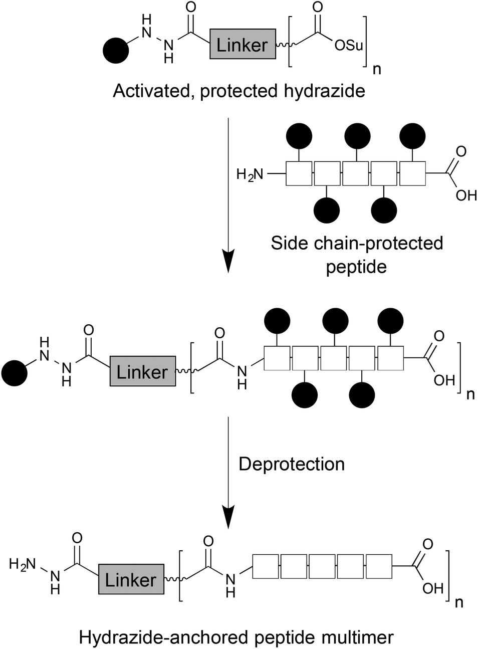 | ||
| Fig. 1 Overview of the strategy for synthesis of a hydrazide-anchored dendron scaffold, allowing multimeric peptide display. | ||
As our interests lie primarily in tissue engineering and regenerative medicine, we chose to demonstrate these scaffolds using the RGD tripeptide. This motif is found in a number of extracellular matrix proteins and is important in cell–matrix adhesion via binding to integrin receptors.51 As a result, RGD and its analogues have been widely studied for improving cell adhesion to poorly adhesive materials.52 In addition, integrin receptors are overexpressed in some cancers, so there has been extensive interest in this tripeptide for the targeting of drugs or imaging agents to tumours.53,54 For monomeric peptide hydrazides, partially protected RGD (H-Arg(Pbf)-Gly-Asp(OtBu)-OH 1), was synthesised by manual Fmoc solid phase peptide synthesis on 2-chlorotrityl resin, followed by cleavage with acetic acid and trifluoroethanol. As a negative control, the partially protected version of the non-adhesive RGE tripeptide (H-Arg(Pbf)-Gly-Glu(OtBu)-OH 2) was synthesised in an analogous manner. These peptides were reacted with succinimide ester 3 to yield the corresponding protected hydrazides 4 and 6 in yields of 45 and 40%, respectively (Scheme 1). Removal of the remaining, acid-labile protecting groups proceeded smoothly via treatment with TFA and triisopropylsilane. However, it was not possible to isolate the resulting peptide hydrazides 5 and 7 using the conventional method of ether precipitation. Instead, excess TFA was removed under vacuum, the crude residue redissolved in 10% acetic acid and extracted with chloroform. Lyophilisation of the aqueous phase yielded the target monosubstituted compounds as crystalline white solids in reasonable yields.
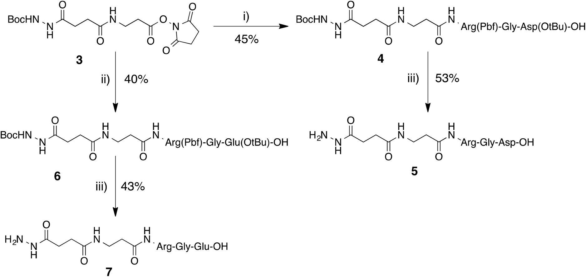 | ||
| Scheme 1 Synthesis of hydrazide-terminated monomers 5 and 7. (i) H-Arg(Pbf)-Gly-Asp(OtBu)-OH (1), DMAP, DMF, rt; (ii) H-Arg(Pbf)-Gly-Glu(OtBu)-OH (2), DMAP, DMF, rt; (iii) TFA–TIS–H2O, rt. | ||
To construct the dimeric scaffold, we chose the commercially available 2-amino-2-methylpropane-1,3-diol 8 as the starting material, enabling the hydrazide functionality to be incorporated via the amine group and the arms of the dendron via extension of the diol (Scheme 2). The amine was initially Boc protected in high yield to furnish the literature compound 9,55,56 which was subsequently converted to the corresponding dinitrile 10 following reaction with acrylonitrile.57 Attempts to hydrolyse 10 directly to diacid 12 proved unsuccessful, so the dinitrile was converted to diester 11, which furnished diacid 12 following hydrolysis with 5 M NaOH at 50 °C.
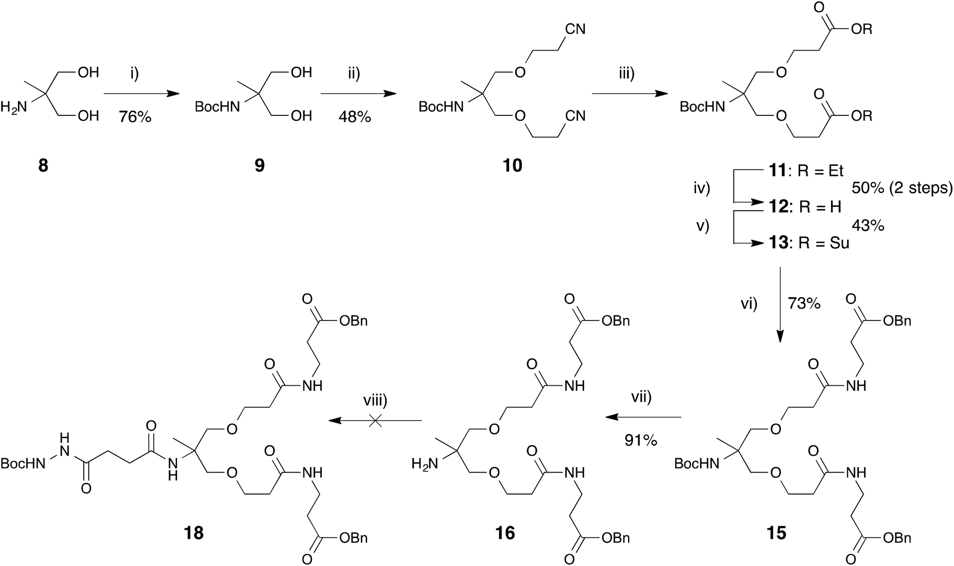 | ||
| Scheme 2 (i) Boc anhydride, NEt3, CH2Cl2; (ii) acrylonitrile, KOH, MeCN, rt; (iii) (a) HCl, reflux, (b) EtOH, reflux; (iv) 5 M NaOH, THF, EtOH, 50 °C; (v) NHS, DIC, DMAP, CH2Cl2, rt; (vi) 3-(benzyloxy)-3-oxopropan-1-aminium 4-methylbenzene-1-sulfonate (14), pyridine, CH2Cl2, rt; (vii) TFA, CH2Cl2, rt; (viii) BocNHNHCO(CH2)2CO2H (17), various conditions. | ||
For eventual insertion of the terminal hydrazide functionality, it was necessary at this point to protect the diacid with a suitable protecting group. With this in mind, β-alanine 14,58,59 protected as a benzyl ester, was introduced on each arm, following activation of 12 as the corresponding disuccinimide ester 13, with the effect of adding a spacer group in addition to a masked carboxylic acid to each arm of protected product 15. To introduce the hydrazide anchor, the Boc group was then cleaved from 15 to expose the free amine of compound 16 for coupling with Boc-protected hydrazide 17.60 However, despite investigating a number of different, standard coupling conditions, the reaction between compounds 16 and 17 did not proceed as expected, possibly due to steric hindrance of the primary amine by the mobile arms of dimer 16. As a result, it was decided to introduce the hydrazide terminus earlier in the synthesis.
This modified approach (Scheme 3) also utilised diol 8 as the starting material but, rather than employing Boc protection at the start of the synthesis, the amine group was left unmodified for earlier introduction of the hydrazide. Analogous to Scheme 2, diol 8 was treated with acrylonitrile to yield dinitrile 19 in good yield of 76% following purification via column chromatography. Conversion of 19 to the corresponding diester 20 proceeded in almost quantitative yield of 94%. In order to introduce the terminal hydrazide, diester 20 was treated with acid 1760 in the presence of a variety of coupling agents (DIC, EDC, HATU, PyBOP), all of which proved unsuccessful. However, when two activating agents (DIC and HOBt) were employed, the reaction proceeded smoothly to furnish compound 21 in good yield. Base hydrolysis of diester 21 led to isolation of the desired diacid 22. As a result of the earlier introduction of the protected hydrazide, further orthogonal protection of the acid dimer was not necessary and conversion to disuccinimide ester 23 yielded our target, activated dimer, capable of further functionalization with suitable nucleophilic species.
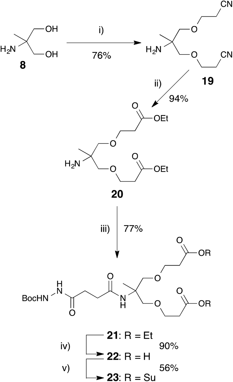 | ||
| Scheme 3 Synthesis of hydrazide-terminated dimer scaffold. (i) Acrylonitrile, 1,4-dioxane, KOH, rt; (ii) HCl, EtOH, reflux; (iii) BocNHNHCO(CH2)2CO2H (17), DIC, HOBt, DMF, rt; (iv) NaOH, EtOH, THF, 50 °C; (v) N-hydroxysuccinimide, DIC, CH2Cl2, rt. | ||
To investigate the application of this dimeric scaffold in chemoselective modification of biomaterials, activated ester 23 was treated with partially protected tripeptides RGD 1 or RGE 2 in DMF. Following reaction for 72 hours at room temperature, the corresponding protected, hydrazide-terminated peptide dimers 24 and 26 were isolated in yields of 11% and 21%, respectively. These low yields can be largely attributed to poor chromatographic separation of the starting materials and products. However, following treatment of 24 and 26 with TFA and TIS, the target deprotected hydrazide dimers 25 and 27 (2 × RGD hydrazide and 2 × RGE hydrazide; Fig. 2) were isolated in good yield as described for their monomer analogues.
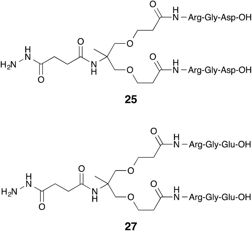 | ||
| Fig. 2 Structures of target peptide hydrazide dimers 25 and 27. | ||
We also wished to investigate whether the methodology we developed could be extended to more complicated dendrimeric species. Hence, we set about preparing a tetramer analogue of ester 21 (Scheme 4). Diacid 22 was treated with EDC and NHS to generate activated ester 23in situ. Reaction with amine 20 under ambient conditions led to target compound 28, which, despite the very similar Rf values of product and starting material, was isolated in reasonable yield of 45% following flash column chromatography. These results suggest that this approach to hydrazide-anchored dendrons has the potential for expansion of the number of branches, enabling the chemoselective decoration of suitable target molecules with multiple bioactive moieties.
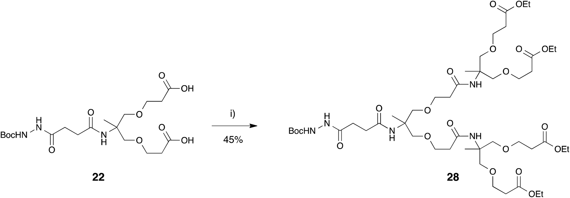 | ||
| Scheme 4 Synthesis of tetramer ester 28. (i) 20, N-(3-dimethylaminopropyl)-N′-ethylcarbodiimide, N-hydroxysuccinimide, DMF, rt. | ||
In order to evaluate the ability of our RGD terminated hydrazides to control cell adhesion to a surface in a chemoselective manner, it was necessary to prepare an aldehyde-rich substrate, which would repel cell adhesion until the introduction of the hydrazide-anchored RGD tripeptides. Films of silk fibroin protein, an extensively studied biomaterial,61 from Bombyx mori silk cocoons were utilized for this purpose. The available carboxylic acid groups present on the surface of the silk fibroin film were activated using carbodiimide chemistry and subsequently reacted with aminated tetraethylene glycol 2962,63 to yield a cell-repulsive surface (Scheme 5).63–65 Aldehyde functionality was then incorporated via reaction of these surfaces with glutaraldehyde. The presence of aldehyde groups on these modified silk films was confirmed by their chemoselective reaction with fluorescein thiosemicarbazide (FTSC) at pH 5.5. Only films that had been treated with glutaraldehyde were found to fluoresce following subsequent incubation with FTSC (Fig. S1†).
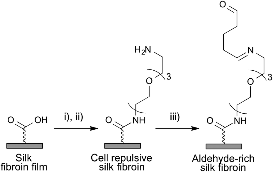 | ||
Scheme 5 Chemical modification of Bombyx mori silk fibroin films (i) EDC, NHS, PBS, rt; (ii) 50 μM NH2(CH2CH2O)3CH2CH2NH229, MeOH–H2O (1![[thin space (1/6-em)]](https://www.rsc.org/images/entities/char_2009.gif) :1 v/v), rt; (iii) 0.01 M glutaraldehyde, PBS, rt. :1 v/v), rt; (iii) 0.01 M glutaraldehyde, PBS, rt. | ||
Following the confirmation of the presence of aldehydes on the silk films, we then investigated the monomeric and dimeric peptide hydrazides for their ability to chemoselectively reintroduce cell-adhesivity to these non-adhesive surfaces. Modified films were incubated with both the RGD and RGE peptide hydrazides for 3 hours at 37 °C (pH 5.5), alongside commercially available RGD as an amine-terminated control. C2C12 myoblasts, as a model cell type, were then seeded on the films and cultured for a period of 72 hours. At 24 hour intervals, relative cell number on the surfaces was assessed using the MTS assay, which detects metabolically active cells (see ESI† for detailed description).
Comparison of the various modified surfaces to tissue culture plastic (TCP) revealed that cells grew selectively on surfaces treated with hydrazides containing RGD peptides, with little or no growth on RGE-containing surfaces, confirming the cell adhesivity of RGD in comparison to RGE (Fig. 3). The chemoselectivity of these hydrazide-anchored peptides towards aldehydes was demonstrated by the lack of significant cell growth on surfaces incubated with native RGD, i.e. the primary amine did not react with the aldehyde-rich surface under physiological conditions. While cells exhibited a reasonable degree of proliferation on unmodified silk, the extent of cell growth at 72 h was significantly less than the growth on TCP, RGD hydrazide and 2 × RGD hydrazide, while the introduction of aldehyde functionality all but eliminated proliferation (Fig. 3). Interestingly, there was no significant difference between cell growth on hydrazide-anchored RGD and 2 × RGD surfaces, despite the increased number of RGD motifs present in the latter. RGD clustering and spacing has been shown to be involved in cell adhesion and spreading.66,67 In this case, the similarity in cell growth on RGD and 2 × RGD surfaces could simply be due to there being sufficient RGD ligands on the monomeric surfaces meaning that doubling the number of ligands had no effect. Alternatively, on the 2 × RGD surfaces, the peptides may have been too close to be distinguished by cell surface integrins and could require additional spacer groups to be added to the scaffold. Nonetheless, both of these species were successfully and selectively ligated to aldehyde-rich surfaces and elicited the expected cellular response confirming the utility of this approach.
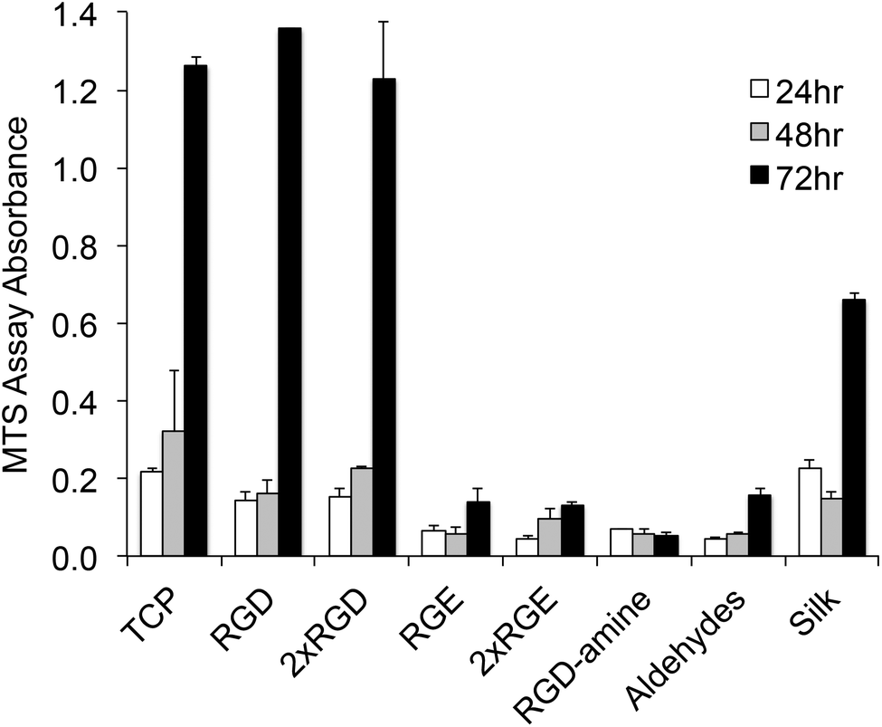 | ||
| Fig. 3 Relative cell number over time following seeding of C2C12 myoblasts on functionalized silk fibroin surfaces in comparison to tissue culture plastic (TCP). Data represent the mean ± standard error of MTS absorbance values (n = 3). | ||
Conclusions
In summary, we have designed and developed a novel, hydrazide-anchored dendron scaffold for the chemoselective functionalization of carbonyl-containing molecules. The terminal NHS ester groups enable this scaffold to be readily decorated with a wide range of biomedically useful molecules. In particular, we have demonstrated that peptides can be coupled to this scaffold following solid phase synthesis and mild cleavage from the resin, leaving side chain protecting groups intact and, thus, preventing non-specific reactions. This approach has potential applications in a number of synthetic and biomedical areas where chemoselective decoration of a target molecule or cell with an increased density of drugs, ligands or labels is required.Acknowledgements
This work was supported by the Engineering and Physical Sciences Research Council [grant number EP/G049572/1].References
- R. Huisgen, Angew. Chem., Int. Ed. Engl., 1963, 2, 565–598 CrossRef PubMed
.
- V. V. Rostovtsev, L. G. Green, V. V. Fokin and K. B. Sharpless, Angew. Chem., Int. Ed., 2002, 41, 2596–2599 CrossRef CAS
- C. W. Tornoe, C. Christensen and M. Meldal, J. Org. Chem., 2002, 67, 3057–3064 CrossRef CAS PubMed
- E. Saxon and C. R. Bertozzi, Science, 2000, 287, 2007–2010 CrossRef CAS
- E. Saxon, J. I. Armstrong and C. R. Bertozzi, Org. Lett., 2000, 2, 2141–2143 CrossRef CAS PubMed
- K. J. Yarema, L. K. Mahal, R. E. Bruehl, E. C. Rodriguez and C. R. Bertozzi, J. Biol. Chem., 1998, 273, 31168–31179 CrossRef CAS PubMed
- E. Rodriguez, L. Marcaurelle and C. Bertozzi, J. Org. Chem., 1998, 63, 7134–7135 CrossRef CAS
- Y. Zhang, Y. Sun, Z. Wang and L. Huang, Glycoconjugate J., 2012, 29, 445–452 CrossRef CAS PubMed
- L. Van Lenten and G. Ashwell, J. Biol. Chem., 1971, 246, 1889–1894 CAS
- P. A. De Bank, B. Kellam, D. A. Kendall and K. M. Shakesheff, Biotechnol. Bioeng., 2003, 81, 800–808 CrossRef CAS PubMed
- E. Saxon, S. Luchansky, H. Hang, C. Yu, S. Lee and C. Bertozzi, J. Am. Chem. Soc., 2002, 124, 14893–14902 CrossRef CAS PubMed
- L. K. Mahal, K. J. Yarema and C. R. Bertozzi, Science, 1997, 276, 1125–1128 CrossRef CAS
- K. L. Kiick, E. Saxon, D. A. Tirrell and C. R. Bertozzi, Proc. Natl. Acad. Sci. U. S. A., 2002, 99, 19–24 CrossRef CAS PubMed
- P. Wu, W. Shui, B. L. Carlson, N. Hu, D. Rabuka, J. Lee and C. R. Bertozzi, Proc. Natl. Acad. Sci. U. S. A., 2009, 106, 3000–3005 CrossRef CAS PubMed
- J. Prescher, D. Dube and C. Bertozzi, Nature, 2004, 430, 873–877 CrossRef CAS PubMed
- P. V. Chang, J. A. Prescher, E. M. Sletten, J. M. Baskin, I. A. Miller, N. J. Agard, A. Lo and C. R. Bertozzi, Proc. Natl. Acad. Sci. U. S. A., 2010, 107, 1821–1826 CrossRef CAS PubMed
- N. Fischer-Durand, M. Salmain, A. Vessières and G. Jaouen, Tetrahedron, 2012, 68, 9638–9644 CrossRef CAS PubMed
- D. Y. Q. Wong, J. Y. Lau and W. H. Ang, Dalton Trans., 2012, 41, 6104–6111 RSC
- J. B. Delehanty, J. B. Blanco-Canosa, C. E. Bradburne, K. Susumu, M. H. Stewart, D. E. Prasuhn, P. E. Dawson and I. L. Medintz, Chem. Commun., 2013, 49, 7878–7880 RSC
- J. B. Blanco-Canosa, I. L. Medintz, D. Farrell, H. Mattoussi and P. E. Dawson, J. Am. Chem. Soc., 2010, 132, 10027–10033 CrossRef CAS PubMed
- W. R. Algar, D. E. Prasuhn, M. H. Stewart, T. L. Jennings, J. B. Blanco-Canosa, P. E. Dawson and I. L. Medintz, Bioconjugate Chem., 2011, 22, 825–858 CrossRef CAS PubMed
- M. Adamczyk, J. C. Gebler, R. E. Reddy and Z. Yu, Bioconjugate Chem., 2001, 12, 139–142 CrossRef CAS PubMed
- J. Lutz and Z. Zarafshani, Adv. Drug Delivery Rev., 2008, 60, 958–970 CrossRef CAS PubMed
- L. Yi, Y.-X. Chen, P.-C. Lin, H. Schröder, C. M. Niemeyer, Y.-W. Wu, R. S. Goody, G. Triola and H. Waldmann, Chem. Commun., 2012, 48, 10829–10831 RSC
- H. Altin, I. Kosif and R. Sanyal, Macromolecules, 2010, 43, 3801–3808 CrossRef CAS
- C. A. DeForest and K. S. Anseth, Nat. Chem., 2011, 3, 925–931 CrossRef CAS PubMed
- L. Beria, T. N. Gevrek, A. Erdog, R. Sanyal, D. Pasini and A. Sanyal, Biomater. Sci., 2013, 2, 67–75 RSC
- P. S. Banerjee, P. Ostapchuk, P. Hearing and I. S. Carrico, J. Virol., 2011, 85, 7546–7554 CrossRef CAS PubMed
- D. Zhao, S.-M. Ong, Z. Yue, Z. Jiang, Y.-C. Toh, M. Khan, J. Shi, C.-H. Tan, J. P. Chen and H. Yu, Biomaterials, 2008, 29, 3693–3702 CrossRef CAS PubMed
- D. Dutta, A. Pulsipher, W. Luo and M. N. Yousaf, J. Am. Chem. Soc., 2011, 133, 8704–8713 CrossRef CAS PubMed
- A. Ciupa, P. A. De Bank and L. Caggiano, Chem. Commun., 2013, 49, 10148–10150 RSC
- M. A. Mintzer and M. W. Grinstaff, Chem. Soc. Rev., 2011, 40, 173–190 RSC
- J. Liu, W. D. Gray, M. E. Davis and Y. Luo, Interface Focus, 2012, 2, 307–324 CrossRef PubMed
- Y. Cheng, L. Zhao, Y. Li and T. Xu, Chem. Soc. Rev., 2011, 40, 2673–2703 RSC
- P. Kesharwani, V. Gajbhiye and N. K. Jain, Biomaterials, 2012, 33, 7138–7150 CrossRef CAS PubMed
- J. Lim and E. E. Simanek, Adv. Drug Delivery Rev., 2012, 64, 826–835 CrossRef CAS PubMed
- P. Wu, M. Malkoch, J. N. Hunt, R. Vestberg, E. Kaltgrad, M. G. Finn, V. V. Fokin, K. B. Sharpless and C. J. Hawker, Chem. Commun., 2005, 5775–5777 RSC
- L. Röglin, E. H. M. Lempens and E. W. Meijer, Angew. Chem., Int. Ed., 2010, 50, 102–112 CrossRef PubMed
- D. Pulido, F. Albericio and M. Royo, Org. Lett., 2014, 16, 1318–1321 CrossRef CAS PubMed
- A. D. Schluter and J. P. Rabe, Angew. Chem., Int. Ed., 2000, 39, 864–883 CrossRef CAS
- J. I. Paez, M. Martinelli, V. Brunetti and M. C. Strumia, Polymers, 2012, 4, 355–395 CrossRef PubMed
- J. Deng, Y. Zhou, B. Xu, K. Mai, Y. Deng and L.-M. Zhang, Biomacromolecules, 2011, 12, 642–649 CrossRef CAS PubMed
- B. Helms, J. L. Mynar, C. J. Hawker and J. M. J. Fréchet, J. Am. Chem. Soc., 2004, 126, 15020–15021 CrossRef CAS PubMed
- O. Gok, S. Yigit, M. M. Kose, R. Sanyal and A. Sanyal, J. Polym. Sci., Part A: Polym. Chem., 2013, 51, 3191–3201 CrossRef CAS PubMed
- D. Bini, L. Russo, C. Battocchio, A. Natalello, G. Polzonetti, S. M. Doglia, F. Nicotra and L. Cipolla, Org. Lett., 2014, 16, 1298–1301 CrossRef CAS PubMed
- X. He, X. Wu, X. Cai, S. Lin, M. Xie, X. Zhu and D. Yan, Langmuir, 2012, 28, 11929–11938 CrossRef CAS PubMed
- A. Battigelli, J. T.-W. Wang, J. Russier, T. Da Ros, K. Kostarelos, K. T. Al-Jamal, M. Prato and A. Bianco, Small, 2013, 9, 3610–3619 CrossRef CAS PubMed
- H. Xiong, P. Leonard and F. Seela, Bioconjugate Chem., 2012, 23, 856–870 CrossRef CAS PubMed
- J. Kalia and R. T. Raines, Angew. Chem., Int. Ed., 2008, 47, 7523–7526 CrossRef CAS PubMed
- P. Agarwal, J. van der Weijden, E. M. Sletten, D. Rabuka and C. R. Bertozzi, Proc. Natl. Acad. Sci. U. S. A., 2013, 110, 46–51 CrossRef PubMed
- M. Barczyk, S. Carracedo and D. Gullberg, Cell Tissue Res., 2010, 339, 269–280 CrossRef CAS PubMed
- U. Hersel, C. Dahmen and H. Kessler, Biomaterials, 2003, 24, 4385–4415 CrossRef CAS
- D. B. Pike and H. Ghandehari, Adv. Drug Delivery Rev., 2010, 62, 167–183 CrossRef CAS PubMed
- U. K. Marelli, F. Rechenmacher, T. R. A. Sobahi, C. Mas-Moruno and H. Kessler, Front. Oncol., 2013, 3 Search PubMed
- T. Tsuji, Y. Iio, T. Takemoto and T. Nishi, Tetrahedron: Asymmetry, 2005, 16, 3139–3142 CrossRef CAS PubMed
- T. Honjo, M. Nakao, S. Sano, M. Shiro, K. Yamaguchi, Y. Sei and Y. Nagao, Org. Lett., 2007, 9, 509–512 CrossRef CAS PubMed
- L. A. Sliedregt, P. C. Rensen, E. T. Rump, P. J. van Santbrink, M. K. Bijsterbosch, A. R. Valentijn, G. A. van der Marel, J. H. van Boom, T. J. van Berkel and E. A. Biessen, J. Med. Chem., 1999, 42, 609–618 CrossRef CAS PubMed
- K. Chakraborty and C. Devakumar, J. Agric. Food Chem., 2005, 53, 7899–7907 CrossRef CAS PubMed
- F. Kloss, T. Lincke and C. Hertweck, Eur. J. Org. Chem., 2011, 1429–1431 CrossRef CAS PubMed
- A. Dirksen, S. Dirksen, T. M. Hackeng and P. E. Dawson, J. Am. Chem. Soc., 2006, 128, 15602–15603 CrossRef CAS PubMed
- G. H. Altman, F. Diaz, C. Jakuba, T. Calabro, R. L. Horan, J. Chen, H. Lu, J. Richmond and D. L. Kaplan, Biomaterials, 2003, 24, 401–416 CrossRef CAS
- M. Numata, K. Koumoto, M. Mizu, K. Sakurai and S. Shinkai, Org. Biomol. Chem., 2005, 3, 2255–2261 CAS
- L. O'Donovan and P. A. De Bank, J. Mater. Chem., 2012, 22, 21878–21884 RSC
- A. Goessl, D. Bowen-Pope and A. Hoffman, J. Biomed. Mater. Res., 2001, 57, 15–24 CrossRef CAS
- M. Shen, Y. V. Pan, M. S. Wagner, K. D. Hauch, D. G. Castner, B. D. Ratner and T. A. Horbett, J. Biomater. Sci., Polym. Ed., 2001, 12, 961–978 CrossRef CAS PubMed
- G. Maheshwari, G. Brown, D. A. Lauffenburger, A. Wells and L. G. Griffith, J. Cell Sci., 2000, 113, 1677–1686 CAS
- E. A. Cavalcanti-Adam, T. Volberg, A. Micoulet, H. Kessler, B. Geiger and J. P. Spatz, Biophys. J., 2007, 92, 2964–2974 CrossRef CAS PubMed
Footnote |
| † Electronic supplementary information (ESI) available: Experimental procedures, characterization data and NMR spectra. See DOI: 10.1039/c4ob00870g |
| This journal is © The Royal Society of Chemistry 2014 |