Issue 4, 2014

Confocal microscopy of cytoplasmic lipid droplets in a live cancer cell: number, polarity, diffusion and solvation dynamics

Abstract

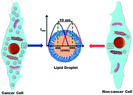
Time resolved confocal microscopy indicates that the cytoplasmic lipid droplets (CLDs) in live cells (normal and cancer lung cells) are less polar, and exhibit slower diffusion (motility) and solvation dynamics than the cytoplasm. The number of CLDs in a human lung cancer cell (A549) is ∼10 times higher than in a non-cancer lung fibroblast cell (WI38). This may result in accumulation of non-polar cell signaling agents in the CLDs of the cancer cell.

Graphical abstract: Confocal microscopy of cytoplasmic lipid droplets in a live cancer cell: number, polarity, diffusion and solvation dynamics

Supplementary files

Article information

Article type
Concise Article
Submitted
20 Sep 2013
Accepted
25 Jan 2014
First published
28 Jan 2014

Med. Chem. Commun., 2014,5, 536-539

Confocal microscopy of cytoplasmic lipid droplets in a live cancer cell: number, polarity, diffusion and solvation dynamics

R. Chowdhury, B. Jana, A. Saha, S. Ghosh and K. Bhattacharyya, Med. Chem. Commun., 2014, 5, 536 DOI: 10.1039/C3MD00269A

To request permission to reproduce material from this article, please go to the Copyright Clearance Center request page.

If you are an author contributing to an RSC publication, you do not need to request permission provided correct acknowledgement is given.

If you are the author of this article, you do not need to request permission to reproduce figures and diagrams provided correct acknowledgement is given. If you want to reproduce the whole article in a third-party publication (excluding your thesis/dissertation for which permission is not required) please go to the Copyright Clearance Center request page.

Read more about how to correctly acknowledge RSC content.

Spotlight

Advertisements