Advances in the production and handling of encoded microparticles
Hector Enrique
Muñoz
,
James
Che
,
Janay Elise
Kong
and
Dino
Di Carlo
*
Department of Bioengineering, California NanoSystems Institute, Jonsson Comprehensive Cancer Center, University of California Los Angeles, 420 Westwood Plaza, 5121 Engineering V, Box 951600, Los Angeles, California 90095, USA. E-mail: dicarlo@seas.ucla.edu
First published on 22nd May 2014
Abstract
Here we highlight emerging technologies in the synthesis, handling, and application of encoded microparticles for multiplexed assays. Traditionally, in drug discovery and life sciences research, multiple reactions will be conducted in parallel using microwell plate formats or microfluidic implementations, in which volumes are confined and reactions annotated by knowledge of what reagents were added to each volume. Microparticle-based information carriers provide an alternative approach to performing such multiplexed reactions, in which reactions and events are instead annotated with unique codes associated with the solid-phase particle. One challenge has been in creating a unique and large enough code set that is also easily readout, and we highlight two approaches that have brought orthogonal optical tagging techniques to bear. Another challenge has been that in such approaches, reactions have usually been confined to the surface of, or within the bulk of the specifically-tagged particle. We also highlight a creative approach and strategy for multiplexing – called “partipetting” – in which the coded particle can be a carrier of a unique fluid reagent.
Highly scalable encoding of microparticles
Microparticle encoding has often been implemented by embedding dyes with complementary spectra at defined ratios,1 but these approaches have had limited capacity to create unique codes when deconvolving a code from few identifying fluorescent or colorimetric channels, and often require complex decoding equipment for low error readouts. Moreover, microparticle fabrication techniques are narrow in design and tend not to translate well for a range of applications, which may require particle biocompatibility and thermal or chemical resilience. Lee et al.2 introduced a novel encoding process that arranges a diverse set of spectrally unique probes in bands along a rod-shaped microparticle. Using stop-flow lithography on laminar co-flowing streams of distinct probe moieties, combination spectral and spatial barcodes are created, with the potential for up to 106 unique particles.2 Importantly, the authors show early proofs of concept across a broad application space towards anti-counterfeiting, pharmaceutical packaging, bioassays, and other massively parallel industrial processes.The encoded microparticles consist of two key components: i) upconversion nanocrystals (UCNs) embedded in ii) an application-specific polymer. First, the UCNs with varying relative ratios of lanthanide ion dopants (Yb3+, Er3+, and Tm3+) were generated to yield ratiometrically unique emission spectra. The production of UCNs is reproducible, yielding high-aspect-ratio (>250 nm) crystals that are insensitive to photobleaching and have high signal-to-noise ratios.3 Moreover, doping with gadolinium allows for magnetization of microparticles and manipulation by an external magnetic field, which could help with handling or orientation of the particles.
The group characterized two different monomers for the microparticle base to enable different applications. Polymerization of hydrophobic poly(urethane) acrylate (PUA) gives rise to particles with high thermal (260 °C) and chemical resistance. Alternatively, the use of poly(ethylene glycol) diacrylate (PEGDA) generates biocompatible, mesoporous particles.
Assembly of the microparticles was achieved using a PDMS microfluidic channel containing co-flowing laminar fluid streams, each containing a unique UCN in a suspension of polymer pre-mix. Photopolymerization across the width of the microchannel generated ~200 μm × 50 μm microparticles with unique spectral barcodes (Fig. 1). Many microparticles were produced using stop-flow lithography at a rate of 18![[thin space (1/6-em)]](https://www.rsc.org/images/entities/char_2009.gif) 000 particles h−1, with the capacity for up to 105 particles h−1 if the laminar streams are instead vertically stacked allowing polymerization of particles in a 2D array.4 High encoding capacity is achieved by varying the number of colors (ratios of Yb/Er/Tm) or stripes (by adding additional co-flowing streams), which gives rise to >106 unique microparticles. A combination with different particle shapes could further increase encoding combinations.
000 particles h−1, with the capacity for up to 105 particles h−1 if the laminar streams are instead vertically stacked allowing polymerization of particles in a 2D array.4 High encoding capacity is achieved by varying the number of colors (ratios of Yb/Er/Tm) or stripes (by adding additional co-flowing streams), which gives rise to >106 unique microparticles. A combination with different particle shapes could further increase encoding combinations.
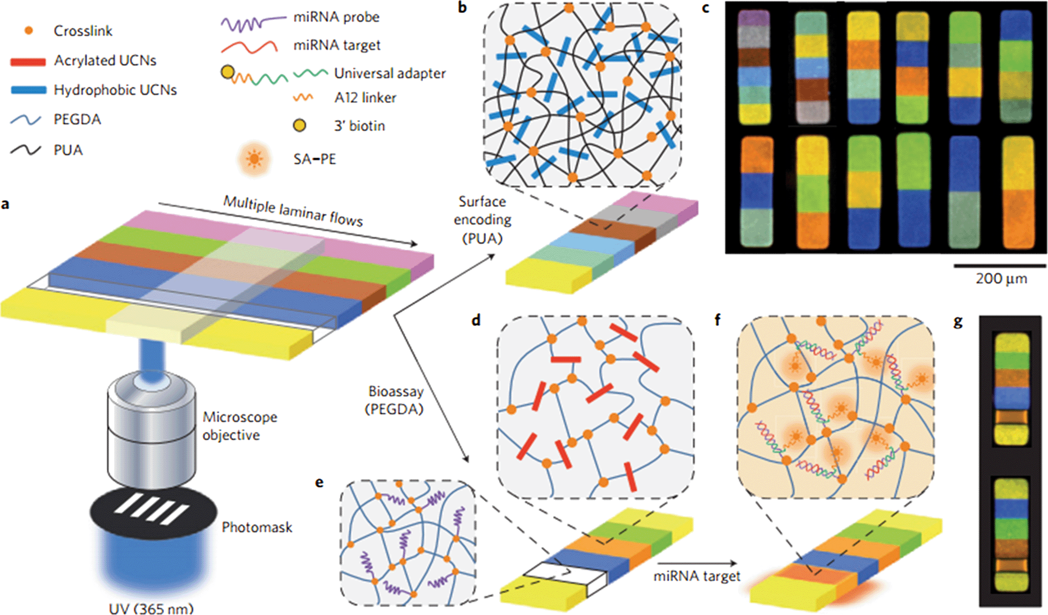 | ||
| Fig. 1 Synthesis of encoded particles by stop-flow lithography. (a) Multiple co-flows of monomer solution (PEGDA or PUA) with UCNs were photopolymerized in a PDMS channel through illumination with photomask-patterned ultraviolet light (365 nm) and collected for future use. (b) The hydrophobic UCNs are physically entrained in the tightly cross-linked PUA matrix. (c) Luminescence images of encoded PUA particles. (d) Acrylated UCNs covalently incorporated into the mesoporous PEGDA matrix. (e) Incorporation of acrylated miRNA probes during flow lithography for bioassay applications. The mesoporous matrix allows diffusion of large (>10 nm) biomolecules through the matrix. (f) Labelling of hydrogel particles after incubation with miRNA targets using a biotinylated universal adapter sequence and streptavidin–phycoerythrin (SA–PE). (g) Luminescence images of encoded PEGDA hydrogel particles after miRNA bioassay (excitation, 1W 980 nm NIR diode laser). Adapted from Lee et al.2 | ||
Importantly, particle codes can be read using simple commercial parts and an affordable optical setup totalling US$3000.2 Lee et al. applied the decoding system toward industrial packaging, in which PUA particles suspended in PUA monomer pre-mix were photopolymerized and laminated onto surfaces. The transparent, embedded microparticles revealed spectral signals that fall within 5 sigma of a training set, which allows for low-error (<10−9 false-alarm rate) anti-counterfeiting measures in pharmaceutical packaging. Similar image clarity was achieved with a 20× objective in conjunction with an Apple iPhone 4S. Finally, in a separate application, hydrophilic PEGDA particles were fabricated with a microRNA probe for either miR-210 or miR-221. Successful detection of miRNA targets was demonstrated by the specific binding of miRNAs to their matching microparticles. Measurement of biomolecule concentrations within the particle was also minimally affected by the coding region of the particle.
In conjunction with high-throughput microparticle handling that can be seamlessly integrated with production plants—such as with roll-to-roll printing or microfluidic control systems—such microparticle based barcodes may see extensive use in industrial labeling, once costs become commercially viable for large scale production.
Photonic crystal encoded microcarriers for biodetection
In an alternative approach to develop simply read and highly multiplexed particle codes, Ye et al. developed photonic crystal (PhC) encoded microcarriers that promise to simplify label-free suspension assays.5 The microcarriers were encoded with an opal PhC core, comprised of packed silica colloidal crystal beads. The packed beads have a characteristic wavelength of light reflected (photonic bandgap), according to the bead size and refractive index. These microcarriers were prepared via self-assembly of silica nanoparticles in microfluidic-generated droplets (Fig. 2a–f). This method achieved highly uniform, packed PhC particles with a diameter of ~200 μm. The microcarriers were further functionalized by introducing an inverse-opal PhC hydrogel shell, which may be functionalized to sense targets of interest. This shell was created by impregnating the network of pores surrounding the silica beads with a hydrogel solution. The solution completely filled the spaces around the beads before being polymerized via UV light. Peripheral silica beads were removed from the microcarriers by mechanical disruption in a buffer path to create regular voids. This results in a uniform hydrogel network that serves as an inverse-opal PhC, with a distinct reflection peak, in addition to that of the core. By varying the size of the highly monodisperse beads, identically-sized microcarriers can be encoded with distinct reflected wavelength peaks. | ||
| Fig. 2 Fabrication of photonic crystal (PhC) microcarriers. (a) The original microcarrier is comprised of an opal PhC core, surrounded by an inverse-opal PhC shell. These PhC regions result in distinct reflection wavelength peaks. After the hydrogel binds with the target, it deforms and its corresponding reflection wavelength undergoes a blue shift. Microcarriers are produced by polymerizing hydrogel within packed silica beads and etching away peripheral beads. (b–d) Reflective images of the process and (e–f) the corresponding reflective wavelengths. (h) Multiple multicarriers that detect Ag+, Hg2+, and Pb selectively react in a solution of Ag+ and Hg2+. Adapted from Ye et al.5 | ||
The hydrogel shell serves as a sensor with the addition of elements that respond to target stimuli and deform the hydrogel shell. The hydrogel's deformation results in a blue shift in the peak reflection wavelength of the PhC, while the packed silica beads remain resilient in changing environments. This was illustrated by including a Hg2+-responsive aptamer in the hydrogel, which undergoes a conformational change when binding. A measurable wavelength shift was recorded for concentrations of Hg2+ ranging from less than 1 nM to 10 μM.
In order to highlight the ability to distinguish different PhC cores (i.e. particle codes), and the selective shifts in the PhC shell wavelengths, Ye et al. produced 3 species of microcarriers with distinct PhC core wavelength peaks and aptamers that target Hg2+, Ag+, and Pb. When exposed to a solution of Hg2+ and Ag+ ions, no significant shift in signal was measured for non-target ions (Fig. 2h).
This same group created similar microcarriers that were completely comprised of silica beads and hydrogel, and had no peripheral PhC shell.6 These PhC microcarriers were designed for use in a multiplex cell suspension assay. Conceivably, the microcarriers used by Ye et al. could be loaded with a cell-responsive hydrogel, and the shell PhC wavelength could describe cell adhesion, traction forces, activity or viability. These microcarriers could be used to measure the cyclic contractions of cardiomyocytes to get real time optical force measurements. In summary, PhC microcarriers have the capability to simplify massively multiplexed assays. This is accomplished by creating several encoded microcarriers that can self-report, eliminating the need for labeling, parallel assays and concern for overlapping fluorescence. The multiplexing ability of this technology is only limited by the ability to precisely fabricate silica beads of varying sizes that are able to produce distinct reflective wavelength peaks, and the introduction of reactive hydrogel components.
One-step pipetting with particles
Drug discovery relies on high-throughput screening to identify therapeutic biomolecules that bind a target or lead to a cellular effect. However, the discovery of such drugs is limited to large pharmaceutical companies or well-funded research labs because of the high cost of automation instrumentation and reagents. A critical bottleneck for assay time and affordability lies in precise liquid handling and transfer, which is typically done through pipetting. To alleviate this challenge, Chung et al. proposed a new technique entitled ‘partipetting’,7 the one-step pipetting of encoded chemical-laden microparticles (ECMs) into microwell arrays for multiplexed and combinatorial assays.The partipetting process starts with the generation of ECMs using optofluidic maskless lithography, a technique developed previously that allows generation of photopolymerized microparticles.8 Each hydrogel particle is loaded with sub-nanoliter quantities of solutions containing chemical substances and encoded with a 2D graphical barcode to identify its contents (Fig. 3a and d). The microparticles are then dried and stored in mineral oil as part of an ECM library. In the presence of a liquid buffer solution, such as cell culture medium, the chemical substances will begin to diffuse out of the ECMs (Fig. 3b and c). When in use, these 180 μm microparticles are mixed at concentrations of the user's choice, singularly pipetted onto the surface of a polydimethyl siloxane (PDMS) chip, and then scraped to fill the 200 μm microwells with individual particles/compounds (Fig. 3e). To ensure the randomness of particle filling, particles numbered from 00–99 were added in the same quantity and the amount of each particle aligned with that expected from a binomial distribution. Therefore, rather than conducting many pipetting steps in the lab, a single step is all that is needed, and all of the automated steps to encode the library is done centrally at a fabrication facility.
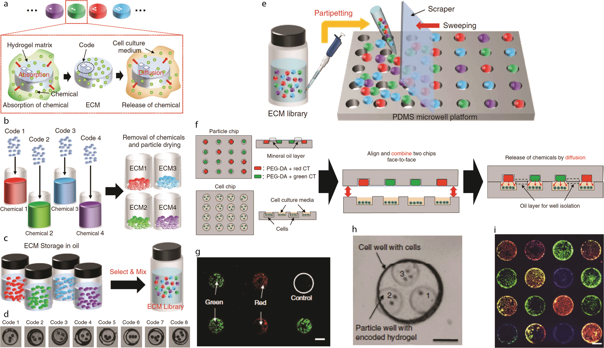 | ||
| Fig. 3 Partipetting. (a) Encoded chemical-laden microparticles (ECMs) loaded with chemical substances and a corresponding 2D graphical barcode. (b) Chemicals are non-specifically absorbed into the ECM hydrogel matrix and then dried. (c) ECMs are stored in mineral oil to prevent cross-contamination. (d) Examples of different ECM 2D graphical barcodes. (e) The partipetting process for filling the microwell particle device. (f) The process for cell-based assays. A microparticle chip is filled via partipetting and then placed face-to-face with a cell microwell device to allow diffusion of chemicals. (g) Proof of concept assay with cell tracker red and green stains. (h) Combinatorial assay with potential for 3 different chemicals to be introduced simultaneously to a cell microwell. (i) Proof of concept combinatorial assay with cell tracker red, green, and blue stains. Adapted from Chung et al.7 | ||
Cell analysis using partipetting requires only two components: a PDMS microwell array that contains individual hydrogel particles, and a microwell array that contains individual cells. These two faces are aligned and placed face-to-face to allow each ECM and its associated compound to come into contact with single cells at 72% probability (Fig. 3f). A proof of concept assay successfully stained cells using particles loaded with cell tracker red and green (Fig. 3g).
The cytotoxicity of ECM-loaded compounds against U2OS human osteosarcoma cells was quantified with Annexin V staining of apoptotic cells, discounting necrotic cells (identified with propidium iodide) and cells that were singularly stained with Hoechst 33342. These results were compared to reference data of the same drug concentrations, obtained using the conventional microtiter well technique. With similar comparisons, PEG-DA hydrogel drugs were shown to retain cytotoxicity within ±17% of the conventional techniques, and remained consistent with 20× scaled devices.
To demonstrate the power of this technology, Chung et al. designed a combinatorial assay combining three drugs by aligning three microparticle microwells with each cell microwell. As a proof-of-concept, three types of encoded microparticles, loaded with cell tracker green, red, and blue, were tested against cells to yield various cocktails of these particles, as shown in Fig. 3h and i. By the same principle, anti-cancer drugs camptothecin (CPT) and sodium salicylate (SS) were used in a combinatorial assay to yield 10 possible combinations of the drugs, which assemble randomly. Cytotoxicity data with the combinations were within ±10% of those obtained using conventional pipetting techniques.
This new partipetting technique for large scale assays benefits from ease of use, low cost, and short processing time. The highly multiplexed and combinatorial attributes of this platform will open new doors in biochemical drug characterization in small research labs, where the costs of producing libraries and screening them is focused at the manufacturing step, rather than at the end user.
References
- S. Birtwell and H. Morgan, Integr. Biol., 2009, 1, 345–362, 10.1039/B905502A.
- J. Lee, P. W. Bisso, R. L. Srinivas, J. J. Kim, A. J. Swiston and P. S. Doyle, Nat. Mater., 2014, 13, 524–529, DOI:10.1038/nmat3938.
- F. Wang, Y. Han, C. S. Lim, Y. Lu, J. Wang, J. Xu, H. Chen, C. Zhang, M. Hong and X. Liu, Nature, 2010, 463, 1061–1065, DOI:10.1038/nature08777.
- K. W. Bong, K. T. Bong, D. Pregibon and P. S. Doyle, Angew. Chem., Int. Ed., 2010, 49, 87–90, DOI:10.1002/anie.200905229.
- B. Ye, H. Ding, Y. Cheng, H. Gu, Y. Zhao, Z. Xie and Z. Gu, Adv. Mater., 2014 DOI:10.1002/adma.201305035.
- W. Liu, L. Shang, F. Zheng, J. Lu, J. Qian, Y. Zhao and Z. Gu, Small, 2014, 10, 88–93, DOI:10.1002/smll.201301253.
- S. E. Chung, J. Kim, D. Y. Oh, Y. Song, S. H. Lee, S. Min and S. Kwon, Nat. Commun., 2014 DOI:10.1038/ncomms4468.
- S. E. Chung, W. Park, H. Park, K. Yu, N. Park and S. Kwon, Appl. Phys. Lett., 2007, 91, 041106, DOI:10.1063/1.2759988.
| This journal is © The Royal Society of Chemistry 2014 |