 Open Access Article
Microfluidic assembly of multistage porous silicon–lipid vesicles for controlled drug release†
Bárbara
Herranz-Blanco
a,
Laura R.
Arriaga
b,
Ermei
Mäkilä
ac,
Alexandra
Correia
a,
Neha
Shrestha
a,
Sabiruddin
Mirza
ab,
David A.
Weitz
b,
Jarno
Salonen
c,
Jouni
Hirvonen
a and
Hélder A.
Santos
*a
aDivision of Pharmaceutical Chemistry and Technology, Faculty of Pharmacy, University of Helsinki, FI-00014 Helsinki, Finland. E-mail: helder.santos@helsinki.fi; Fax: +358 9 191 59144; Tel: +358 9 191 59661
bDepartment of Physics, School of Engineering and Applied Science, Harvard University, MA 02138, USA
cLaboratory of Industrial Physics, Department of Physics and Astronomy, University of Turku, FI-20014 Turku, Finland
First published on 6th January 2014
Abstract
A reliable microfluidic platform for the generation of stable and monodisperse multistage drug delivery systems is reported. A glass-capillary flow-focusing droplet generation device was used to encapsulate thermally hydrocarbonized porous silicon (PSi) microparticles into the aqueous cores of double emulsion drops, yielding the formation of a multistage PSi–lipid vesicle. This composite system enables a large loading capacity for hydrophobic drugs.
Microfluidic technologies are becoming widely used methods for many applications ranging from material science1–3 to biology.4 Particularly relevant for drug delivery applications is the controlled production of capsules5–7 or vesicles8,9 using microfluidic methods; these not only provide a higher encapsulation efficiency10–12 than conventional production techniques, but also endow the delivery vehicles with extremely uniform sizes3 and very controlled chemical compositions. Widely used templates for capsules or vesicles are water-in-oil-in-water (W/O/W) double emulsion drops: their aqueous cores provide an ideal environment for the dissolution of hydrophilic drugs, whereas their oil shells can be loaded with hydrophobic molecules.13 After careful removal of the oil from the emulsion shells, double emulsion drops turn into capsules or vesicles, and their membranes protect the encapsulated material from the external environment.14
Due to their high biocompatibility, lipid vesicles are long-lasting recognized as promising drug delivery vehicles.15 If prepared using microfluidic technologies,16 they can efficiently encapsulate large amounts of drugs within their aqueous cores, particularly hydrophilic drugs.17 Unfortunately, due to the ultrathin nature of the lipid bilayer, the loading capacity of lipid vesicles for hydrophobic drugs is rather limited.14 Porous silicon (PSi) particles, which are also highly biocompatible delivery vehicles and widely studied for biomedical applications,18–25 can overcome the limited hydrophobic loading capacity of the lipid vesicles. With typical pore sizes ranging from 2 to 50 nm, drug molecules confined within the mesopores maintain an amorphous conformation, avoiding extensive crystallization of the drugs, thereby leading to an increase in the drug dissolution rates.18,21,26–28 However, PSi microparticles typically show a much faster release of their cargo,29,30 which may cause a premature degradation of drugs in the body and a decrease in their therapeutic effect. The development of a new delivery platform that preserves the high biocompatibility of the lipid vesicles and PSi microparticles, provides a high loading capacity for both hydrophilic and hydrophobic drugs, and allows a sustained release of cargo, is therefore highly desired.1,3,31–35
Herein, we propose the combination of lipid vesicles with PSi microparticles as a multistage drug delivery system. Using a glass-capillary flow-focusing droplet generation device,10,36 thermally hydrocarbonized PSi microparticles (THCPSiMPs)19,23,30,37 were encapsulated within the aqueous cores of water-in-oil-in-water (W/O/W) double emulsion drops with ultrathin shells. Dewetting of the oil from the shell of the double emulsions induced the formation of a lipid bilayer.10 The presence of THCPSiMPs within the aqueous cores of these lipid vesicles not only provided the vesicles with higher loading capacity for hydrophobic drugs, as a result of the high porosity of the PSi microparticles, but also their synergistic effect endowed the vehicle with a sustained drug delivery capacity.
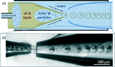
To fabricate lipid-stabilized double emulsion drops with ultrathin shells and simultaneously encapsulate THCPSiMPs within their cores, we used a glass-capillary microfluidic device that consisted of two tapered cylindrical capillaries with an outer diameter of 1 mm, carefully sanded to a tip diameter of 80 and 120 μm for injection and collection, respectively, and inserted into the opposite ends of a square capillary with an outer diameter of 1 mm, as illustrated schematically in Fig. 1a. An additional capillary inserted into the injection capillary allowed both, the inner water and middle oil phases, to co-flow within the injection capillary. The injection capillary was hydrophobic; this favored the contact of the middle oil phase with its wall. Large plug-like water drops containing the THCPSiMPs were thus formed within the injection capillary and broken-up at the tip into double emulsion drops stabilized by the thin layer of oil that was wetting the capillary wall (Fig. 1a). The collection capillary was hydrophilic; this prevented the wetting of the oil shell of the emulsion on the wall of the collection capillary.
As the inner water phase, we used an aqueous suspension of THCPSiMPs with typical particle sizes of ca. 15 μm. To facilitate the dispersion of the THCPSiMPs, they were first wetted with ethanol and later suspended in an aqueous solution that contained 8 wt-% polyethylene glycol (PEG, 6 kDa) and 2 wt-% polyvinyl alcohol (PVA, 13–23 kDa) at a final concentration of 3 mg mL−1. The suspension was sonicated and poured into a syringe together with a magnetic stirrer allowing vigorous shaking of the suspension, thereby avoiding microparticle aggregation and sedimentation during the injection of the suspension within the microfluidic chip. Importantly, our microfluidic approach provides a hundred percent encapsulation efficiency as all the particles in the inner water phase ended-up in the cores of the double emulsions as shown in Fig. 1b. The middle oil phase contained 4.6 mg mL−1 of 1,2-dioleyl-sn-glycero-3-phosphocholine (DOPC, Avanti) dissolved in a mixture of chloroform and hexane at a volume ratio of 1![[thin space (1/6-em)]](https://www.rsc.org/images/entities/char_2009.gif) :
:![[thin space (1/6-em)]](https://www.rsc.org/images/entities/char_2009.gif) 1.8, eventually containing 0.25 mole-% of 1,2-dioleoyl-sn-glycero-3-phosphoethanolamine-N-(lissamine rhodamine B sulfonyl) (DHPE-Rh, Molecular Probes) to fluorescently label the lipid bilayer. The outer aqueous phase consisted of a 10 wt-% PVA (13–23 kDa) aqueous solution.
1.8, eventually containing 0.25 mole-% of 1,2-dioleoyl-sn-glycero-3-phosphoethanolamine-N-(lissamine rhodamine B sulfonyl) (DHPE-Rh, Molecular Probes) to fluorescently label the lipid bilayer. The outer aqueous phase consisted of a 10 wt-% PVA (13–23 kDa) aqueous solution.
The double emulsion droplets were collected in an aqueous solution of sucrose with the same osmolarity as the inner water phase (100 mOsm L−1); this avoided any osmotic stress that may induce destabilization of the double emulsion droplets. Under these conditions, the chloroform in the middle oil phase easily evaporated from the shell of the double emulsions, thereby increasing the ratio of hexane (which is a poor solvent for the lipids) within the oil shell of the double emulsion. This reduction in solvent quality induces an attraction between the two monolayers of lipids at the oil/water interfaces of the double emulsions, which leads to the dewetting of the hexane from the inner cores of the double emulsion droplets; this ultimately results in the formation of the lipid bilayer. The dewetting process occurs in about a couple of minutes.10 The small amount of residual solvent present in the vesicle after dewetting remains concentrated in a very small region of the bilayer, allowing the bilayer to behave similarly to vesicles produced by conventional approaches.10 Moreover, the residual solvent evaporates further in the next days.
The resultant THCPSiMPs–lipid vesicle system had a mean diameter of 114 ± 8.4 μm, as shown in the optical microscope image in Fig. 2a. Each delivery vehicle consisted of a lipid bilayer, which appears fluorescently labeled in the microscope image shown in Fig. 2b, encapsulating the THCPSiMPs within its aqueous core. A detailed scanning electron microscope (SEM) image of a THCPSiMP such as those within the vesicles, is shown in Fig. 2c. The residual oil after the dewetting process is also visible by optical microscopy as pointed out by the white region in Fig. 2b.
To test the potential of THCPSiMPs–lipid vesicles for drug delivery applications, the THCPSiMPs were loaded with a model drug, piroxicam. This drug belongs to the Biopharmaceutical Classification System class II, i.e. it is highly hydrophobic, poorly water-soluble and highly permeable across biological membranes. To load piroxicam, the THCPSiMPs were stirred in an acetone solution of piroxicam at 15 mg mL−1 for 2 h. Then, the suspension was centrifuged at 11![[thin space (1/6-em)]](https://www.rsc.org/images/entities/char_2009.gif) 300 ×g for 4 min and the pellet was washed 3 times with 0.5 mL of MilliQ-water; water allows the removal of the excess piroxicam from the surface of the particles, while avoiding a premature release of the drugs from the pores. Finally, the loaded microparticles were suspended into the inner water phase and the production proceeded as described previously.
300 ×g for 4 min and the pellet was washed 3 times with 0.5 mL of MilliQ-water; water allows the removal of the excess piroxicam from the surface of the particles, while avoiding a premature release of the drugs from the pores. Finally, the loaded microparticles were suspended into the inner water phase and the production proceeded as described previously.
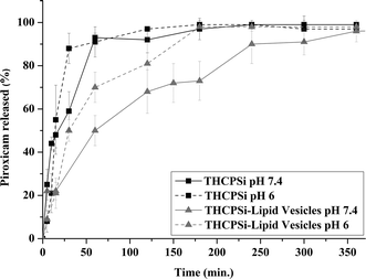
The drug release tests were performed by introducing the resultant THCPSiMPs–lipid vesicles in a dialysis bag, immersed into a buffer solution. We estimate that the number of THCPSiMPs–lipid vesicles loaded in each dialysis bag is approximately 90![[thin space (1/6-em)]](https://www.rsc.org/images/entities/char_2009.gif) 000, and each droplet contains a THCPSiMP average mass of 0.0023 μg. The solution was placed in an orbital incubator shaker at 37 °C and 45 rpm. Ten milliliters of two saline phosphate buffer solutions, with different pH values of 7.4 and 6, were used as the release media. Sample aliquots of 200 μL were taken at different time points, and the same volume of a pre-warmed fresh release medium was immediately replaced to keep constant the dissolution volume. The concentration of piroxicam in the samples was analyzed by spectrophotometry at a maximum absorbance wavelength of 333 nm (NanoDrop, Thermo Scientific) and the results are shown in Fig. 3. The drug (piroxicam) loading degree in the system was 19%. THCPSiMPs–lipid vesicles showed sustained release compared to the bare THCPSiMPs. Moreover, while at pH 6 the whole payload was released after 3 h, at pH 7.4 the drug release was completed only after 6 h. These differences could be due to the heterogeneous stability behavior of the lipid bilayer towards different pH values. Although no morphological changes were observed on the vesicles over time (ESI 1†), breakage of vesicles was significant after approximately 4 h; this explains the faster release observed after 200 min in Fig. 3.
000, and each droplet contains a THCPSiMP average mass of 0.0023 μg. The solution was placed in an orbital incubator shaker at 37 °C and 45 rpm. Ten milliliters of two saline phosphate buffer solutions, with different pH values of 7.4 and 6, were used as the release media. Sample aliquots of 200 μL were taken at different time points, and the same volume of a pre-warmed fresh release medium was immediately replaced to keep constant the dissolution volume. The concentration of piroxicam in the samples was analyzed by spectrophotometry at a maximum absorbance wavelength of 333 nm (NanoDrop, Thermo Scientific) and the results are shown in Fig. 3. The drug (piroxicam) loading degree in the system was 19%. THCPSiMPs–lipid vesicles showed sustained release compared to the bare THCPSiMPs. Moreover, while at pH 6 the whole payload was released after 3 h, at pH 7.4 the drug release was completed only after 6 h. These differences could be due to the heterogeneous stability behavior of the lipid bilayer towards different pH values. Although no morphological changes were observed on the vesicles over time (ESI 1†), breakage of vesicles was significant after approximately 4 h; this explains the faster release observed after 200 min in Fig. 3.
In order to evaluate the cell viability of the particles, mucus-secreting intestinal HT-29 (5.0 × 10⁵ cells per well)29,30 cells were selected and the cells were cultured as described in the ESI 2.† Briefly, a suspension of cells in Dulbecco's modified Eagle medium was seeded into 96-well plates (100 μL per well, Perkin Elmer Inc.) and allowed to attach overnight. The THCPSiMPs and THCPSiMPs−lipid vesicles suspensions at concentrations of 0.05–0.5 mg mL−1 were added into the wells (100 μL per well). After 3 h incubation, the viability was assayed with CellTiter-Glo (Promega Corporation) using a Varioskan Flash fluorometer (Thermo Fisher Scientific).18–20,24–26,28–30
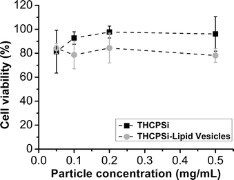
As a result of the micrometric size of these microvehicles, they are envisaged to be applied for oral drug delivery, and thus, we evaluated their cytocompatibility with gastrointestinal tract-related HT-29 cancer cell line (Fig. 4). In general, and as expected,29,30 we observed a decrease in the cell viability as the concentration of the particles increased. There were no statistically significant differences (p < 0.05) in cell viability between the bare THCPSiMPs and THCPSiMPs–lipid vesicles, which demonstrates the cytocompatibility of both systems in mucus-secreting intestinal HT-29 cells.
Conclusions
In conclusion, we have successfully produced a multistage drug delivery system consisting of THCPSiMPs, with high drug loading capacity, embedded within the aqueous core of lipid vesicles. The microfluidic technology made possible the production of THCPSiMPs–lipid vesicles in a highly reliable, reproducible, and efficient manner. The delivery system was characterized by optical microscopy and the drug release profiles were obtained at different pH values. The results showed that the encapsulation of the THCPSiMPs within lipid vesicles with a mean diameter of 114 μm is highly efficient and stable. The drug loading degree of the system was 19%, and the encapsulated drug showed a sustained release from the THCPSiMPs–lipid vesicles compared to the bare THCPSiMPs, particularly at pH 7.4, at which the whole payload was released only after 6 h. Due to its potential biocompatibility, production reliability, large loading capacity for hydrophobic drugs, and sustained drug delivery capacity, we propose the THCPSiMPs–lipid vesicle composite as a promising drug delivery system for multiple biomedical applications.Acknowledgements
Support for this work by Academy of Finland (decisions no. 252215 and 256394), the University of Helsinki Funds and the European Research Council under the European Union's Seventh Framework Programme (FP/2007–2013) grant no. 310892 is greatly acknowledged. LRA acknowledges financial support from Marie Curie Program (FP7-PEOPLE-2012-IOF).Notes and references
- A. R. Abate, S. Seiffert, A. S. Utada, A. Shum, R. Shah, J. Thiele, W. J. Duncanson, A. Abbaspourad, M. H. A. Lee, I. D. Lee, A. Rotem and D. A. Weitz, Microfluidic techniques for synthesizing particles. Available online: http://weitzlab.seas.harvard.edu/publications/Bookchapter_Microfluidic_techniques.pdf (accessed on 06 November 2013) Search PubMed.
- R. Karnik, F. Gu, P. Basto, C. Cannizzaro, L. Dean, W. Kyei-Manu, R. Langer and O. C. Farokhzad, Nano Lett., 2008, 8, 2906–2912 CrossRef CAS PubMed.
- Q. Xu, M. Hashimoto, T. T. Dang, T. Hoare, D. S. Kohane, G. M. Whitesides, R. Langer and D. G. Anderson, Small, 2009, 5, 1575–1581 CrossRef CAS PubMed.
- M. T. Guo, A. Rotem, J. A. Heyman and D. A. Weitz, Lab Chip, 2012, 12, 2146–2155 RSC.
- A. Abbaspourrad, N. J. Carroll, S. H. Kim and D. A. Weitz, J. Am. Chem. Soc., 2013, 135, 7744–7750 CrossRef CAS PubMed.
- A. M. DiLauro, A. Abbaspourrad, D. A. Weitz and S. T. Phillips, Macromolecules, 2013, 46, 3309–3313 CrossRef CAS.
- W. J. Duncanson, T. Lin, A. R. Abate, S. Seiffert, R. K. Shah and D. A. Weitz, Lab Chip, 2012, 12, 2135–2145 RSC.
- E. Amstad, S. H. Kim and D. A. Weitz, Angew. Chem., Int. Ed., 2012, 51, 12499–12503 CrossRef CAS PubMed.
- S. H. Kim, J. W. Kim, D. H. Kim, S. H. Han and D. A. Weitz, Small, 2013, 9, 124–131 CrossRef CAS PubMed.
- L. R. Arriaga, S. S. Datta, S.-H. Kim, E. Amstad, T. E. Kodger, F. Monroy and D. A. Weitz, Small, 2013 DOI:10.1002/smll.201301904.
- S. H. Kim, H. C. Shum, J. W. Kim, J. C. Cho and D. A. Weitz, J. Am. Chem. Soc., 2011, 133, 15165–15171 CrossRef CAS PubMed.
- H. C. Shum, Y. J. Zhao, S. H. Kim and D. A. Weitz, Angew. Chem., Int. Ed., 2011, 50, 1648–1651 CrossRef CAS PubMed.
- T. Nii and F. Ishii, Int. J. Pharm., 2005, 298, 198–205 CrossRef CAS PubMed.
- R. Delgado-Rivera, R. Rosario-Meléndez, W. Yu and K. E. Uhrich, J. Biomed. Mater. Res., Part A, 2013 DOI:10.1002/jbm.a.34949.
- V. P. Torchilin, Nat. Rev. Drug Discovery, 2005, 4, 145–160 CrossRef CAS PubMed.
- D. van Swaay and A. deMello, Lab Chip, 2013, 13, 752–767 RSC.
- K. Hettiarachchi, S. Zhang, S. Feingold, A. P. Lee and P. A. Dayton, Biotechnol. Prog., 2009, 25, 938–945 CrossRef CAS PubMed.
- L. M. Bimbo, O. V. Denisova, E. Mäkilä, M. Kaasalainen, J. K. De Brabander, J. Hirvonen, J. Salonen, L. Kakkola, D. Kainov and H. A. Santos, ACS Nano, 2013, 7, 6884–6893 CrossRef CAS PubMed.
- L. M. Bimbo, M. Sarparanta, H. A. Santos, A. J. Airaksinen, E. Mäkilä, T. Laaksonen, L. Peltonen, V. P. Lehto, J. Hirvonen and J. Salonen, ACS Nano, 2010, 4, 3023–3032 CrossRef CAS PubMed.
- P. J. Kinnari, M. L. Hyvönen, E. M. Mäkilä, M. H. Kaasalainen, A. Rivinoja, J. J. Salonen, J. T. Hirvonen, P. M. Laakkonen and H. A. Santos, Biomaterials, 2013, 34, 9134–9141 CrossRef CAS PubMed.
- H. A. Santos, L. M. Bimbo, V. P. Lehto, A. J. Airaksinen, J. Salonen and J. Hirvonen, Curr. Drug Discovery Technol., 2011, 8, 228–249 CrossRef CAS.
- H. A. Santos, J. Riikonen, J. Salonen, E. Mäkilä, T. Heikkilä, T. Laaksonen, L. Peltonen, V. P. Lehto and J. Hirvonen, Acta Biomater., 2010, 6, 2721–2731 CrossRef CAS PubMed.
- M. Sarparanta, L. M. Bimbo, J. Rytkonen, E. Mäkilä, T. J. Laaksonen, P. Laaksonen, M. Nyman, J. Salonen, M. B. Linder, J. Hirvonen, H. A. Santos and A. J. Airaksinen, Mol. Pharmaceutics, 2012, 9, 654–663 CrossRef CAS PubMed.
- M. P. Sarparanta, L. M. Bimbo, E. M. Mäkilä, J. J. Salonen, P. H. Laaksonen, A. M. Helariutta, M. B. Linder, J. T. Hirvonen, T. J. Laaksonen, H. A. Santos and A. J. Airaksinen, Biomaterials, 2012, 33, 3353–3362 CrossRef CAS PubMed.
- M.-A. Shahbazi, M. Hamidi, E. M. Mäkilä, H. Zhang, P. V. Almeida, M. Kaasalainen, J. J. Salonen, J. T. Hirvonen and H. A. Santos, Biomaterials, 2013, 34, 7776–7789 CrossRef CAS PubMed.
- P. Kinnari, E. Mäkilä, T. Heikkilä, J. Salonen, J. Hirvonen and H. A. Santos, Int. J. Pharm., 2011, 414, 148–156 CrossRef CAS PubMed.
- M. Tahvanainen, T. Rotko, E. Mäkilä, H. A. Santos, D. Neves, T. Laaksonen, A. Kallonen, K. Hamalainen, M. Peura, R. Serimaa, J. Salonen, J. Hirvonen and L. Peltonen, Int. J. Pharm., 2012, 422, 125–131 CrossRef CAS PubMed.
- N. Vale, E. Mäkilä, J. Salonen, P. Gomes, J. Hirvonen and H. A. Santos, Eur. J. Pharm. Biopharm., 2012, 81, 314–323 CrossRef CAS PubMed.
- D. Liu, L. M. Bimbo, E. Mäkilä, F. Villanova, M. Kaasalainen, B. Herranz-Blanco, C. M. Caramella, V. P. Lehto, J. Salonen, K. H. Herzig, J. Hirvonen and H. A. Santos, J. Controlled Release, 2013, 170, 268–278 CrossRef CAS PubMed.
- D. Liu, B. Herranz-Blanco, E. Mäkilä, L. R. Arriaga, S. Mirza, D. A. Weitz, N. Sandler, J. Salonen, J. Hirvonen and H. A. Santos, ACS Appl. Mater. Interfaces, 2013, 5, 12127–12134 CAS.
- A. Abbaspourrad, S. S. Datta and D. A. Weitz, Langmuir, 2013, 29, 12697–12702 CrossRef CAS PubMed.
- X. Gong, S. Peng, W. Wen, P. Sheng and W. Li, Adv. Funct. Mater., 2009, 19, 292–297 CrossRef CAS.
- R. K. Shah, J.-W. Kim and D. A. Weitz, Adv. Mater., 2009, 21, 1949–1953 CrossRef CAS.
- A. S. Utada, L.-Y. Chu, A. Fernandez-Nieves, D. R. Link, C. Holtze and D. A. Weitz, MRS Bull., 2007, 32, 702–708 CrossRef CAS.
- W. Wang, M.-J. Zhang, R. Xie, X.-J. Ju, C. Yang, C.-L. Mou, D. A. Weitz and L.-Y. Chu, Angew. Chem., Int. Ed., 2013, 52, 8084–8087 CrossRef CAS PubMed.
- S.-H. Kim, J. W. Kim, J.-C. Cho and D. A. Weitz, Lab Chip, 2011, 11, 3162–3166 RSC.
- M. Sarparanta, E. Mäkilä, T. Heikkilä, J. Salonen, E. Kukk, V. P. Lehto, H. A. Santos, J. Hirvonen and A. J. Airaksinen, Mol. Pharmaceutics, 2011, 8, 1799–1806 CrossRef CAS PubMed.
Footnote |
| † Electronic supplementary information (ESI) available: ESI 1: morphological changes over time of the THCPSiMPs–lipid vesicles during release experiments. ESI 2: cell viability experiments. See DOI: 10.1039/c3lc51260f |
| This journal is © The Royal Society of Chemistry 2014 |


