
A special JAAS issue devoted to the second International Glow Discharge Spectroscopy Symposium
The 2nd International Glow Discharge Spectroscopy Symposium (IGDSS2014) was held at the Czech Technical University in Prague, Czech Republic, over a three-day period from April 7–9, 2014. This conference, and the previous one in Albi, France, in 2010, were organized by the European Working Group for Glow Discharge Spectroscopy (EW-GDS) and headed by the EW-GDS Chairman Peter Robinson. The event was organized in the exceptional environment of the ancient city of Prague and was very successful. Nearly 100 delegates from over 20 different countries worldwide attended the conference, confirmation of the truly international research community and evidence of its growing importance. The presentations covered the traditional areas of optical emission and mass spectrometry for materials research and bulk analysis, as well as some more recent and innovative research. The diverse scientific program highlighted the latest research on glow discharge and associated analytical techniques.Program highlights
The keynote welcome lecture was given by Professor Ken Shimizu of Keio University, Japan, and described a research journey from surface science dominated by secondary-ion mass spectrometry (SIMS) to the present day, when glow discharge spectroscopies are an accepted, and critically important, analytical tool in the discipline. Professor Shimizu described an epiphany occasioned by a visit from a GD instrumentation vendor, when he ran his first elemental depth profiling experiment using glow discharge optical emission spectroscopy (GD-OES). “Everything that happened in the next 10 minutes after arrival was unbelievable.” Coming from the world of SIMS, surface analysis by GD-OES was comparatively simple, very rapid, and provided exceptional depth profiling resolution. These themes: ease of experiment, rapidity of analysis, and incredible analytical results were well represented at the IGDSS 2014 meeting.Depth profile analysis was a popular topic at the IGDSS, and it seems clear that GD-OES and GD-MS techniques have indeed reached a level of analytical proficiency that rivals, and often bests, analysis by TOF-SIMS. In this issue, one such example is provided in the manuscript of Di Sabatino and coworkers1 in describing the depth analysis of impurities in photovoltaic solar cells by GD-MS. Here, impurities were intentionally added to multicrystalline silicon samples by high-energy ion implantation, producing materials with which implantation depth and elemental solid diffusion were measured. Over depths up to 6 μm from the sample surface, B, Fe, Ti, Cu, and P concentrations were monitored at levels often below several ppbw. The author's conclusions are exemplary of many reports at IGDSS; GD-MS provides depth profile spatial resolution that rivals TOF-SIMS, and limits of detection that are often significantly better. The lower limits of detection are especially prominent for elements of lower mass and higher ionization potential (e.g., P).
Glow discharge spectroscopy continues to expand the arena of applications outside of conventional materials analysis, as evidenced by a large number of diverse reports at IGDSS. Qian and coworkers report on the use of GD-MS for the analysis of nephrite geological samples in this issue, assaying for 14 rare earth elements by employing a tantalum pin to establish a direct electrical connection to the geological samples.2 Multielemental composition was then used to establish the origin of the material within China by principle components analysis (PCA) and rare earth elemental ratio classification. Indeed, multielemental (and isotopic) information coupled with excellent limits of detection and wide dynamic range facilitate a wide breadth of analytical applications.
Glow discharge spectrometry relies on accurate spectroscopic data for accurate and reproducible analytical results. While much is known about the mechanisms operating in the analytical GD, it still holds many intriguing spectroscopic mysteries. For instance, glow discharges can be operated using different gases (e.g., Ar, He, Ne) or gas mixtures (e.g., Ne + O2), and interesting variations on the emission spectra are observed as a result.
The detailed analysis of spectra belonging to the specific plasma gas employed is of primary importance when compiling line wavelength and intensity assignments, and also provides valuable spectrochemical insights. Here, Konjevic and co-workers3 investigated the Ne I spectral line shapes in a Grimm-type glow discharge, using “side-on” and “end-on” optical emission detection. The side-on spectra showed a spectral line shift, sometimes combined with a line split, in the cathode fall region of the glow discharge. These spectroscopic data, together with available data for the dc Stark effect, were used to estimate the electric field strength in the cathode fall region. Additionally, in the negative glow region, plasma broadening was found to contribute to the line profile resulting in larger line widths. The end-on spectra were observed to be the result of integral radiation coming from regions from the cathode surface to the far end of the negative glow.
Studies of GD emission spectra from a Cu sample in a neon GD plasma containing small amounts of oxygen were reported at IGDSS by Mushtaq and co-workers.4 These data support interpretation of the excitation and ionization processes taking place in the discharge (e.g., Penning ionization (PI) of analyte atoms, asymmetric charge transfer (ACT) and Penning excitation of analyte ions). The results showed that the presence of small quantities of oxygen in the GD effected the populations of Ne metastable levels and ions. Emission yields of copper ionic lines excited by Ne-ACT were found to fall considerably when oxygen was added, probably due to a fall in the Ne+ population. Moreover, lines predominantly excited by PI exhibited a smaller fall than ACT-excited lines with oxygen addition. Additional investigations on enhanced excitation of various analytes, when using small amounts of hydrogen or oxygen in either argon or neon GD plasmas, have been reported by Mushtaq and co-workers.5 In this communication the authors argue that this enhancement is due to three body collisions involving two hydrogen or oxygen atoms and a sample atom.
The emission spectra of iron observed using a Grimm-type glow discharge in Ar, Ar with 0.3% v/v hydrogen, and Ne were studied by Zdeněk Weiss and co-workers6 in order to identify the major excitation and ionization processes of iron ions in the plasma. Transition rate diagrams versus Boltzmann plots were used to describe the excitation processes in the low pressure GD. The following processes were identified to be responsible for excitation of Fe II glow discharge spectra: charge transfer from argon/neon ions to ground state and metastable iron atoms, Penning excitation of ground state and metastable iron ions by argon metastables, and collisional excitation of metastable iron ions by electrons. Moreover, this study showed that the process that dominates excitation of the Fe II spectrum both in Ar and Ne GDs is the ACT reaction between ions of the discharge gas and neutral iron atoms.
The breadth of GD spectrometry research continues to expand, as evidenced by the number of presentations describing new GD sources and the use of GD in unconventional spectrometry. For example, this issue contains a manuscript by Wang and coworkers7 that describes design modifications to a novel solution-cathode glow discharge (SCGD) instrument for OES. The SCGD is a direct current atmospheric pressure GD operated in the ambient atmosphere and in a manner that places the GD plasma in direct contact with the solution sample under analysis. Here, the GD plasma is responsible for sampling the solution under analysis and also desolvating, vaporizing, and exciting the constituent atoms. The SCGD, therefore, holds a number of advantages over traditional ICP-OES approaches in terms of simplicity, affordability, and portability. The authors directly compare the analysis of trace element impurities in TiO2 samples by ICP-OES and SCGD-OES and report that for identical samples containing many elements of interest, the limits of detection (1–10 μg L−1), accuracy (1–2% RSE), and precision (1–5% RSD) achieved by the two techniques were comparable. Indeed, the limits of detection achieved for some elements using the SCGD were lower than those achieved by ICP-OES. These results lend credence to claims that the SCGD may replace ICP-OES instrumentation in some targeted analyses in the not too distant future. However, for this prognosis to occur, a fuller understanding of the mechanism of this intriguing GD must be achieved. In these studies, the authors report an enhancement of the emission of Pb in the presence of formic acid that is not observed when acetic or nitric acid are used. The reason for this, and several other similar element-specific matrix effects reported elsewhere, are currently unknown but are surely linked to the unique sampling, atomization and desolvation mechanisms of the SCGD. For the SCGD to find analytical utility for routine applications, further fundamental spectrochemical investigations are required.
Other unconventional GD research efforts at IGDSS reported exploitation of the GD plasma itself; one of the most popular approaches being the use of the GD as an ionization source for ambient desorption/ionization mass spectrometry (ADI-MS). This recent innovation permits molecules to be desorbed and ionized directly from the surface of virtually any sample of interest, permitting very rapid MS experiments to be conducted with high sensitivity and selectivity and without the usual sample pre-treatment requirements associated with more rigorous analyses. Here, the flowing afterglow of GDs or other plasmas are used both to desorb molecules from the surface of the sample and to create reagent ions (e.g., (H2O)nH+) through reaction with components of the ambient atmosphere. These reagent ions then ionize the analyte molecules through gas-phase reactions. A number of ADI-MS experiments based on atmospheric pressure GDs were presented at IGDSS, and in this issue Guillot and coworkers8 report on the use of a dielectric barrier discharge (DBD) as a source for ADI-MS. Although the GD and DBD are different plasma discharges, they do exploit many of the same ion reactions to produce reagent ions. In this work, the authors report OES and MS experiments used to study mechanistic pathways of ion formation in the DBD. Of course, the plasma structures of GD and DBD are fundamentally very different, as evidenced by the formation of plasma bullets in the DBD. These moving bullet-like luminous structures travel through the afterglow of the DBD at astounding rates (∼50 km s−1), often three orders of magnitude more rapid than the surrounding plasma gas flow rate. As the authors note, relatively little is known of the composition or formation mechanism of these intriguing structures, but they may provide an opportunity for energy focusing in plasma systems.
2014 Payling Prize
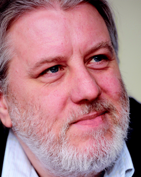
Sohail Mushtaq
The IGDSS meeting hosts the Payling Prize, an award dedicated to the memory of Dr Richard Payling and presented to the most meritorious presentation of the conference given by a young scientist. We are pleased to report that the 2014 Richard Payling Award was presented to Sohail Mushtaq, of Imperial College London. Dr Mushtaq is a postdoctoral fellow in the laboratory of Professor Edward Steers, and he was recognized for his outstanding contributions to fundamental spectroscopic understanding of GD plasmas, as evidenced by three contributions to this issue. He joins a legacy of excellent awardees.
Steven Ray, Jorge Pisonero, Peter Robinson and Cornel Venzago.
Steven Ray
Jorge Pisonero
Peter Robinson
Cornel Venzago
References
- M. Di Sabatino, C. Modanese and L. Arnberg, J. Anal. At. Spectrom., 2014 10.1039/c4ja00175c.
- B. Siqin, R. Qian, S. Zhuo, J. Gao, J. Jin and Z. Wen, J. Anal. At. Spectrom., 2014 10.1039/c4ja00172a.
- N. M. Sisovic, N. V. Ivanovic, G. L. Majstorovic and N. Konjevic, J. Anal. At. Spectrom., 2014 10.1039/c4ja00162a.
- S. Mushtaq, E. B. M. Steers, J. C. Pickering and V. Weinstein, J. Anal. At. Spectrom., 2014 10.1039/c4ja00192c.
- S. Mushtaq, E. B. M. Steers, J. C. Pickering and P. Smid, J. Anal. At. Spectrom., 2014 10.1039/c4ja00193a.
- Z. Weiss, E. B. M. Steers, J. C. Pickering and S. Mushtaq, J. Anal. At. Spectrom., 2014 10.1039/c4ja00214h.
- Z. Wang, R. Gai, L. Zhou and Z. Zhang, J. Anal. At. Spectrom., 2014 10.1039/c4ja00173g.
- L. Chauvet, L. Thérèse, B. Caillier and P. Guillot, J. Anal. At. Spectrom., 2014 10.1039/c4ja00255e.
| This journal is © The Royal Society of Chemistry 2014 |