Morphological and compositional features of blue and yellow pigments used in Portuguese glazed ceramics by SEM/EDX – unravelling manufacturing differences
A.
Guilherme
a,
V.-D.
Hodoroaba
*b,
S.
Benemann
b,
J.
Coroado
c and
M. L.
Carvalho
a
aAtomic Physics Centre, University of Lisbon, 1649-003 Lisbon, Portugal
bBAM Federal Institute for Materials Research and Testing, Division 6.8 Surface Analysis and Interfacial Chemistry, Berlin, Germany. E-mail: Dan.Hodoroaba@bam.de; Fax: +49 30 8104 1827; Tel: +49 30 8104 3144
cDepartment of Arts, Conservation & Restoration, Polytechnic Institute of Tomar, 2300-313 Tomar, Portugal
First published on 9th October 2013
Abstract
Morphological and compositional features regarding blue and yellow pigments used in glazed ceramics from Portugal are investigated by means of a SEM/EDX system. Fragments of samples produced in the main pottery-making centres between the XVI–XVIII centuries (Lisbon and Coimbra) were the objects of study. The main question to be answered is whether there are differences in the manufacturing procedures employed. For this purpose, two types of samples were analysed: faiences and wall-tiles (the famous “Azulejos”, in Portuguese). Differences between the manufacturing techniques of the two production centres were veritably found. The expected linkage between Fe–Co–Ni–As–Bi was challenging to investigate. Thus, Co was found in Fe- or Ni-rich structures, but with no structural connection to As or Bi. However, the As-needle-like structures were always identified in blue regions. One interesting finding related particularly to the blue pigment was the presence of SnO2 agglomerates throughout the whole glaze, which suggests its intentional addition for “lightening” the well known dark Co blue. Concerning the yellow pigment differences established in the crystal shape and size are indicative of different firing temperatures used.
Introduction
The lack of scientific data regarding manufacturing techniques used in Portuguese traditional glazed ceramics has triggered the present study. The problems of mislabelling ceramic objects due to the lack of consistent data, especially concerning the glazing and surface decoration procedures, need to be overcome. The literature on Portuguese glazed ceramics is rather scarce and focused on the technological aspects of bulk and glaze of wall-tiles.1,2 As far as the pigments are concerned, some investigations have been carried out,3 but no systematic microstructural characterization of pigments has been reported, to our knowledge. In particular for faiences from Coimbra the only existing one is written in Portuguese.4 According to that, one learns that the final objects may have been submitted to either two or three firing procedures: one for the ceramic body, one for the glaze (together with surface decoration) and/or a third one for the surface decoration. Depending on the procedure selected together with the particular crystal structure of the pigment itself, different diffusion mechanisms involving the pigment take place throughout the glaze.Literature reporting Italian and Spanish glazed ceramics is vast.5,6 However, the knowledge provided by these studies does not apply to the objects of the present work, as a comparison between them is not the purpose here.
Previous investigations on such pieces have provided chemical information about the glazes and the different pigments used. X-ray fluorescence (XRF) analyses on entire museum objects have indicated a lead-tin-based glaze. This holds true for faiences and wall-tiles as well.7 Additionally, the elemental identification of blue and yellow decorative motifs – both on museum objects and fragments – has raised further questions about their content. XRF spectra from blue regions have revealed the presence of iron (Fe), cobalt (Co), nickel (Ni), arsenic (As) and in some cases manganese (Mn) and bismuth (Bi).7,8 By literature inspection on blue pigments used for glazed ceramics decoration, one finds that the use of Co-pigments until about the 12th century AD is related to two production regions only: (i) Qamsar and Anarak in Persia and (ii) the Erzgebirge – Ore Mountains – border between Saxony (Germany) and Bohemia (Czech Republic). These were geologically rich enough to support a trade route over Europe, North Africa and the East (until China). The main mining industry in the Erzgebirge was silver (Ag), whose minerals were found admixed in the so-called ‘five-element’ veins (Co–Ni–As–Ag–Bi). Since then, the Erzgebirge has supplied the Co-pigment market to a large extent.9,10
Regarding the yellow pigments, a linkage between antimony (Sb) and lead (Pb) was found,7,8 which again by literature research is related to the common use of the yellow pigment Naples yellow (Pb2Sb2O7).11
Further investigations were aimed to evaluate the pigment diffusion throughout the glaze. For this purpose, highly resolved XRF scans on the cross-section of the samples were performed.12 This study demonstrates that one of the main differences is the glaze thickness, namely thinner (up to 200 μm) for faience samples and thicker (up to 400 μm) for wall tiles. In the case of faiences the blue pigment is homogeneously distributed throughout the entire glaze, while in wall-tiles it goes down to 200 μm in depth. Most interesting is that the yellow pigment is retained until the first tens of micrometers below the surface in faiences and wall-tiles.8,12
Taking all these features into account, a deeper knowledge about the morphological and compositional properties of these samples at the micrometer scale became necessary in order to complement and understand the reason for the differences obtained so far. For this purpose systematic SEM/EDX analyses on glaze, blue and yellow decorations were carried out.
Experimental and materials
The experiments have been carried out with a scanning electron microscope (SEM) Zeiss Supra 40 (Carl Zeiss AG, Oberkochen, Germany) with a Schottky field emitter having attached to a silicon drift detector energy dispersive X-ray spectrometer (SDD-EDS) Quantax 400 from Bruker (Bruker Nano GmbH, Berlin, Germany) – with an energy resolution at Mn Kα of 124 eV and 10 mm2 crystal active area – as well as a Si(Li)-EDS from Thermo Fisher Scientific (Thermo Fisher Scientific Inc., Waltham, MA, USA) – with an energy resolution at Mn Kα of 129 eV and 10 mm2 crystal active area – see ref. 13−15 for more details. Various beam voltages from 5 to 30 kV have been applied, in relation to the size of the structural details to be analysed as well as the excitation probability of the X-ray lines of the elements of interest. EDX spectra have been taken mostly in the “point and shoot” mode in order to pick up as much as possible information from representative structural features observed with the SEM.The samples have been analysed by SEM/EDX both onto their surface and in the depth after cross-sectioning them. Depending on the size of the structural features selected for investigation various magnifications have been used. The real magnification (periodically calibrated) can be addressed over the micrometer marker on each SEM micrograph. Note that some micrographs have been taken in the conventional (high-resolution) mode of the SEM and some micrographs having associated selected areas for EDX “point and shoot” analyses have been taken over the EDS system in the low-resolution mode. This lies in the need for increasing the efficiency of analysis for the rather high number of samples and investigated areas of interest.
A total of fourteen samples were analysed: (i) six faiences from Coimbra, (ii) four wall-tiles from Coimbra, and (iii) four wall-tiles from Lisbon. They all possess a ceramic support, a base glaze and a surface decoration – which can be more or less dispersed into the base glaze. This latter point is also key in this study.
Results and discussion
The blue pigment
It is known that in addition to the choice and preparation of the base materials, the firing and cooling conditions play an important role in the granularity and differences in the spatial distributions of the finished composition.11 Therefore, it is crucial to evaluate the stratigraphy of these objects, as there is no typical expected layered-system in such items. Hence, it becomes necessary to gain knowledge about the micro-morphology not only of the glazes but also of the pigments. In the case of blue pigments although Co is the colour-carrier, a link between several elements exists: Fe–Co–Ni–As–Bi, as mentioned above. Moreover, tracing the Co-signal throughout the glaze, one realizes how finely distributed this pigment is. In addition to what has been explained up to now, the fact that cobalt is expensive and, therefore, “scarcely” added make the characterization of the morphologic features of the structures to which Co is attached quite difficult.In Fig. 1a a cross-section of the blue-coloured glaze from a faience sample from Coimbra is presented. Several cassiterite (SnO2) agglomerates throughout the whole glaze – from top to bottom (marked by white circles) – are observed. Fig. 1b represents an enlargement of one of the areas containing SnO2 agglomerates. The presence of Sn in agglomerates as those in Fig. 1b is clearly proven by the EDX spectrum shown in Fig. 1c. The Fe–Co–Ni structures (marked by blue circles) are rather scarce in the whole region. Moreover, they are found somewhat “buried” in the glaze (around 100 μm in depth according to Fig. 1a), in agreement with ref. 9. This means that given the heterogeneity in these objects together with the fact that the excitation volume of the X-ray electron probe microanalysis in such matrices at the high-voltages employed is about 1–3 μm, the EDX analyses of the Fe–Co–Ni structures become a real challenge in this case.
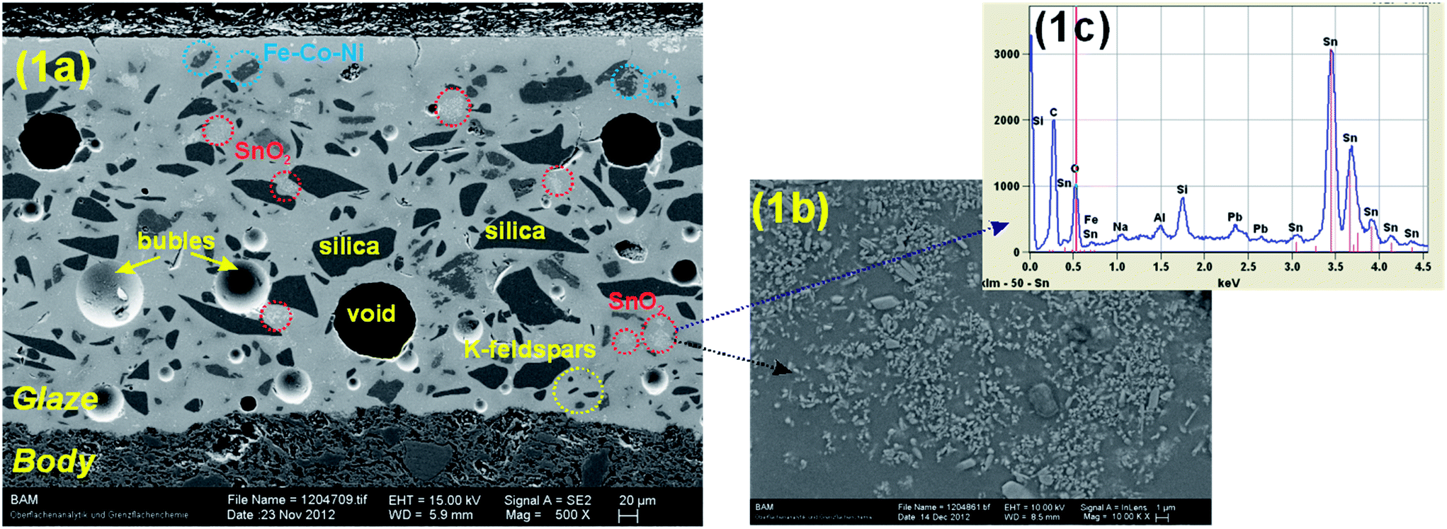 | ||
| Fig. 1 SEM micrographs on the “blue” sample areas: (a) cross-section of a faience sample from Coimbra (low magnification) with visualization of cassiterite (SnO2) agglomerates (white marks) and Fe–Co–Ni structures (blue marks); (b) enlargement of one area of the SnO2 agglomerates and (c) EDX spectrum taken from the area shown in (b) demonstrating the massive presence of Sn. | ||
From the structures found in Fig. 1, the amount of Sn-agglomerates in the blue regions is a hint for a particular manufacturing process. Co-blue is in its natural state i.e. a very strong and dark blue, which is not the hue in the blue surface motifs, by naked-eye observation. So, since Sn is a natural white-opacifier, one can deduce that the potter added it intentionally to the pigment in order to obtain a lighter blue hue.
The micrograph in Fig. 2a corresponds to a blue-painted surface of a faience sample from Coimbra. Some needle-like structures (see Fig. 2b) correspond to arsenic compounds, which, markedly, were found in blue regions only. The EDX spectra in Fig. 2c correspond to the areas marked in Fig. 2a. The analytical challenges described above are here demonstrated. The EDX spectra taken from area #1 and area #2 in Fig. 2a show no significant Co peaks (Kα @ 6.93 keV and Lα @ 0.77 keV). However, the presence of other elements that are linked with the Co-structure, such as Fe, is observed. Moreover, the L-line of arsenic is well detected, especially in the spectrum corresponding to area #3. This is easily observed by the higher concentration of “bright” As-structures (needle-like). Fig. 2b corresponds to an enlargement of area #3 from Fig. 2a. Point #1 in Fig. 2c is obviously rich in Fe – as proved by the corresponding EDX spectrum. Furthermore, this structure is presumably rather thin in comparison to the needle-like structures, as one can see by the “shade” it provokes on the As-structure in Fig. 2b. The EDX spectra of points #2, 3 and 4 show the presence of the As-L line (1.28 keV), while for point #1 the line which partially overlaps this one is Mg–K (1.25 keV).
 | ||
| Fig. 2 SEM micrographs of a surface area of a faience from Coimbra (low magnification (2a) and high magnification (2b)) with visualization of arsenic needle-like structure and (c) EDX spectra correspondent to the areas marked in (2a) demonstrating the presence of As in the bright needle-like structures; note that a Fe–Ni-rich structure could also be identified (see point “1” in 2c) by EDX. | ||
A comparison between Coimbra faiences and Lisbon wall-tiles micrographs on blue painted regions is made. Fig. 3 shows examples of blue surfaces from two faiences from Coimbra. In Fig. 3a a smooth mixture of dark and bright zones is observable, which correspond to the Fe-containing structures and Sn-crystals, respectively (Fig. 3b). Fig. 3c corresponds to another Coimbra faience sample and in this case, the Fe-containing structures are more dispersed at the surface level.
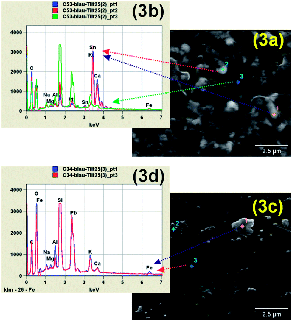 | ||
| Fig. 3 SEM micrographs: (a) surface of a faience from Coimbra with visualization of the mixture between Sn-crystals and Fe–Ni-rich structures; (b) EDX spectra corresponding to the point analysis marked in (a); (c) surface of a faience from Coimbra with visualization of Fe–Ni-rich structures, dispersed throughout the whole coloured area; (d) EDX spectra corresponding to the points marked in (c). | ||
Regarding wall-tile samples, Fig. 4 shows a micrograph of the surface of a wall-tile from Lisbon at a low magnification. In Fig. 4a relatively darker and brighter large regions are observed. Comparing this surface micrograph with the one from Fig. 2a and b (faience from Coimbra), one can conclude that As-structures (needle-like) are not observable in the wall-tile sample at similar magnifications. This is also confirmed by the EDX spectra in Fig. 4b, where arsenic X-ray lines were not detected. According to ref. 9, in the glaze, arsenic was found as being linked to Ca and Pb only. Furthermore, Pb–M and Ca–K lines of lower intensity are present in the spectrum in Fig. 4b. There is, however, a higher areal concentration of Sb-compounds – see e.g. area #3 in Fig. 4a and the respective EDX spectrum in red (Fig. 4b) – in comparison to the faience surface in Fig. 2a and b, which indicates the presence of yellow painted regions close to the blue regions. Fig. 4c is an enlargement of area #3 in Fig. 4a. Some micro- and sub-micro-structures are observed and the blurry appearance indicates that these are somewhat “buried” in the depth of the blue-glaze. However, one of these structures seems to “touch” the surface (blue point in Fig. 4c), see the respective EDX spectrum in Fig. 4d. Ni, Co and Mn were clearly detected in this structure. The line intensity ratios corresponding to these elements suggest a Ni-rich structure enclosing the Co-blue pigment, in comparison to the Coimbra samples, where a Fe-rich environment is found. A quantification of the EDX results of the small and buried structures corresponding to the blue pigment (see Fig. 3a and c) leads to faulty results.
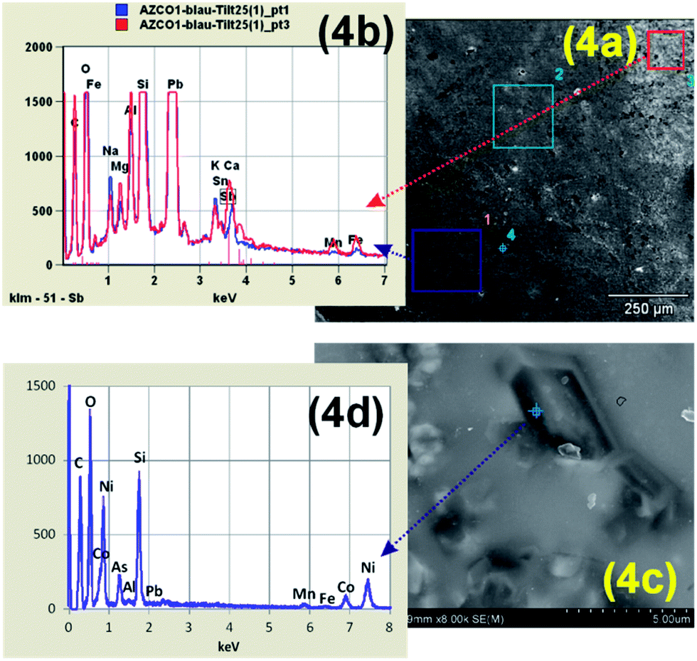 | ||
| Fig. 4 SEM micrographs of two Lisbon wall-tile samples in the blue painted areas together with EDX spectra (b and d) on selected spots demonstrating the presence of Fe–Mn-rich or Ni-rich micro-structures. | ||
Comparing the SEM/EDX results corresponding to faiences and wall-tiles, it could be established that Co-structures are in a Fe-rich environment in faiences and in a Ni-rich environment in wall-tiles.
The yellow pigment
Representative SEM micrographs taken on the “yellow” surface of one of each object type are displayed in Fig. 5. The faience from Coimbra exhibits localized very bright areas (for example area #3 in Fig. 5a) which correspond to a local higher density of Sb-crystals – as shown by the respective EDX spectra in Fig. 5b. The wall-tile from Coimbra appears in Fig. 5d relatively darker in general with lower local density of Sb-compounds – as indicated by the EDX spectra (Fig. 5e). The wall-tile from Lisbon in Fig. 5g shows a more uniform mixture of bright and darker regions, which indicates a quite homogeneous distribution of the Sb-crystals – as proved by the EDX spectra of all marked regions on the micrograph (Fig. 5h).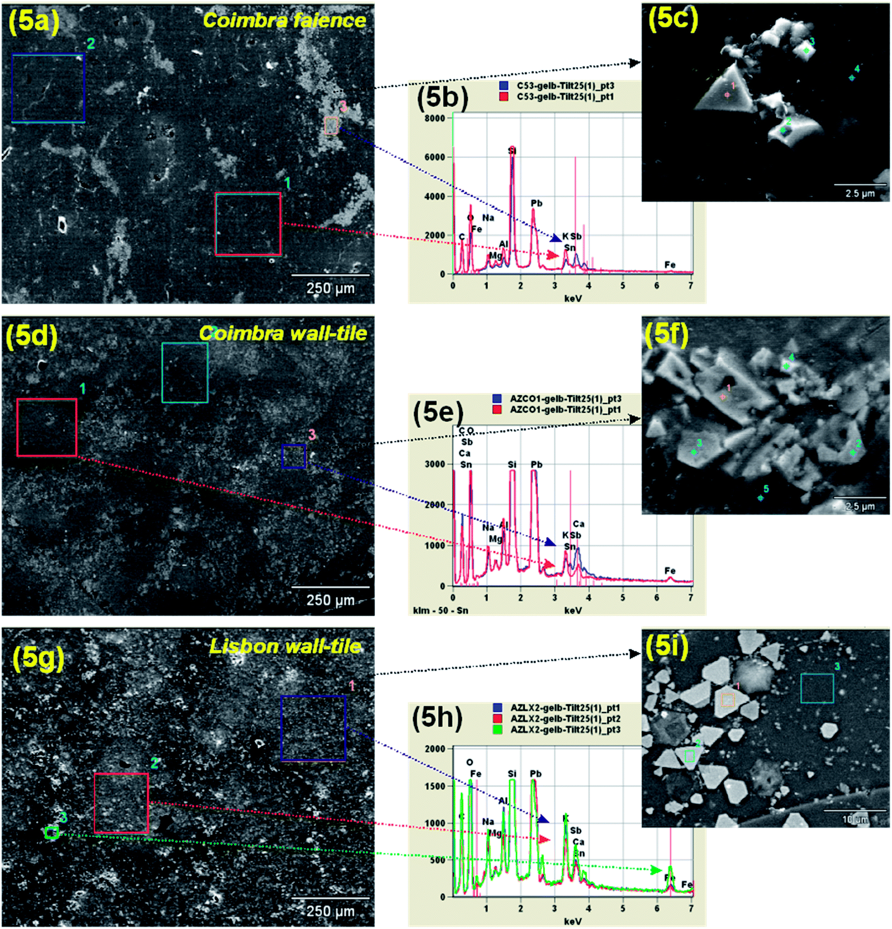 | ||
| Fig. 5 SEM micrographs of yellow painted areas of the surface of (a) a Coimbra faience sample, (d) a Coimbra wall-tile sample, and (g) a Lisbon wall-tile sample. On the right side EDX spectra of selected spots and further high-magnification micrographs are shown. | ||
The rough concentration of the elements present in yellow regions as detected by EDX for all analysed samples was calculated by using the commercial standardless quantification routines offered by one of the EDS systems employed (NSS 300 Thermo Fisher Scientific). The corresponding measurements were taken at a low magnification, integrating over larger areas, and the morphology of the cross-section was checked as well in order to better evaluate the concentrations. The analyses with wide-area mode on yellow decorations (Table 1) reveal that the wall-tiles have a lower content of Sb, in comparison to faiences. Furthermore, the presence of Sn (see Table 1) indicates that the Sn-ions may participate in the crystalline structure that forms the yellow pigment – possibly by replacing Sb.16 Other elements, which are embedded in the glaze matrix, such as Si, are also identified in these spectra due to the rather large analyzed volume.
| Samples | Na | Mg | Si | K | Ca | Fe | Sn | Sb | Pb | O |
|---|---|---|---|---|---|---|---|---|---|---|
| Coimbra faiences (N = 4) | 1–3 | ≤1 | 13–15 | 2–4 | 1–2 | 2–4 | 2–4 | 11–14 | 36–39 | 25–27 |
| Coimbra wall-tiles (N = 2) | 1–2 | <1 | 16–17 | 1–3 | 2–3 | 1–3 | 3–4 | 6–8 | 35–37 | 25–27 |
| Lisbon wall-tiles (N = 4) | 1–2 | <1 | 12–16 | 3–4 | ≤1 | 4–6 | 1–2 | 4–6 | 12–15 | 45–48 |
Another interesting perspective to observe the way the Sb-crystals are spread through the glaze is shown in Fig. 6. As marked with yellow circles, the Sb-crystals “stay” localized into the first tens of micrometers beneath the surface. This constitutes also a reason why the quantification of the EDX spectra could be applied.
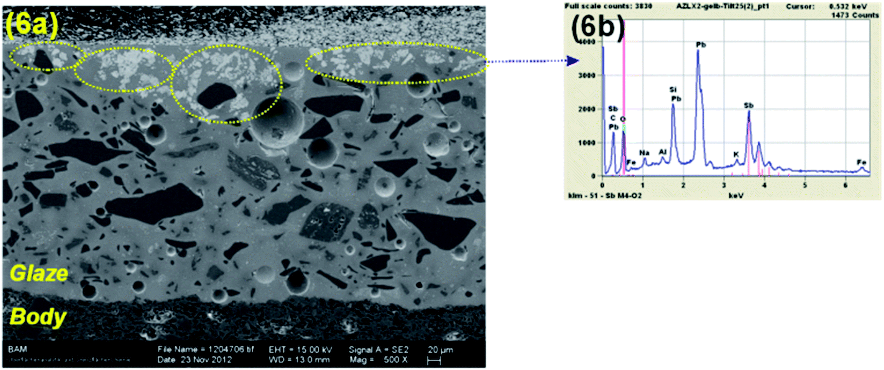 | ||
| Fig. 6 (a) SEM micrograph of a cross-sectioned Coimbra faience sample in an yellow painted area; (b) EDX spectrum corresponding to the areas marked with yellow in (a) demonstrating the presence of Sb. | ||
High-magnification SEM micrographs were also taken onto the yellow surface areas for all three types of samples (see Fig. 5c, f and i) together with EDX spectra on selected areas. The size of the crystals is generally smaller in the sample from Coimbra (faiences and wall-tiles) than in the samples from Lisbon. The crystal size is about 1–2 μm for the former and about 5–10 μm for the latter ones. This finding together with the particular crystal shape is a hint for the firing temperatures used. According to ref. 11, the hexagonal shape is typical for the so-called Naples yellow pigment. From 950 °C up some crystals start forming agglomerates, but in an irregular way (as can be recognized in Fig. 7a for the samples from Coimbra), and only from 1100 °C the hexagonal phase starts to appear11 – as it is rather the case for the Lisbon samples. Fig. 7 shows the differences between the morphology of the Sb-crystals between wall tiles from Coimbra and Lisbon. As explained above, the shape of the crystal is directly related to the firing temperature to which the samples have been submitted. The crystals corresponding to the sample from Coimbra reveal rather triangular-like shapes with a tendency for hexagonal-shape, while in samples from Lisbon a clear hexagonal shape is observed.
 | ||
| Fig. 7 SEM micrographs showing Sb-crystals at the surface of a wall tile from Coimbra (a) and a wall tile from Lisbon (b). | ||
Conclusions
Looking at the micro-morphology of the objects selected for investigations in the form of faiences from Coimbra and wall-tile samples from both Coimbra and Lisbon, it is definitely fascinating how much detail and careful manufacturing processes lie behind.Regarding the blue pigments interesting new results were obtained. A higher concentration of Sn-agglomerates was found in blue regions by analyzing the SEM-micrographs mainly of the cross-sectioned samples. This is a hint that the potter intentionally added Sn as an opacifying white-agent to the blue pigment in order to produce a lighter hue of blue – being known that Co-blue is very dark by nature. The expected linkage between Fe–Co–Ni–As–Bi could be investigated just partially using the techniques employed in this study. Co was difficult to observe but the other elements that participate on the pigment structure were investigated. Co was found in Fe- or Ni-rich structures but with no structural connection to As or Bi. However, the As-needle-like structures were always identified in blue regions. A difference between faiences and wall-tiles was observed: in faiences Co is found in Fe-rich structures while in wall-tiles Co is found in Ni-rich structures.
Concerning the yellow pigment, the micrographs have shown that the Sb-crystals are mainly concentrated at the surface of the samples (over the first tens of micrometers in depth). Moreover, differences noticed in the crystal shape and size are indicatives for the same firing cycle and different firing temperatures, respectively.11 The rather hexagonal shape found for the microstructures in the wall-tiles from Lisbon indicates that, in this context, a higher firing temperature was used, in comparison to the samples from Coimbra (faiences and wall-tiles), whose micro-structure shape is rather irregular and triangular-like. Thus, one can conclude that the firing cycle plays an important role in this simple linear model, which means that similar crystal-morphologies could be obtained for both short-firing cycles at high temperatures and for long-firing cycles at lower temperatures. The term “firing cycle” refers to the whole process of firing – the piece is submitted to a continuous heating up process until a plateau, followed by a cooling process, down to room temperature.
Acknowledgements
The authors would like to thank Dr Alexandre Pais for having supplied the samples, property of the “Museu Nacional do Azulejo (MNAz)” (National Tile Museum, in Lisbon), and the “Museu Machado de Castro (MMC)” (in Coimbra) for having provided the faiences. Further acknowledgments go to Mrs Olivia Netzband (BAM-6.7) for the careful cross-section preparations.Notes and references
- J. Coroado and C. Gomes, Proceedings of the 7th European Meeting of Ancient Ceramics, IPA, Lisboa, 2005, pp. 33–39 Search PubMed.
- http://emac2013.geoscienze.unipd.it/EMAC2013_book_of_abstracts.pdf, accessed 30 September 2013.
- S. Coentro, J. M. Mimoso, A. M. Lima, A. S. Silva, A. N. Pais and V. S. F. Muralha, J. Eur. Ceram. Soc., 2012, 32, 37 CrossRef CAS PubMed.
- A. Pais, A. Pacheco and J. Coroado, Cerâmica de Coimbra, INAPA, Lisboa, 2007 Search PubMed.
- D. Barilaro, V. Crupi, S. Interdonato, D. Majolino, V. Venuti, G. Barone, M. F. La Russa and F. Bardelli, Appl. Phys. A, 2008, 92, 91 CrossRef CAS.
- V. Crupi, D. Majolino, V. Venuti, G. Barone, P. Mazzoleni, A. Pezzino, M. F. La Russa, S. A. Ruffolo and F. Bardelli, Appl. Phys. A, 2010, 100, 845 CrossRef CAS.
- A. Guilherme, S. Pessanha, M. L. Carvalho, J. M. F. Dos Santos and J. Coroado, Spectrochim. Acta, Part B, 2010, 65, 328 CrossRef PubMed.
- A. Guilherme, J. Coroado, J. M. F. Dos Santos, L. Lühl, T. Wolff, B. Kanngießer and M. L. Carvalho, Spectrochim. Acta, Part B, 2011, 66, 297 CrossRef CAS PubMed.
- A. Zucchiatti, A. Bouquilloni, A. Katona and A. D'Allessandro, Archaeometry, 2006, 48, 131 CrossRef CAS.
- S. Coentro, J. M. Mimoso, A. M. Lima, A. S. Silva, A. N. Pais and V. S. F. Muralha, J. Eur. Ceram. Soc., 2012, 32, 37 CrossRef CAS PubMed.
- K. Sakellariou, C. Miliani, A. Morresi and M. Ombelli, J. Raman Spectrosc., 2004, 35, 61 CrossRef CAS.
- A. Guilherme, G. Buzanich, M. Radtke, U. Reinholz, J. Coroado, J. M. F. Dos Santos and M. L. Carvalho, J. Anal. At. Spectrom., 2012, 27, 966 RSC.
- M. Procop and V.-D. Hodoroaba, Microchim. Acta, 2008, 161, 413 CrossRef CAS.
- V.-D. Hodoroaba, M. Radtke, L. Vincze, V. Rackwitz and D. Reuter, Nucl. Instrum. Methods Phys. Res., Sect. B, 2010, 268, 3568 CrossRef CAS PubMed.
- V. Rackwitz, S. Bjeoumikhova, U. Panne and V.-D. Hodoroaba, J. Anal. At. Spectrom., 2011, 26, 499 RSC.
- F. Rosi, V. Manuali, C. Miliani, B. G. Brunetti, A. Sgamellotti, T. Grygar and D. Hradil, J. Raman Spectrosc., 2009, 40, 107 CrossRef CAS.
| This journal is © The Royal Society of Chemistry 2014 |