 Open Access Article
 This Open Access Article is licensed under a
 Creative Commons Attribution 3.0 Unported Licence
Hysteretic behaviour in a vacuum deposited submonolayer of single ion magnets†
David
Klar
*a,
Andrea
Candini
b,
Loïc
Joly
c,
Svetlana
Klyatskaya
d,
Bernhard
Krumme
a,
Philippe
Ohresser
e,
Jean-Paul
Kappler
ce,
Mario
Ruben
cd and
Heiko
Wende
a
aFaculty of Physics and Center for Nanointegration Duisburg-Essen (CENIDE), University of Duisburg-Essen, Lotharstraße 1, D-47048 Duisburg, Germany. E-mail: david.klar@uni-due.de; Fax: +49 (0)203 379 3601; Tel: +49 (0)203 379 1662
bCentro S3, Istituto Nanoscienze – CNR, via Campi 213/a, 41125 Modena, Italy
cInstitut de Physique et Chimie des Matériaux de Strasbourg, UMR 7504 UdS-CNRS, 67034 Strasbourg Cedex 2, France
dInstitute of Nanotechnology, Karlsruhe Institute of Technology (KIT), D-76344 Eggenstein-Leopoldshafen, Germany
eSynchrotron SOLEIL, L'Orme des Merisiers, Saint-Aubin - BP 48, 91192 Gif-sur-Yvette, France
First published on 13th May 2014
Abstract
With element-specific X-ray absorption spectroscopy and X-ray magnetic circular dichroism we have investigated submonolayer coverages of TbPc2 and DyPc2 molecules sublimated on highly ordered pyrolytic graphite. We have studied the field dependence of the magnetization of the central lanthanide ion at very low temperatures. Even in zero applied magnetic field we still observe a remanence in the magnetization. Since there are neither intermolecular coupling nor magnetic interactions with the substrate, this remanent behaviour results just from single-ion anisotropy. On the very inert surface of graphite at temperatures between 0.5 K and 2 K the spin relaxation is slow enough to observe a memory effect in the timescale of the experimental measurements.
Integrating organic materials into spintronic devices has become an important part of research over the last few years.1–6 Organic molecules offer new technological possibilities concerning small and effective data storage devices. Single molecule magnets (SMMs) are predestined for such very small devices because of their magnetic hysteresis without the need for long range ordering and have been the subject of intense studies over the last two decades. Most of the SMMs consist of several magnetic ions, e.g. Mn12 complexes,7–9 while in bis(phthalocyaninato) lanthanide complexes (LnPc2) there is only a single Ln ion (Dy3+, Tb3+) centered at the molecule between two phthalocyanine (Pc) rings (Fig. 1c). The remanent magnetization arises from the single ion anisotropy.10–12 Both molecules show uniaxial anisotropy, with the energy barriers of TbPc2 being much higher than that of the DyPc2 molecule. Spin and charge dynamics of powder samples of these two molecules were studied by Branzoli et al.13 The general problem with regard to applications in devices is the conservation of their SMM behaviour at high enough temperatures. The spin relaxation of TbPc2 molecules above 4 K is already too fast to detect a remanent magnetization by X-ray absorption spectroscopy (XAS), a rather slow experimental method.14 Magnetization curves of TbPc2 molecules show typically butterfly shaped hysteresis even at very low temperatures, originating from the spin quantum tunneling at zero magnetic fields.15–17 At temperatures below 4 K the relaxation is slow enough to identify these quantum tunneling effects with ac susceptibility measurements.18 One possibility to block relaxation at higher temperatures is the coupling on a ferromagnetic substrate.19,20 The magnetic anisotropy and field dependence of TbPc2 molecules have been investigated by XAS on non-magnetic metallic surfaces,14,17 on antiferromagnetic surfaces21 and ferromagnetic surfaces.22 Here we investigate submonolayer coverages of TbPc2 and DyPc2 molecules on highly ordered pyrolytic graphite (HOPG) with XAS, and in particular with X-ray magnetic circular dichroism (XMCD). Over the last decade, XAS and XMCD have been successfully used as powerful methods to determine the electronic and magnetic properties of submonolayers of different magnetic molecules interacting with surfaces.23–27 The deposition of less than a monolayer leads to well separated and oriented molecules on the graphite surface. In contrast to a metallic surface the hybridization between the molecules and the graphite is very weak and this enables the observation of the undisturbed molecules’ properties. Recently it has been demonstrated that the TbPc2 molecules are strongly affected by their environment28: the opening of the magnetization loop disappears on a conducting substrate, whereas remanence appears in bulk systems at T = 2 K (see also ESI†). We show here that at temperatures between 0.5 K and 2 K the spin relaxation is slow enough to observe a completely open magnetization curve with only slight butterfly shape. At 0.5 K we are able to observe remanence even for DyPc2, where the blocking temperature is reported to be considerably lower than for TbPc2.18 By time-dependent measurements of the magnetization we could determine that the relaxation time at this temperature is of the order of one hour and therefore detectable by our experimental technique.
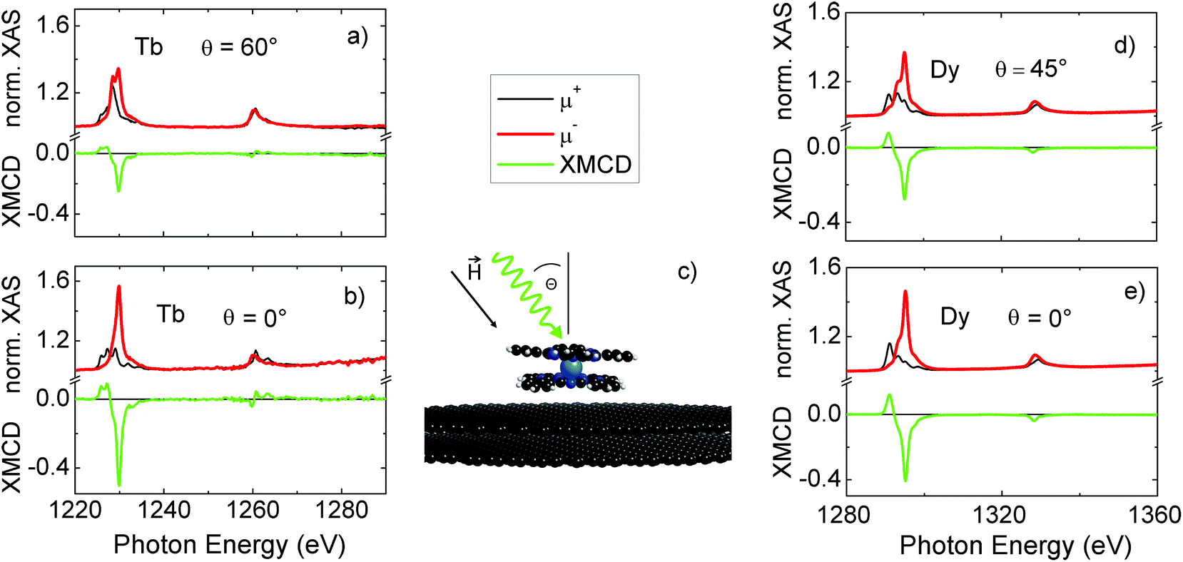 | ||
| Fig. 1 (a) and (b) Normalized XA spectra at the Tb M4,5 edges for left (red) and right (black) circular polarized X-rays and the dichroic spectra (green) for the angles of 60° and 0° recorded in a magnetic field of 6 T and at a temperature of 2 K. (c) Schematic illustration of the sample: the top two layers of the HOPG crystal with the LnPc2 molecule on top are shown and X-rays are irradiated under the angle θ. (d) and (e) Normalized XA spectra at the Dy M4,5 edges for left (red) and right (black) circular polarized X-rays and the dichroic spectra (green) for the angles of 45° and 0° recorded in a magnetic field of 1 T and at a temperature of 0.5 K. | ||
DyPc2 and TbPc2 were synthesized according to an earlier established procedure13 and analytical data confirm their intact structure (see ESI†). A submonolayer (0.5 ± 0.2 ML) coverage of LnPc2 was prepared in situ by thermal evaporation of the molecules onto the freshly cleaved HOPG crystal held at room temperature (for characterization details, see ESI†). The measurements relative to the TbPc2 molecule were performed at the DEIMOS29 beamline at the synchrotron SOLEIL, while DyPc2 molecules were studied at the SIM beamline at the SLS synchrotron, Paul Scherrer Institute, Villigen, Switzerland with the unique possibility to perform XMCD experiments at sub-Kelvin temperatures.30 The temperature of 0.5 K is crucial for the detection of the open hysteresis curve of DyPc2 because of its very low blocking temperature. Field-dependent measurements were performed to determine the SMM properties of the molecules. To study the spin relaxation after turning off the magnetic field, we carried out time-dependent XMCD measurements. With angle-dependent XMCD we studied the molecule orientation and the magnetic anisotropy.
Fig. 1 shows the angle-dependent spectra at the Tb and the Dy M4,5 edges for different X-ray incidence angles, normal (0°) and grazing (60° or 45°) at T = 2 K or 0.5 K and external magnetic field B = 6 T or 1 T. The black line represents the circular right polarization μ+, the red line the circular left polarization μ− and the green line the difference of the two spectra, the XMCD signal. The integrated white line (μ+ + μ−)/2 intensity at the M5 edge is normalized to 1. This normalization enables a direct comparison of the magnitude of the XMCD signals corresponding to the different angles. In the recorded spectra there are clear structural differences visible, especially at the M5 edge. However, the shape of the XMCD does not change with variation of the angle. Only the magnitude of the XMCD changes exactly with the cosine of the incidence angle. This behaviour of the magnetic anisotropy was observed for the TbPc2 molecules also on other substrates.14 The large anisotropy prevents the magnetic moments of Tb from turning into the plane of the Pc ring. The magnetization of the LnPc2 is still perpendicular, even in an external magnetic field of 6 T applied in the Pc-plane direction. Since we detect the magnetization just along the incident photons, the spectra only give the projection of the magnetization that is scaled with cos(θ).
The field dependence of the Tb magnetization is given in Fig. 2(a). The curves show the maximum of the M5 edge (1229.8 eV) normalized to the pre-edge (1220 eV). At a temperature of 2 K the magnetization of the Tb ion reaches the saturation value at about 1 T. Between the positive and negative saturation the magnetization curve is clearly open. The inset in this figure demonstrates that the curves do not cross at zero field. There is a remanent signal with a small coercive field of about 50 mT. The measurement of one complete curve takes more than one hour, but the relaxation at this temperature is slow enough to observe the remanent magnetization. The spin quantum tunneling that causes the typical butterfly shape is strongly reduced, but apparently the hysteresis curve is a bit more narrow at B = 0 T, indicating that spin tunneling is still present. The detailed origin of this behaviour is still unclear and falls beyond the scope of the present work. We note that a similar behaviour has been recently reported by another group for a crystallite sample of the same molecule.15 The spin relaxation is very sensitive to the temperature, leading to a vanishing remanence above 4 K.
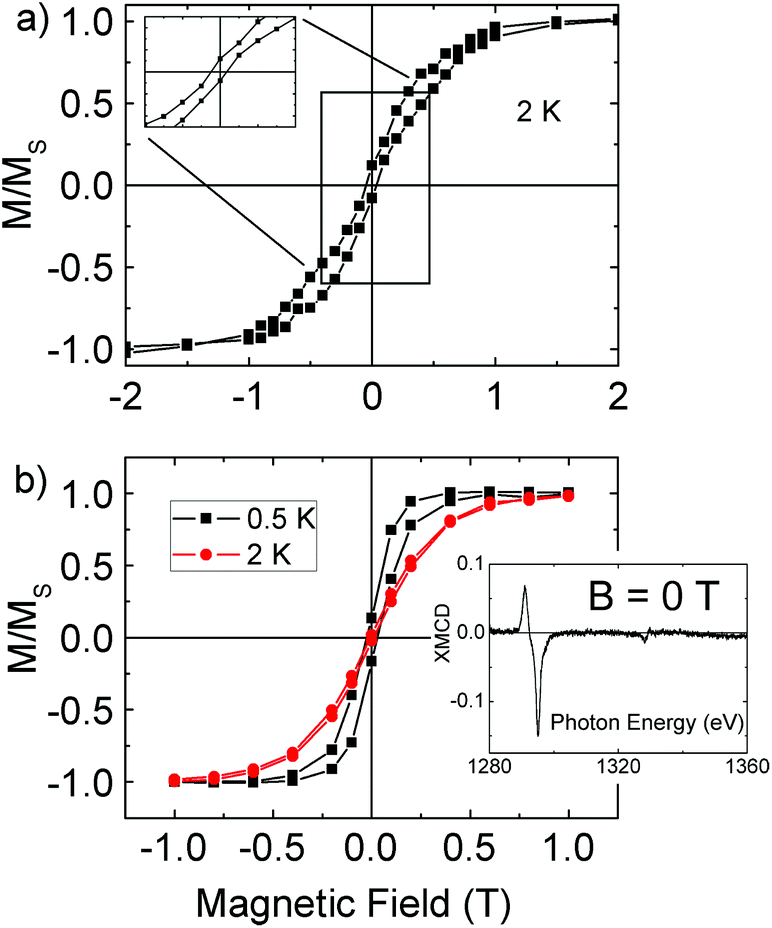 | ||
| Fig. 2 Field-dependent magnetization measured at the maximum of (a) the Tb M5 edge XMCD at T = 2 K and (b) the Dy M5 XMCD edge at T = 0.5 K (black) and T = 2 K (red), normalized to the saturation magnetization. | ||
Each curve in Fig. 2(b), showing the field dependence of the Dy magnetization, consists of only 26 data points. Each point belongs to an XMCD maximum at 1295.1 eV, taken from completely recorded μ+ and μ− spectra. In this way, each data point took approximately 30 minutes. Nevertheless, the opening of the magnetization curve is clearly visible for the black magnetization curve, demonstrating the long relaxation times at this low temperature. The remanent magnetization is also verified by the XMCD signal (Fig. 2b, inset) at B = 0 T. However, at T = 2 K (red line), no remanent magnetization is visible, confirming the lower blocking temperature of DyPc2 compared to TbPc2.
To make a quantitative statement about the relaxation time of the DyPc2 magnetization, we measured the time-dependent magnetization without an applied magnetic field (Fig. 3). Each triangle in this figure represents the normalized difference μ+ − μ− at the maximum XMCD intensity of 1295.1 eV. Approximately one data point was recorded every 90 seconds. The functional correlation between the XMCD intensity and time was fitted with a simple exponential function, while the maximum at the beginning was normalized to 1. The red curve fits the data points very well, demonstrating that the relaxation follows an exponential behaviour with a half-life of τ = 48 minutes. This time is in the range of typical XMCD measurements, giving us the opportunity to observe the magnetic remanence.
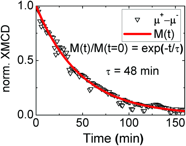 | ||
| Fig. 3 Time dependence of the normalized XMCD intensity at the maximum of the Dy M5 edge without external magnetic field at a temperature of 0.5 K. The red curve is an exponential function with a half life of 48 minutes. | ||
In conclusion, we have observed the remanent magnetization of both TbPc2 and DyPc2 molecules. Submonolayers of these molecules on the very inert HOPG substrate were deposited under UHV conditions to ensure that the interaction with the molecules’ environment is kept as low as possible. For the first time we could show the open hysteresis of vacuum deposited and well oriented LnPc2 molecules in an X-ray absorption study due to the very low temperature at which the measurements have been performed. This is a fundamental step for the possibility to use molecular memories in future devices. Because of the strongly suppressed spin relaxation at this temperature we were also able to determine a half life of the magnetization of 48 minutes.
Experiments were performed on the DEIMOS beamline at SOLEIL Synchrotron, France (proposal number 20110125) and on the SIM BEAMLINE (proposal number 20110200) at the Swiss Light Source, Paul Scherrer Institute, Villigen, Switzerland. We are grateful to the SOLEIL staff for smoothly running the facility. We thank Armin Kleibert at Swiss Light Source whose outstanding efforts have made these experiments possible.
References
- A. R. Rocha, V. M. Garcia-Suarez, S. W. Bailey, C. J. Lambert, J. Ferrer and S. Sanvito, Nat. Mater., 2005, 4, 335–339 CrossRef CAS PubMed.
- L. Bogani and W. Wernsdorfer, Nat. Mater., 2008, 7, 179 CrossRef CAS PubMed.
- M. Mannini, F. Pineider, P. Sainctavit, C. Danieli, E. Otero, C. Sciancalepore, A. M. Talarico, M.-A. Arrio, A. Cornia and D. Gatteschi, et al. , Nat. Mater., 2009, 8, 194–197 CrossRef CAS PubMed.
- A. Candini, S. Klyatskaya, M. Ruben, W. Wernsdorfer and M. Affronte, Nano Lett., 2011, 11, 2634–2639 CrossRef CAS PubMed.
- M. Urdampilleta, S. Klyatskaya, J. Cleuziou, M. Ruben and W. Wernsdorfer, Nat. Mater., 2011, 10, 502–506 CrossRef CAS PubMed.
- R. Vincent, S. Klyatskaya, M. Ruben, W. Wernsdorfer and F. Balestro, Nature, 2012, 488, 357–360 CrossRef CAS PubMed.
- R. Sessoli, D. Gatteschi, A. Caneschi and M. Novak, Nature, 1993, 365, 141–143 CrossRef CAS.
- G. Christou, D. Gatteschi, D. N. Hendrickson and R. Sessoli, MRS Bull., 2000, 67 Search PubMed.
- D. Gatteschi and R. Sessoli, Angew. Chem., Int. Ed., 2003, 42, 268–297 CrossRef CAS PubMed.
- J. D. Rinehart and J. R. Long, Chem. Sci., 2011, 2, 2078–2085 RSC.
- N. Ishikawa, M. Sugita, T. Okubo, N. Tanaka, T. Iino and Y. Kaizu, Inorg. Chem., 2003, 42, 2440–2446 CrossRef CAS PubMed.
- F. Branzoli, P. Carretta, M. Filibian, G. Zoppellaro, M. J. Graf, J. R. Galan-Mascaros, O. Fuhr, S. Brink and M. Ruben, J. Am. Chem. Soc., 2009, 131, 4387–4396 CrossRef CAS PubMed.
- F. Branzoli, P. Carretta, M. Filibian, M. J. Graf, S. Klyatskaya, M. Ruben, F. Coneri and P. Dhakal, Phys. Rev. B: Condens. Matter, 2010, 82, 134401 CrossRef.
- S. Stepanow, J. Honolka, P. Gambardella, L. Vitali, N. Abdurakhmanova, T.-C. Tseng, S. Rauschenbach, S. L. Tait, V. Sessi and S. Klyatskaya, et al. , J. Am. Chem. Soc., 2010, 132, 11900–11901 CrossRef CAS PubMed.
- M. Gonidec, E. S. Davies, J. McMaster, D. B. Amabilino and J. Veciana, J. Am. Chem. Soc., 2010, 132, 1756–1757 CrossRef CAS PubMed.
- M. Gonidec, R. Biagi, V. Corradini, F. Moro, V. De Renzi, U. del Pennino, D. Summa, L. Muccioli, C. Zannoni and D. B. Amabilino, et al. , J. Am. Chem. Soc., 2011, 133, 6603–6612 CrossRef CAS PubMed.
- L. Margheriti, D. Chiappe, M. Mannini, P. Car, P. Sainctavit, M. A. Arrio, F. B. de Mongeot, J. C. Cezar, F. M. Piras, A. Magnani, E. Otero, A. Caneschi and R. Sessoli, Adv. Mater., 2010, 22, 5488 CrossRef CAS PubMed.
- N. Ishikawa, M. Sugita and W. Wernsdorfer, Angew. Chem., Int. Ed., 2005, 44, 2931–2935 CrossRef CAS PubMed.
- A. Lodi Rizzini, C. Krull, T. Balashov, J. Kavich, A. Mugarza, P. Miedema, P. Thakur, V. Sessi, S. Klyatskaya, M. Ruben, S. Stepanow and P. Gambardella, Phys. Rev. Lett., 2011, 107, 177205 CrossRef CAS.
- D. Klar, S. Klyatskaya, A. Candini, B. Krumme, K. Kummer, P. Ohresser, V. Corradini, V. de Renzi, R. Biagi, L. Joly, J.-P. Kappler, U. del Pennino, M. Affronte, H. Wende and M. Ruben, Beilstein J. Nanotechnol., 2013, 4, 320–324 CrossRef PubMed.
- A. Lodi Rizzini, C. Krull, T. Balashov, A. Mugarza, C. Nistor, F. Yakhou, V. Sessi, S. Klyatskaya, M. Ruben and S. Stepanow, et al. , Nano Lett., 2012, 12, 5703–5707 CrossRef CAS PubMed.
- L. Malavolti, L. Poggini, L. Margheriti, D. Chiappe, P. Graziosi, B. Cortigiani, V. Lanzilotto, F. B. de Mongeot, P. Ohresser and E. Otero, et al. , Chem. Commun., 2013, 49, 11506–11508 RSC.
- A. Scheybal, T. Ramsvik, R. Bertschinger, M. Putero, F. Nolting and T. A. Jung, Chem. Phys. Lett., 2005, 411, 214 CrossRef CAS PubMed.
- H. Wende, M. Bernien, J. Luo, C. Sorg, N. Ponpandian, J. Kurde, J. Miguel, M. Piantek, X. Xu, P. Eckhold, W. Kuch, K. Baberschke, P. M. Panchmatia, B. Sanyal, P. M. Oppeneer and O. Eriksson, Nat. Mater., 2007, 6, 516 CrossRef CAS PubMed.
- M. Bernien, J. Miguel, C. Weis, M. E. Ali, J. Kurde, B. Krumme, P. M. Panchmatia, B. Sanyal, M. Piantek, P. Srivastava, K. Baberschke, P. M. Oppeneer, O. Eriksson, W. Kuch and H. Wende, Phys. Rev. Lett., 2009, 102, 047202 CrossRef CAS.
- M. Bernien, D. Wiedemann, C. F. Hermanns, A. Krüger, D. Rolf, W. Kroener, P. Müller, A. Grohmann and W. Kuch, J. Phys. Chem. Lett., 2012, 3, 3431–3434 CrossRef CAS.
- D. Klar, B. Brena, H. Herper, S. Bhandary, C. Weis, B. Krumme, C. Schmitz-Antoniak, B. Sanyal, O. Eriksson and H. Wende, Phys. Rev. B: Condens. Matter, 2013, 88, 224424 CrossRef.
- L. Malavolti, M. Mannini, P.-E. Car, G. Campo, F. Pineider and R. Sessoli, J. Mater. Chem. C, 2013, 1, 2935–2942 RSC.
- P. Ohresser, E. Otero, F. Choueikani, K. Chen, S. Stanescu, F. Deschamps, T. Moreno, F. Polack, B. Lagarde and J.-P. Daguerre, et al. , Rev. Sci. Instrum., 2014, 85, 013106 CrossRef CAS PubMed.
- I. Letard, P. Sainctavit, J.-P. Kappler, P. Ghigna, D. Gatteschi and B. Doddi, et al. , J. Appl. Phys., 2007, 101, 113920–113920 CrossRef PubMed.
Footnote |
| † Electronic supplementary information (ESI) available: Bulk reference measurements and characterization of the molecules and the submonolayer. See DOI: 10.1039/c4dt01005a |
| This journal is © The Royal Society of Chemistry 2014 |