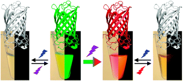
Fluorescent proteins (FPs) from the GFP family have become indispensable as marker tools for imaging live cells, tissues and entire organisms. A wide variety of these proteins have been isolated from natural sources and engineered to optimize their properties as genetically encoded markers. Here we review recent developments in this field. A special focus is placed on photoactivatable FPs, for which the fluorescence emission can be controlled by light irradiation at specific wavelengths. They enable regional optical marking in pulse-chase experiments on live cells and tissues, and they are essential marker tools for live-cell optical imaging with super-resolution. Photoconvertible FPs, which can be activated irreversibly via a photo-induced chemical reaction that either turns on their emission or changes their emission wavelength, are excellent markers for localization-based super-resolution microscopy (e.g., PALM). Patterned illumination microscopy (e.g., RESOLFT), however, requires markers that can be reversibly photoactivated many times. Photoswitchable FPs can be toggled repeatedly between a fluorescent and a non-fluorescent state by means of a light-induced chromophore isomerization coupled to a protonation reaction. We discuss the mechanistic origins of the effect and illustrate how photoswitchable FPs are employed in RESOLFT imaging. For this purpose, special FP variants with low switching fatigue have been introduced in recent years. Despite nearly two decades of FP engineering by many laboratories, there is still room for further improvement of these important markers for live cell imaging.
