 Open Access Article
 This Open Access Article is licensed under a Creative Commons Attribution-Non Commercial 3.0 Unported Licence
Peptide–perylene diimide functionalized magnetic nano-platforms for fluorescence turn-on detection and clearance of bacterial lipopolysaccharides†
Fang
Liu
a,
Jing
Mu
a,
Xiangyang
Wu
a,
Surajit
Bhattacharjya
*b,
Edwin Kok Lee
Yeow
*a and
Bengang
Xing
*a
aDivision of Chemistry and Biological Chemistry, School of Physical & Mathematical Sciences, Nanyang Technological University, Singapore, 637371, Singapore. E-mail: Bengang@ntu.edu.sg; Edwinyeow@ntu.edu.sg
bDivision of Structural Biology & Biochemistry, School of Biological Sciences, Nanyang Technological University, Singapore, 637371, Singapore. E-mail: Surajit@ntu.edu.sg
First published on 25th March 2014
Abstract
A simple and unique strategy has been successfully designed for sensitive detection and rapid clearance of bacterial lipopolysaccharides (LPS) by integration of core–shell Fe3O4@SiO2 magnetic nanoparticles with a perylene-diimide (PDI) conjugated LPS-recognition peptide.
Bacterial lipopolysaccharides (LPS), key components in the outer membrane of Gram-negative bacteria, are well known as bacterial endotoxins that may cause serious septic shock or many other health problems in the intensive care.1 The extreme toxicity of bacterial LPS has thus urgently initiated extensive research efforts toward the development of novel and specific strategies to sensitively identify, and more importantly, to rapidly remove such highly toxic endotoxins from biological and pharmaceutical products. One commonly used FDA-approved technique for LPS detection is the enzymatic limulus amoebocyte lysate (LAL) assay. However, this assay requires a rather long and complicated testing procedure, and moreover, the results from the LAL assay are also susceptible to environmental variations, especially in pH and temperature.2 Very recently, several fluorescent, colorimetric or electrochemical sensors based on small molecules like,3 CD14-derivatized peptides,4 liposomes,5 or polymers6 have been reported for sensitive determination of bacterial LPS in aqueous solutions. Although all of these attempts have shown great promise with their individual detection limit range (from micromolar to picomolar level),5a,6 none of these assays indicated the capacity for the effective separation of LPS from the detection mixtures. Given the clinical importance of LPS, a simple and specific sensing strategy with more promising functions to clear bacterial endotoxins remains a big challenge in current research. Although several conventional techniques such as ultrafiltration, two-phase extraction, chromatography and functionalized nanoparticles have been utilized to remove LPS contaminants from certain biological preparations,7 these techniques may not report the specific and sensitive determination of LPS directly, and their LPS clearance efficiency could be potentially affected by environmental changes (e.g. pH).7b,c Therefore, the development of a stable and easily controlled method with a combination of sensitive detection and efficient clearance of LPS in complex biological samples such as bacterial cell lysates and human serums will be of great importance in the pharmaceutical industry, and such highly desirable studies have not been fully explored yet.
Here we first present simple and novel dual-functional magnetic nanoparticles (MNPs) for effective detection and removal of bacterial LPS by introducing a specific perylene diimide (PDI) conjugated LPS-binding peptide onto the surface of nanoparticles. PDI derivatives were chosen mainly because of their high thermal and photostability, high quantum yield, and more importantly, their favorable self-assembly properties in aqueous solutions.8 Along with our continuous efforts in selective bacterial imaging and photo-inactivation of pathogens,9 a set of designed LPS recognition peptides were screened that exhibited promising binding affinity for the bacterial surfaces. In this rational design, a simple and high-affinity LPS binding peptide with the YVLWKRKRKFCFI–NH2 sequence was selected as the targeting ligand to conjugate with magnetic Fe3O4@SiO2 core–shell structures through a short linker containing PDI as a fluorescent reporting group. In the absence of LPS, the fluorescence of the PDI molecules may undergo self-quenching due to the strong π⋯π stacking interactions.10 However, in the presence of LPS, the specific LPS binding would trigger conformational changes in the peptide9 that may lead to the disruption of the hydrophobic stacking between adjacent PDI molecules and induce a significant fluorescence recovery (Fig. 1). Furthermore, the LPS captured by the recognition peptide on MNPs can be easily removed by using a simple magnet, therefore separating the toxic bacterial LPS from the contaminated biological mixture, thus lowering the risk of LPS toxicity to both the environment and human healthcare units.
 | ||
| Fig. 1 LPS recognition peptide functionalized Fe3O4@SiO2 core–shell MNPs for fluorescence turn on detection and clearance of LPS. | ||
In this study, the affinity ligand engineered MNPs were chosen as solid platforms because they have demonstrated great promise in biomedical applications including cancer imaging and therapy,11 bio-separation,12 targeted drug or gene delivery,13 and the sorting of living cells and bio-molecules.14 The Fe3O4@SiO2 core–shell MNPs were prepared according to the method reported previously.11,15 To achieve effective particle functionalization, an azido-dPEG4 linker was first coupled to the Fe3O4@SiO2 surface followed by conjugation with the alkyne modified PDI peptide moieties through "Click-chemistry" (Fig. 1). The formation of the PDI-peptide functionalized MNP (MNPPDI-peptide) with the size distribution of about 25 nm was finally confirmed by spectroscopic and TEM analysis. The optimal amount of the peptide conjugate on MNPPDI-peptide was determined to be 10 nmol mg−1 (Fig. S1–S4, ESI†).
We further examined the possibility of MNPPDI-peptide responding to bacterial LPS and the induced fluorescence change in buffer solutions. Typically, 100 μg mL−1 of MNPPDI-peptide were incubated with 100 μg mL−1 of LPS in Tris-buffered saline solution (TBS, 50 mM, pH 7.4). As expected, very weak fluorescence was detected before addition of LPS in MNPPDI-peptide solutions (Fig. 2a), mainly due to the self-quenching caused by the π⋯π stacking of PDI on particle surfaces.10 However, upon the addition of LPS for 15 min, there was over 7-fold fluorescence enhancement at the emission wavelength of 539 nm, and no obvious further change was observed as the incubation time was increased (Fig. 2a, Fig. S5, ESI†). As a control, the MNPPDI conjugate without the LPS recognition peptide was also applied to respond to LPS (Fig. 2a). No obvious fluorescence change was observed, clearly confirming that the LPS binding peptide functioned as an indispensable moiety to specifically target bacterial LPS, hence changing the conformation of stacked PDI molecules and eventually restoring the fluorescence from MNPPDI-peptide. In addition, the analysis also showed that the fluorescence enhancement was LPS concentration dependent. The plot of fluorescence intensity at 539 nm versus the LPS concentration displayed a good linear relationship (R2 = 0.996) with a LPS concentration range up to 20 μg mL−1 (Fig. 2b), and the detection limit of LPS in TBS buffer was determined to be 280 ng mL−1 (Fig. S6, ESI†), which was comparable to most of the LPS sensing systems reported previously.3a,4,5 Furthermore, the fluorescence intensity of MNPPDI-peptide itself at 539 nm did not change dramatically in a wide pH range from 4 to 11. Upon addition of LPS, the fluorescence intensity was increased about 4–7 times under all tested pH conditions (Fig. 2c), clearly suggesting that the as-prepared MNPPDI-peptide could serve as an environmentally stable and reliable platform for sensitive LPS detection.
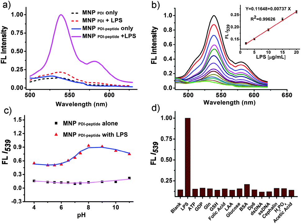 | ||
| Fig. 2 (a) Fluorescence spectra (λex = 450 nm) of MNPPDI-peptide and MNPPDI with or without LPS (100 μg mL−1) in TBS buffer; (b) fluorescence of MNPPDI-peptide (100 μg mL−1) after incubation with LPS at different concentrations (from 0, 1 to 250 μg mL−1) in TBS buffer at 37 °C. The inset shows the plot and linear fitting of normalized fluorescence at 539 nm vs. the concentration of LPS (2–20 μg mL−1); (c) the fluorescence of 100 μg mL−1 MNPPDI-peptide in the absence or presence of LPS (100 μg mL−1) under different pH conditions; (d) the selective fluorescence increase upon addition of LPS against various biologically important species (all at 10 μM). | ||
Furthermore, a selectivity study was also performed by incubating MNPPDI-peptide with various small or macro-biomolecules (Fig. 2d, Fig. S7 and S8, ESI†). Compared to the results obtained from MNPPDI-peptide with LPS, no significant fluorescence response was detected against all the tested analytes at concentrations up to 10 μM including the highly negatively charged BSA or DNA, which has been reported to potentially interfere with LPS detection,5a confirming that the specific LPS-peptide interaction could easily replace the nonspecific electrostatic attraction between the negatively charged bio-molecules and LPS affinity peptides. The conformational change in the activated peptide9 will greatly induce the recovery of the quenched fluorescence by disrupting π⋯π stacking of PDI on the particle surface. These results indicated that the MNPPDI-peptide could work as a highly selective fluorescent turn-on sensor for toxic LPS determination.
More importantly, we also exploited the feasibility of using MNPPDI-peptide to remove the toxic bacterial components. Typically, the procedure to remove LPS consisted of two simple steps: (1) mixing of MNPPDI-peptide with suspension of lysed bacterial cells (e.g. E. coli. DH5α etc.) or LPS contaminated human serum samples for 15 minutes and (2) applying a small magnet to attract MNPPDI-peptide to the wall of the vials to capture LPS (Fig. 3a). The optimum concentration of LPS and protein components from mixtures were determined by the LAL assay or the standard Bradford method, which were used to evaluate the LPS clearance efficiency.1,2 As shown in Fig. 3b, the removal of LPS was strongly dependent on the concentration of MNPPDI-peptide and effective LPS removal (e.g. over 87% from lysate of 109 cfu mL−1E. coli. cells) could be easily achieved when 400 μg mL−1 of MNPPDI-peptide was used, while no obvious loss of the total proteins was observed during the process of LPS removal. Moreover, after a simple multi-cycle removal (e.g. 3 or 4 cycles) with a fixed amount of MNPPDI-peptide (e.g. 200 μg mL−1) each time, the LPS could be completely cleared from about 6800 endotoxin units (EU) per mL and there was even less than 1 EU mL−1 left in the bacterial lysates (Fig. S9a and S10, ESI†). In contrast, when similar clearance was conducted with Gram-positive B. subtilis cell lysates, only non-specific background signals were observed before and after treatment, suggesting that MNPPDI-peptide could selectively remove the LPS components from Gram-negative bacteria. Moreover, we also examined the feasibility of MNPPDI-peptide to detoxify LPS contamination by efficient removal of LPS endotoxins from human serum samples. Similarly, LPS removal strongly depends on the concentrations of MNPPDI-peptide and more efficient LPS clearance (e.g. >89%) could be easily achieved at higher concentrations of MNPPDI-peptide (e.g. 2.0 mg mL−1), even only upon a single cycle particle separation (Fig. S9b, ESI†). Meanwhile, the detoxification effect after removal of LPS was also evaluated in macrophage cultures by measuring the concentration of nitrite,16 a stable metabolite of NO that has been well established as a pre-inflammatory stimulus to reflect the cell stress upon stimulation by bacterial LPS (Fig. 3c). Compared to the control study without any LPS in the serum, the macrophage RAW 264.7 cells incubated with the LPS contaminated serum for 24 hours would obviously induce the generation of a high level of nitrite, indicating the occurrence of cell stress stimulated by LPS. In contrast, incubation of serum under similar conditions but after efficient LPS removal resulted in a low level of nitrite generation. The decreasing nitrite concentration observed in a macrophage culture medium was consistent with the lower level of remaining LPS after effective separation using MNPs (Fig. 3). Importantly, the MNPPDI-peptide itself did not demonstrate obvious cytotoxicity (e.g. cell viability of 87%) even at the concentration of nanoparticle conjugate up to 400 μg mL−1 (Fig. S11, ESI†), suggesting that a trace amount of the residue MNPPDI-peptide in the whole cell environment during the magnetic LPS removal would not be sufficient to induce obvious cellular toxicity.
 | ||
| Fig. 3 (a) Scheme of LPS removal based on MNPPDI-peptide, (b) quantified magnetic LPS removal and protein recovery from E. coli cell lysates, (c) NO generated from RAW 264.7 cells pre-treated with serum before and after LPS detoxification. Blank: serum treated cells without LPS. | ||
In summary, by combining Fe3O4@SiO2 core–shell MNPs with a specific affinity ligand based on a PDI conjugated LPS recognition peptide, we first developed a simple and novel strategy for effective fluorescence detection of LPS and rapid clearance of such toxic bacterial endotoxins. Based on the specific interactions between LPS and affinity peptides on the surface of MNPs, self-stacking of adjacent PDI molecules was disrupted, which broke self-fluorescence quenching and thus restored the fluorescence. This peptide functionalized MNPs can sensitively identify LPS with the promising detection limit down to the nano-molar level and it is highly selective for bacterial LPS when compared to other interfering bio-analytes. More importantly, such simple and effective LPS detection nanoparticles also indicated the low risk of LPS toxicity to the environment and human healthcare by rapidly clearing bacterial LPS from complicated bacterial lysates and LPS contaminated serum samples. We envision that such simple and novel development would hold great potentials for the future treatment of bacteria associated sepsis with minimum complicated sample pre-treatment in extensive clinical practice.
The authors acknowledge the Start-Up Grant (SUG), the A*STAR PSF Grant (SERC1121202008), RG64/10, and a COS research collaboration award, Nanyang Technological University, Singapore.
Notes and references
- C. R. H. Raetz and C. Whitfield, Annu. Rev. Biochem., 2002, 71, 635 CrossRef CAS PubMed.
- B. Beutler and E. T. Rietschel, Nat. Rev. Immunol., 2003, 3, 169 CrossRef CAS PubMed.
- (a) L. Zeng, J. Wu, Q. Dai, W. Liu, P. Wang and C. Lee, Org. Lett., 2010, 12, 4014 CrossRef CAS PubMed; (b) G. Jones and H. Jiang, Bioconjugate Chem., 2005, 16, 621 CrossRef CAS PubMed; (c) V. Ganesh, K. Bodewits, S. J. Bartholdson, D. Natale, D. J. Campopiano and J. C. Mareque-Rivas, Angew. Chem., Int. Ed., 2009, 48, 356 CrossRef CAS PubMed.
- S. Voss, R. Fischer, G. Jung, K. H. Wiesmuller and R. Brock, J. Am. Chem. Soc., 2007, 129, 554 CrossRef CAS PubMed.
- (a) J. Wu, A. Zawistowski, M. Ehrmann, T. Yi and C. Schmuck, J. Am. Chem. Soc., 2011, 133, 9720 CrossRef CAS PubMed; (b) M. Rangin and A. Basu, J. Am. Chem. Soc., 2004, 126, 5038 CrossRef CAS PubMed.
- M. Lan, J. Wu, W. Liu, W. Zhang, J. Ge, H. Zhang, J. Sun, W. Zhao and P. Wang, J. Am. Chem. Soc., 2012, 134, 6685 CrossRef CAS PubMed.
- (a) P. O. Magalhaes, A. M. Lopes, P. G. Mazzola, C. Rangel-Yagui, T. C. Penna and A. Pessoa Jr., J. Pharm. Pharm. Sci., 2007, 10, 388 Search PubMed; (b) J. Li, G. Shang, M. You, S. Peng, Z. Wang, H. Wu and G. Chen, Biomacromolecules, 2011, 12, 602 CrossRef CAS PubMed; (c) M. Sakata, K. Uezono, K. Kimura and M. Todokoro, Anal. Biochem., 2013, 443, 41 CrossRef CAS PubMed; (d) I. K. Herrmann, M. Uener, S. Graf, C. M. Schumacher, B. Roth-Z'graggen, M. Hasler, W. J. Stark and B. Beck-Schimmer, Adv. Healthcare Mater., 2013, 2, 829 CrossRef CAS PubMed.
- (a) D. Görl, X. Zhang and F. Würthner, Angew. Chem., Int. Ed., 2012, 51, 6328 CrossRef PubMed; (b) D. Ding, K. Li, B. Liu and B. Tang, Acc. Chem. Res., 2013, 46, 2241 Search PubMed.
- (a) F. Liu, A. Ni, Y. Lim, H. Mohanram, S. Bhattacharjya and B. Xing, Bioconjugate Chem., 2012, 23, 1639 CrossRef CAS PubMed; (b) A. Bhunia, H. Mohanram, P. N. Domadia, J. Torres and S. Bhattacharjya, J. Biol. Chem., 2009, 284, 21991 CrossRef CAS PubMed; (c) B. Xing, C. Yu, P. Ho, K. Chow, T. Cheung, H. Gu, Z. Cai and B. Xu, J. Med. Chem., 2003, 46, 4904 CrossRef CAS PubMed; (d) Q. Shao and B. Xing, Chem. Commun., 2012, 48, 1739 RSC.
- (a) Z. Chen, A. Lohr, C. R. Saha-Moller and F. Würthner, Chem. Soc. Rev., 2009, 38, 564 RSC; (b) A. C. Grimsdale and K. Müllen, Angew. Chem., Int. Ed., 2005, 44, 5592 CrossRef CAS PubMed; (c) J. Zhao, Y. Ruan, R. Zhou and Y. Jiang, Chem. Sci., 2011, 2, 937 RSC; (d) F. Biedermann, E. Elmalem, I. Ghosh, W. M. Nau and O. A. Scherman, Angew. Chem., Int. Ed., 2012, 51, 7739 CrossRef CAS PubMed.
- (a) H. Na, I. Song and T. Hyeon, Adv. Mater., 2009, 21, 2133 CrossRef CAS PubMed; (b) E. S. Shibu, S. Sugino, K. Ono, H. Saito, A. Nishioka, S. Yamamura, M. Sawada, Y. Nosaka and V. Biju, Angew. Chem., Int. Ed., 2013, 52, 10559 CrossRef CAS PubMed.
- (a) J. Gao, H. Gu and B. Xu, Acc. Chem. Res., 2009, 42, 1097 CrossRef CAS PubMed; (b) D. Ho, X. Sun and S. Sun, Acc. Chem. Res., 2011, 44, 875 CrossRef CAS PubMed.
- F. M. Z. Kievit and M. Zhang, Acc. Chem. Res., 2011, 44, 853 CrossRef CAS PubMed.
- (a) Y. Pan, X. Du, F. Zhao and B. Xu, Chem. Soc. Rev., 2012, 41, 2912 RSC; (b) J. Lee, K. Jeong, M. Hashimoto, A. Kwon, A. Rwei, S. Shankarappa, J. Tsui and D. Kohane, Nano Lett., 2014, 14, 1 CrossRef CAS PubMed.
- Y. Yang, J. Aw, K. Chen, F. Liu, P. Padmanabhan, Y. Hou, Z. Cheng and B. Xing, Chem. – Asian. J., 2011, 6, 1381 CrossRef CAS PubMed.
- S. M. Zughaier, W. M. Shafer and D. S. Stephens, Cell. Microbiol., 2005, 7, 1251 CrossRef CAS PubMed.
Footnote |
| † Electronic supplementary information (ESI) available: Synthesis and characterization of magnetic nanoparticles and PDI-peptide conjugates and additional experimental details and figures. See DOI: 10.1039/c4cc01266f |
| This journal is © The Royal Society of Chemistry 2014 |