Jin Zhou,
Xiaoyue Shan,
Juanjuan Ma,
Yamin Gu,
Zhaosheng Qian,
Jianrong Chen and
Hui Feng*
College of Chemistry and Life Science, Zhejiang Normal University, Jinhua 321004, China. E-mail: fenghui@zjnu.cn; Fax: +86-579-82282269; Tel: +86-579-82282269
First published on 11th December 2013
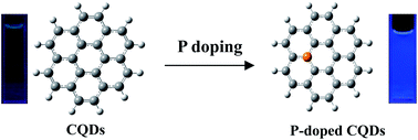
P-doped carbon quantum dots (PCQDs) were synthesized by a solvent-thermal method using phosphorous tribromide and hydroquinone as precursors. The as-prepared PCQDs present strong visible fluorescence with quantum yield up to 25%. The toxicity and bioimaging experiments showed that PCQDs have low cell toxicity and excellent biolabeling ability.
Usually, doping carbon nanomaterials with heteroatoms is an effective method which can tune their intrinsic properties.6 There has been much progress in doping carbon nanomaterials with heteroatoms such as nitrogen and boron that the doping of heteroatoms promoted their properties.7,8 The N atom has been used for chemical doping with CQDs.9 Although phosphorus atom is larger than carbon atom, it has been shown that phosphorous can form substitutional defects in diamond sp3 thin films, behaving as an n-type donor and thereby modifying the electronic and optical properties.10 In view of the remarkable quantum-confinement and edge effects of carbon quantum dots, doping CQDs with P atoms could alter their electronic characteristics and offer more active sites, thus producing new and unexpected properties.
In the communication, we proposed a simple and efficient route to prepare phosphorus atom doped carbon quantum dots (PCQDs) with strong photoluminescence through solvent-thermal reaction. The PCQDs were synthesized by solvent-thermal reaction between phosphorous bromide and hydroquinone with different ratios under 200 °C in a Teflon-lined vessel. The as-prepared PCQDs present strong blue fluorescence with quantum yield up to 25%. In order to explore the role of doped phosphorus in the CQDs, pristine CQDs without doped phosphorus were also prepared using the same method with CCl4 and hydroquinone as precursors. Fig. 1 showed the typical TEM images of PCQDs as well as the as-prepared pristine CQDs for comparison. It is found that the diameters of PCQDs are mainly distributed in the range of 5–15 nm while the CQDs is well-dispersed with a narrow size distribution of 3–5 nm. No substantial differences in shape between them could be detected. HR-TEM image of PCQDs shown in Fig. 1C displayed the discernible crystalline lattice structures. The spacing of adjacent lattice planes is about 3.0 Å, close to the (002) diffraction planes of graphite.9 Its crystal-like structure was also verified by Raman spectroscopy (Fig. S1†), in which the peak at 1590 cm−1 is very close to that resulted from E2g vibration mode of graphite.
As shown in Fig. 2, both XPS spectra of pristine CQDs and PCQDs displayed predominant C1s peak at 284.8 eV and O1s peak at 531.8 eV. There are obvious P2s and P2p peaks in the XPS spectra of PCQDs located at 190.8 eV and 133.8 eV respectively, indicating the successful doping of P atoms. As shown in Fig 2C, the peak of P2p at 133.7 eV and 134.9 eV can be assigned to P–O and P–Br bonds respectively according to the ref. 11 and 12. In Fig. 2D, oxygen 1s core level peaks appeared at 531.7 eV and 533 eV, 534 eV which are assigned to the oxygen components in adsorbed oxygen, P–O bond and –OH.11,12 These XPS results confirmed the incorporation of P atoms into CQDs. The elemental analysis indicated the composition of the P 8.5%, O 24.2% and C 65.4% for PCQDs (Table 1).
![]() | ||
Fig. 2 Selected regions of XPS spectra of CQDs (A) and PCQDs (B). The enlarged regions for P2p and O1s of PCQDs (C and D). |
The PCQDs obtained at different reaction times and different ratios of precursors exhibited strong blue fluorescence whereas pristine CQDs showed a weak green fluorescence under UV lamp. As shown in Fig. 3, pristine CQDs presented an emission band with the central wavelength of 440 nm. PCQDs synthesized with the radio of 1:
1 PBr3 and quinol and one hour's reaction time have a narrow emission band with the central wavelength of 368 nm. When the ratio of PBr3 and quinol was changed to 2
:
1, the position of emission band of obtained PCQDs has no obvious shift comparing with the radio of 1
:
1 PBr3 and quinol. The photoluminescence spectroscopy of PCQDs obtained with the radio of 2
:
1 PBr3 and quinol with different reaction times of 1, 3, 5 and 9 hours were also determined. Fluorescence and UV-Vis spectra of all samples were displayed in Fig. S2–S6.† From Fig. 3, one can see that the emission peaks shifted obviously with the change of reaction times, which indicated the distinct influence of reaction time on the obtained PCQDs. Comparing the different samples, the PCQDs-3 has the strong fluorescence with quantum yield of approximately 25.1% in ethanol which is much higher than that of the pristine CQDs of 3.4%. The quantum yield of PCQDs-3 is 17.7% in water solution and thus is expected to be used in biological imaging. According to their lifetimes listed in Table 2, it is inferred that the photoluminescence of PCQDs belongs to fluorescence. Their fluorescence time-resolved decay curves are displayed in Fig. S7.† The stability in water solution and performance of resistance to photobleaching in water solution was also investigated as shown in Fig. S8 and S9.† It can be seen that PCQDs show good stability to photodegradation, but their performance of resistance to photobleaching is not as good as CQDs in water due to possible chemical activity of P atoms.
Comparing with pristine CQDs, quantum yield of PCQDs was greatly improved, which indicated doping P atoms into CQDs is an effective method to achieve the fluorescence enhancement of CQDs. However, the underlying fluorescence enhancement mechanism by P atoms is still an open question. Until now, the intrinsic luminescence mechanism of carbon dots has not been entirely understood.14 We discussed the possible fluorescence mechanism of carbon-based nanomaterials with combined experimental and theoretical methods.15 It is supposed that the coexistence of defect sites and isolated sp2 carbon clusters may more efficiently increase the band gap into UV-Vis light regions, and give rise to stronger visible fluorescence than the existence of single sp2 carbon clusters. The fluorescence enhancement may also originate from the isolated sp2 carbon clusters because addition of phosphorus into CQDs causes more isolated sp2 carbon clusters, and the number of these clusters increases with the introduction of more phosphorus atoms. However, there may be another possibility that the formed defects give rise to fluorescence and more defects enhance the fluorescence. Our results suggested that doping of P atoms into CQDs provided a simple and effective approach to tune the emission and elevate the quantum yield of carbon dots.
To assess the possible cellular toxicity brought by doped P atoms, in vitro cytotoxicity of PCQDs was evaluated with human Hela cells by methylthiazolyl-diphenyltetrazolium bromide (MTT) assay as shown in Fig. 4. Results suggested that PCQDs showed relative low toxicity to human Hela cells with cell viability of higher than 75% when their contents remain below 100 μg mL−1. Comparing with toxicity effect of CQDs on human Hela cells shown in Fig. S10,† the biocompatibility of PCQDs is not as good as that of CQDs in this work, however, their low toxicity is comparable to that of carbon dots reported in the previous papers.16 We also tested their ability to label the cells as shown in Fig. 5. The Hela cells without CQDs are dark and shows nearly no fluorescence signal as shown in Fig. S11.† Results showed that PCQDs easily went into cytoplasm through cell wall and clearly labeled the area of cytoplasm. Comparing with bioimaging ability of CQDs depicted in Fig. S12,† the labelled area with PCQDs is much brighter than that with CQDs because of higher emission efficiency of PCQDs than CQDs, suggesting that stronger capability of bioimaging of PCQDs than CQDs.
![]() | ||
Fig. 5 Laser scanning confocal microscopy images of human Hela cells labeled by PCQDs. Images for A and B were captured in bright field and at excitation wavelength of 355 nm respectively. |
In summary, we developed a simple and effective method to synthesize phosphorus atom doped carbon quantum dots with highly efficient fluorescence. The doping of phosphorus atoms can tune the emission band and improve the emission efficiency of carbon dots. The toxicity and bioimaging experiments showed that P-doped CQDs have low cell toxicity and excellent biolabeling ability. P-doped CQDs could alter their electronic characteristics and offer more active sites, thus are expected to produce new properties and potential applications in catalysis and photovoltaic device.
We are thankful for finding projects (21005073, 21275131, 21275130, LY13B050001, LQ13B050002 and Y201225601).
Footnote |
† Electronic supplementary information (ESI) available: XPS, fluorescence spectra and experimental details. See DOI: 10.1039/c3ra45294h |
This journal is © The Royal Society of Chemistry 2014 |