Narrowly dispersed imprinted microspheres with hydrophilic polymer brushes for the selective removal of sulfamethazine

Jiangdong Dai a, Yongli Zou c, Zhiping Zhou *a, Xiaohui Dai b, Jianming Pan b, Ping Yu d, Tianbian Zou a, Yongsheng Yan b and Chunxiang Li *b
aSchool of Material Science and Engineering, Jiangsu University, Zhenjiang 212013, China. E-mail: Zhouzp@ujs.edu.cn; Fax: +86-0511-88791800; Tel: +86-0511-88790683
bSchool of Chemistry and Chemical Engineering, Jiangsu University, Zhenjiang 212013, China. E-mail: ujs2013txh@163.com
cSchool of Environment and Safety Engineering, Jiangsu University, Zhenjiang 212013, China
dSchool of Computer Science, Jilin Normal University, 1301 Haifeng Street, Siping 136000, China

Received 22nd August 2013 , Accepted 10th October 2013

First published on 14th October 2013


Abstract

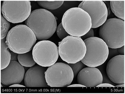
In this work, a facile and general protocol to synthesize molecularly imprinted microspheres (MIMs), combining reverse atom transfer radical polymerization with precipitation polymerization, is described. Well-tuned surface properties in the as-obtained microspheres were achieved through the “living” nature of the active ATRP-initiators present on the surface in order to further functionalize the microspheres with hydrophilic poly(2-hydroxyethyl methacrylate) (PHEMA) brushes. The presence of a PHEMA shell on the MIMs was confirmed by SEM, FT-IR and water contact angle studies, and some quantitative information is also provided. Batch adsorption experiments were carried out in pure water to investigate the equilibrium, kinetics and selectivity properties of the imprinted materials. The adsorption data of the ungrafted imprinted materials for sulfamethazine (SMZ) were fitted well to the Freundlich isotherm model. However, after grafting, the Langmuir isotherm fits the data better, mainly because of the changes to the surface properties that increase the hydrophilicity and suppress the hydrophobically driven non-specific interactions. The imprinted microspheres exhibit a large adsorption capacity, and are also specific to SMZ but non-specific to other antibiotics. The obtained materials also show rapid kinetics and great reusability and stability, displaying potential for practical applications for the selective removal of pollutants from aqueous environments.


1. Introduction

Owing to their superior physical and chemical stability, low cost, and easy preparation, molecularly imprinted polymers (MIPs), as a new type of synthetic receptors with predetermined molecular recognition sites, present remarkable potential applications in various aspects, including separation, sensors, catalysis, drug delivery, and so on.1–8 Typically, the target molecule or a derivative is employed as the template and self-assembled to form a complex with a functional monomer via covalent or non-covalent interactions, such as hydrogen bonds, ionic and/or hydrophobic interactions, and around which cross-linking monomers are arranged and co-polymerized to form a rigid polymer. Subsequently, removal of the template from the three-dimensional polymer network leads to active cavities, complementary in size, shape, and functionality with respect to the template, and so now the material has the ability to specifically recognize and rebind the target. Among the main mechanisms developed to form MIPs, free radical polymerization is the most widely employed, generally including bulk polymerization, emulsion polymerization, suspension polymerization, and precipitation polymerization.9–16

Over the past decade, and even with the rapid progress of a research hotspot such as the molecular imprinting technology, a number of challenges still remain to be addressed. Particularly, the previously synthesized MIPs can be successfully used for efficient molecular recognition of a large range of target molecules, predominantly non-aqueous solvents.17 However, current protocols often fail to generate MIPs exhibiting the capacity for capturing specific templates from pure aqueous media, due to the significant hydrophobically driven non-specific binding between the molecule and the exposed material's surface.18,19 Besides, excessive cross-linkers are commonly employed to fix and stabilize the imprinted cavities, thereby making it difficult to adjust and control their binding properties. However, there are still few versatile approaches for the development of new MIPs applicable in pure aqueous systems.

In order to suppress the non-specific binding, two useful strategies have been developed by improving the surface hydrophilicity of the MIPs closer to that of biological receptors, which can be potentially applicable in separation or sensors in aqueous environments. The first one is based on the direct addition of appropriate amounts of hydrophilic co-monomers in the imprinting process, including 2-hydroxyethyl methacrylate (HEMA),20 acrylic acid (AA),21N-isopropylacrylamide (NIPAAm),22 or acrylamide (AAM)23 as functional monomers, as well as poly(ethylene glycol diacrylate) (PEGDA),24 and N,N-methylenebisacrylamide (MBAA)25 as cross-linkers. In spite of the simplicity and directness for the above methods, the optimization of the formulation components during the imprinting process is rather complicated and time-consuming, because the materials' properties are largely influenced by many parameters (e.g. the type and amount of the hydrophilic co-monomers added) or have a limited range of application. The second one focuses on post-modifications of already preformed MIPs by chemical modification of the surface functional groups (e.g. epoxide ring opening26 or covalent coupling of ligands27) or surface-grafting of hydrophilic shielding polymers.28 The preparation of hydrophilic MIPs is thus more versatile and flexible, by involving the independent processes of a first normal imprinting method and the subsequent modification. The grafting of hydrophilic polymers from the MIPs surfaces has draw great attraction, due to the hydrophilicity of the surfaces and functional layers which prevents biological macromolecules from blocking the imprinted cavities.29

Well-defined polymers with various tailored architectures were prepared via controlled “living” radical polymerization techniques (CRPs) in the past decade,30 including atom transfer radical polymerization (ATRP), nitroxide-mediated radical polymerization (NMP), reversible addition-fragmentation chain transfer (RAFT) polymerization, and iniferter-induced “living” radical polymerization. With respect to the free radical polymerization, the use of CRPs in the synthesis of highly cross-linked 3D polymers is considered to generate more homogeneous structures, because of the improved match in chain growth and chain relaxation.31 Compared to other CRPs, ATRP is chemically versatile and compatible with a broad range of usable monomers and functional groups, and presents a good tolerance towards a relatively high degree of impurities, whose controllability lies in a fast, dynamic equilibrium established between the dormant species (alkyl halides) and active radicals, with transition-metal complexes acting as reversible halogen atom transfer reagents.32 Recently, owing to the existence of “living” fragments, ATRP, as one of most powerful CRPs, has been utilized in molecular imprinting for the controlled synthesis of advanced MIPs with well-tuned architectures and improved binding properties, including bulk polymers,33 microspheres,34 MIP nanotube membranes,35 surface-imprinted core–shell particles.36 To the best of our knowledge, however, the preparation of MIPs with pure water-compatible properties bearing functional polymer brushes by a facile grafting-from method via ATRP, and especially by reverse atom transfer radical polymerization (RATRP) taking advantage of the “living” nature of the alkyl halide fragments, has not been reported to date.

The aim of this study was therefore to obtain imprinted materials capable of selective recognition and separation of the analytes from pure aqueous media with minimum non-specific binding. As a consequence of the abuse in the use of sulfamethazine (SMZ), herein selected as the template molecule, it has been found in aquatic environments, increasing the public concern on the potential human health risks due to serious side effects such as allergies, carcinogenesis, and possible development of antibiotic resistance.37,38 Imprinted microspheres with surface-immobilized “living” fragments were prepared via a method combining RATRP with precipitation polymerization in a methanol–water mixture, and then used as macro-ATRP agents for further modification with hydrophilic polymer brushes (PHEMA). The physical and chemical properties of the as-prepared materials were characterized by FT-IR, SEM, water contact angle tests, and water dispersion stability studies. The binding properties of the ungrafted MIPs and grafted MIPs in pure water was investigated in detail.

2. Experimental section

2.1. Materials

Aqueous ammonia (NH3·H2O, 28 wt%), acetic acid, methanol, copper(I) bromide (CuBr, ≥98%) and copper(II) bromide (CuBr2, ≥98%) of analytical grade were purchased from Sinopharm Chemical Reagent Co., Ltd. (Shanghai, China). 4-Vinylpyridine (4-VP, ≥96%), 2-hydroxyethyl methacrylate (HEMA, 98%), sulfamethazine (SMZ), tetracycline (TC, 98%), cefalexin (CFX, 98%), azo-bis-isobutyronitrile (AIBN, ≥98%), and N, N, N′, N′,N′-pentamethyl diethyenetriamine (PMDETA, ≥98%) were purchased from Aladdin Reagent Co., Ltd. (Shanghai, China) and used as received. Ethylene glycol dimethacrylate (EGDMA, ≥98%) (Aladdin Reagent Co. Ltd., Shanghai, China) was washed consecutively with 10% aqueous NaOH, water and brine, dried over CaH2, filtered and then distilled under reduced pressure. Deionized ultrapure water was purified with Purelab Ultra (Organo, Tokyo, Japan).

2.2. Synthesis of molecularly imprinted microspheres with “living” fragments

SMZ-imprinted microspheres with surface-immobilized alkyl-halide groups (MIMs) were prepared via a robust method, combining reverse atom transfer radical polymerization with precipitation polymerization. In brief, 4-VP (0.034 mL, 0.32 mmol), SMZ (27.8 mg, 0.04 mmol), EGDMA (0.317 g, 1.60 mmol), and 30 mL of a mixture of methanol and water (4.0[thin space (1/6-em)]:[thin space (1/6-em)]1.0; v/v) were successively added into a 50 mL one-neck round-bottom flask. After a long self-assembly reaction time at room temperature, CuBr2 (11.2 mg, 0.05 mmol), PMDETA (0.021 mL, 0.1 mmol), and AIBN (24.63 mg, 0.15 mmol) were added, and the reaction mixture was then deoxygenized with nitrogen for 0.5 h and sealed. The flask was then submerged in an incubator shaker at a rate of 160 rpm and the polymerization was carried out at 60 °C for 24 h. The as-synthesized products were collected by centrifugation and subsequently eluted through a Soxhlet extraction with methanol–acetic acid (9.0[thin space (1/6-em)]:[thin space (1/6-em)]1.0; v/v) until no template could be detected in the extraction solution. Non-imprinted microspheres (NIMs) were prepared under the same conditions, except for the absence of the template.

2.3. Synthesis of molecularly imprinted microspheres with PHEMA brushes

The molecularly imprinted microspheres with hydrophilic polymer brushes (HMIMs) were synthesized by the surface-initiated ATRP technique, where MIMs with the surface-immobilized “living” fragment were used as macro-ATRP agents. The detailed polymerization procedure is presented as follow: 100 mg of MIMs with alkyl-halide groups, HEMA (2.0 mL, 9.0 mmol), CuBr (10.47 mg, 0.073 mmol), PMDETA (37.95 mg, 0.219 mmol), and 2.0 mL of methanol–water (1.0[thin space (1/6-em)]:[thin space (1/6-em)]1.0, v/v) were successively added into a one-neck round-bottom flask (25 mL). After being degassed under ultrasonication and exchanged with nitrogen for 0.5 h, the flask was sealed and immersed in a water bath at 40 °C and stirred for 24 h. After centrifugation, the resulting solid products were thoroughly washed with methanol until no white sediments were detected when ether was added to the washing solutions; and then dried at room temperature under vacuum to a constant weight, giving MIPs with polymer brushes with a yield of 110.5 mg. Non-imprinted microspheres with PHEMA brushes (HNIMs) were also prepared following the above procedure, with NIMs with alkyl-halides groups instead of MIMs, with a yield of 109.8 mg.

2.4. Apparatus and characterization

Fourier transform infrared (FT-IR) spectra of the obtained materials were recorded using a Nicolet NEXUS-470 spectrophotometer using KBr pellets (U.S.A.). A PHS-2 acidimeter (The Second Analytical Instrument Factory of Shanghai, China) was employed for the pH measurements. The morphology, particle size, and size distribution of the samples were observed with a scanning electron microscope (SEM, JEOL, JSM-7001F).

The calculated SEM size data reflect an average of more than 200 particles in a representative area from the SEM images, which were obtained according to the following formulas:

image file: c3ra44602f-t1.tif
where Dn and Dw are the number-average and weight-average diameters, respectively. k is the total number of particles, Di is the diameter of the particles, ni is the number of the particles with a diameter Di, and U is the size distribution index.

Dynamic light scattering (DLS) measurements were also carried out in a thermostat bath regulated at room temperature on a Bettersize 2000 apparatus (Dandong Baite Instrument Co., China) in order to measure the autocorrelation functions.

The MIMs, NIMs, HMIMs and HNIMs were dispersed in ethanol under ultrasonication, with a concentration of 15 mg mL−1, and then dropped on clean glass surfaces to prepare the polymer thin films. After the solvent was allowed to evaporate at ambient temperature overnight, a KSV CM200 contact angle instrument (Finland) was utilized to determine their static water contact angles. Two measurements were taken for each sample, with the mean being used for analysis.

A UV-2450 UV-vis spectrophotometer (Shimadzu Corporation, Japan) was used to detect the concentration of the samples by their corresponding absorbance. A reverse-phase HPLC system (Agilent 1200 series, U.S.A.) was equipped with a UV-vis detector to achieve the simultaneous detection of several samples.

Suspensions of MIMs, NIMs, HMIMs and HNIMs in pure water (1.0 mg mL−1) were first dispersed by ultrasonication, respectively, and then allowed to settle down for 2.0 h at 25 °C to check their dispersion stability.

2.5. Batch adsorption experiments

Equilibrium adsorption experiments were carried out by adding 5.0 mg of MIMs, NIMs, HMIMs or HNIMs into a 10 mL SMZ aqueous solution, with different concentrations, from 10 to 180 μmol L−1, at 25 °C for 12 h. The supernatants obtained by centrifugation were measured to quantify the amount of remaining templates, and then the amount of SMZ adsorbed on the polymer materials could be calculated. The adsorption experiments were performed by incubating a tetracycline (TC) water solution (10 mL, 100 μmol L−1) with 5.0 mg of HMIMs or HNIMs at different mixing times at 25 °C, to study the kinetic properties in detail. The adsorption selectivity of HMIMs or HNIMs was evaluated by measuring their competitive binding capacities towards SMZ and reference antibiotics, including TC and CFX. Typically, 5.0 mg of the HMIMs or HNIMs were incubated with 10 mL of single-antibiotic solutions with a concentration of 100 μmol L−1 at 25 °C for 12 h, respectively. Furthermore, double-antibiotic solutions containing SMZ and one other antibiotic were also used, and the amount of SMZ bound to HMIMs and HNIMs was quantified by HPLC, in order to investigate the effect of the presence of competitive antibiotics. All the above-mentioned adsorption experiments were carried out in duplicate and the mean values were calculated.

3. Results and discussion

3.1. Preparation of molecularly imprinted microspheres with hydrophilic polymer brushes

Herein, we present a general method to prepare MIMs with hydrophilic polymer brushes to improve their pure water-compatible binding properties, taking full advantage of ATRP. The schematic illustration for the preparation process is shown in Fig. 1. In the first step, the “living” MIMs/NIMs with surface-immobilized alkyl-halide groups were prepared via reverse atom transfer radical precipitation polymerization, utilizing CuBr2/PMEDTA/AIBN as the initiation system, which is an improvement over normal ATRP. In the polymerization system, SMZ was used as the template, 4-VP as the functional monomer, EGDMA as the cross-linker, and a mixture of methanol and water as the porogenic solvent (the volume percentage being about 99%), respectively, where hydrophobic interactions and hydrogen bond interactions are formed between 4-VP and SMZ. Subsequently, PHEMA was grafted on the “living” groups immobilized on the surface of the MIMs/NIMs using surface-initiated ATRP.
image file: c3ra44602f-f1.tif
Fig. 1 Schematic illustration of the preparation of MIMs with hydrophilic polymers brushes.

3.2. Characterization

FT-IR analysis was used to check the successful synthesis and surface-functionalization of the molecularly imprinted microspheres. Fig. 2 shows the FT-IR spectra of MIMs, NIMs, HMIMs and HNIMs. The three significant peaks at 1728 (C[double bond, length as m-dash]O stretching), 1258 and 1150 cm−1 (C–O–C stretching) confirm the existence of EGDMA in the polymers.39 Moreover, the small peak at 1638 cm−1 belongs to some unreacted C[double bond, length as m-dash]C double bonds of EGDMA molecules used as cross-linkers in the polymers. The characteristic peaks arising from the C[double bond, length as m-dash]N stretching (1600 and 1558 cm−1) and C[double bond, length as m-dash]C stretching (1460 cm−1) of the pyridine rings reveal that the 4-VP monomer participates in the imprinting polymerization.40 The C–H stretching vibration of the alkyl chain appears at 2958 and 2989 cm−1 for the –CH2 and –CH3 groups, respectively. The peak at 651 cm−1 is attributed to the C–Br group, demonstrating the presence of “living” groups. After the surface-initiated polymerization of HEMA, the characteristic broad band at 3400 cm−1 is assigned to the O–H stretching of HEMA. The vibration intensity of C–Br and C[double bond, length as m-dash]N decreases appreciably. All the results indicate the successful synthesis of imprinted polymers and the subsequent grafting of the hydrophilic monomer on the “living” initiator on the surface.
image file: c3ra44602f-f2.tif
Fig. 2 The FT-IR spectra of MIMs, HMIMs, NIMs and HNIMs.

Scanning electron microscopy was used to characterize the morphology, size, and size distribution of the obtained polymer microspheres. Fig. 3 shows the SEM images of MIMs, NIMs, HMIMs, and HNIMs. All four samples are smoothly spherical with a narrowly dispersed size distribution. The number-average diameter (Dn) obtained from the images for the MIMs and NIMs is 3.529 μm and 3.994 μm, respectively. In Table 1, the polydispersity indices (U) calculated for the MIMs and NIMs are 1.093 and 1.112, respectively. Compared to the ungrafted MIMs and NIMs, the thickness of the grafted hydrophilic polymer shells for HMIMs and HNIMs is 22 and 18.5 nm (according to ΔDn/2), respectively, with low U values. The statistical data from the SEM images is basically in accordance with the results obtained through dynamic light scattering (DLS) measurements (in Fig. S1). These results further indicate the successful grafting of PHEMA brushes onto the MIMs/NIMs (Fig. 3).


image file: c3ra44602f-f3.tif
Fig. 3 SEM images of MIMs (a and b), HMIMs (d and e), NIMs (c), and HNIMs (f).
Table 1 Characterization data for the MIMs, NIMs, HMINs and HNIMs
Samples ΔW% D n (μm) U L (nm) Contact angle (°) Yield %
MIMs 3.994 1.093 128.8 ± 2.6 77.47
HMIMs 10.5 4.036 1.099 22.0 60.2 ± 1.5
NIMs 3.529 1.112 122.7 ± 2.1 72.48
HNIMs 9.8 3.568 1.101 19.5 59.3 ± 1.8


Water contact angle experiments were performed to investigate the surface hydrophilic properties of the prepared materials. The profiles of water drops on MIM/NIM and HMIM/HNIM films are shown in Fig. 4. The static water contact angles were determined to be 128.8° and 122.7° for the MIM and NIM films, respectively. After surface-grafting the hydrophilic polymer brushes, the static water contact angles of the HMIM and HNIM films were significantly reduced to 60.18° and 59.27°, respectively, exhibiting a significantly higher hydrophilicity than the ungrafted ones, and demonstrating the presence of PHEMA brushes on the surfaces of the HMIMs and HNIMs. Moreover, the dispersion stability of the materials prepared in water has been efficiently improved by the surface-grafting of hydrophilic polymer brushes.41Fig. 4c and f show the dispersion of the MIMs/NIMs and HMIMs/HNIMs in a concentration of 1.0 mg mL−1 in pure water at ambient temperature after resting for 2.0 h. It is clear that the ungrafted materials present much more sedimentation in pure water compared to the grafted ones, which demonstrate that the PHEMA chains are anchored on the surface of the MIMs/NIMs by covalent binding.


image file: c3ra44602f-f4.tif
Fig. 4 The profiles of the water contact angle tests for MIMs (a), HMIMs (b), NIMs (d), and HNIMs (e); the photographs for the dispersion of the MIMs/HMIMs (c) and NIMs/HNIMs (f) in pure water (1.0 mg mL−1) at room temperature (after resting for 2.0 h).

3.3. Adsorption isotherms

The adsorption isotherms of MIMs, NIMs, HMIMs and HNIMs towards SMZ are shown in Fig. 5. For all the four samples, the adsorption capacity increases rapidly with the increasing initial concentration and then gradually becomes stable. The common phenomenon can be explained by the adsorption–desorption equilibrium between the polymers and the initial solution. The adsorbed amount is small at low initial concentrations, due to the low occupancy of the empty recognition sites. And then, more and more recognition sites are utilized until complete saturation as a result of the increase in the initial concentration, also providing the necessary driving force to overcome the resistance to the mass transfer of SMZ between the liquid and the solid phases. From Fig. 5, it is clearly seen that both the ungrafted and grafted MIMs bind more SMZ molecules than their corresponding NIMs, indicating the presence of selective recognition sites in the MIMs. Besides, both the ungrafted MIMs and NIMs exhibit high adsorption capacities, mainly due to their high surface hydrophobicity. Grafting PHEMA chains on the surface of the MIMs significantly improves their surface hydrophilicity, as demonstrated by their static water contact angle, but reduces their adsorption capacity towards SMZ. However, they present an enhanced specific adsorption in pure aqueous media, according to the increase in the value of the imprinting factor in a wide concentration range (calculated by the equation, IF = Qimprinted/Qnon-imprinted) (shown in Fig. 5b).
image file: c3ra44602f-f5.tif
Fig. 5 Adsorption isotherms of MIMs, NIMs, HMIMs and HNIMs with SMZ in pure water (a), the imprinted factors for the ungrafted and grafted microspheres (b).

Two classic isotherm models, namely Langmuir and Freundlich, were applied to simulate the equilibrium characteristics of the adsorption. The Langmuir model describes an idealized adsorption taking place at specific homogeneous sites within the adsorbent, not involving any intermolecular interaction of the adsorbate. In most cases, the Freundlich model, as an empirical equation, can be considered as a generalization of the Langmuir model suitable for a heterogeneous surface, removing the limitations of the idealization.

In general, the linear forms of the Langmuir42 and Freundlich43 models are expressed as:

 
image file: c3ra44602f-t2.tif(1)
 
image file: c3ra44602f-t3.tif(2)
where Qe and Qm (μmol g−1) are the equilibrium and maximum amount of SMZ adsorbed on the adsorbent, respectively, Ce (μmol L−1) is the equilibrium concentration of the SMZ solution, and KL (L μmol−1) and KF ((μmol g−1) (L μmol−1)1/n) are the Langmuir and Freundlich equilibrium constants, respectively.

As shown in Table 2 and Fig. 6, the experimental data were found to fit well the Freundlich isotherm in terms of the R2 value for the ungrafted microspheres. However, after the surface-grafting with the PHEMA brushes, the adsorption data of the HMIMs and HNIMs towards SMZ fitted better the Langmuir model compared to the Freundlich model. These results may be caused by the change in the hydrophilicity of the exposed surface, which decreases the intermolecular interactions of SMZ with the template molecule occupying a recognition site.

Table 2 Parameters of the Langmuir and Freundlich isotherm models
Samples The Langmuir model The Freundlich model
Q e,exp (μmol g−1) Q m,c (μmol g−1) K L (L μmol−1) R 2 K F ((μmol g−1) (L μmol−1)1/n) 1/n R 2
MIMs 47.532 74.074 0.0138 0.9760 2.017 0.7018 0.9966
HMIMs 38.664 54.945 0.0182 0.9975 1.981 0.6838 0.9848
NIMs 36.974 60.241 0.0113 0.9623 2.076 0.7306 0.9967
HNIMs 25.782 36.364 0.0191 0.9954 1.990 0.6880 0.9742


image file: c3ra44602f-f6.tif
Fig. 6 The Langmuir (a) and Freundlich model (b) fittings for the adsorption of SMZ on MIMs, NIMs, HMIMs and HNIMs, respectively.

3.4. Adsorption kinetic properties

In order to determine the necessary equilibrium times, the adsorption of SMZ onto HMIMs and HNIMs at an initial concentration of 100 μmol L−1 was studied. Fig. 7 presents the effect of time on the removal of SMZ at 298 K. Initially, because of the large number of vacant recognition sites, the adsorption uptake increases rapidly with time. After 90 min, the small amount of remaining vacant sites is more difficult to occupy, due to the repulsion between the SMZ molecules located on the surface and the bulk, and the process reaches an equilibrium state.
image file: c3ra44602f-f7.tif
Fig. 7 Adsorption kinetic data of SMZ on HMIMs and HNIMs and the linear fitting to the pseudo-second-order model (inset).

The rate of adsorbate uptake on the adsorbent is well described by adsorption kinetics. In this work, two kinetic models were used to analyze the kinetics of the adsorption data, the pseudo-first-order44 and pseudo-second-order45 models, the linear form of which can be expressed as eqn (3) and (4), respectively.

 
image file: c3ra44602f-t4.tif(3)
 
image file: c3ra44602f-t5.tif(4)

Here, Qe (μmol g−1) and Qt (μmol g−1) are the SMZ adsorbed amount at equilibrium and at time t, respectively, k1 (min−1) is the pseudo-first-order kinetics constant and k2 (g μmol−1 min−1) is the constant of the pseudo-second-order kinetics equation.

The kinetic parameters for the adsorption of SMZ on HMIMs and HNIMs according to the two models are tabulated in Table 3. The low R2 values and notable differences between the experimental and theoretical data for both HMIMs and HNIMs clearly demonstrate the poor fitting to the pseudo-first-order kinetics equation. From Fig. 7 and Table 3, it can be clearly seen that the pseudo-second-order model fits the experimental data quite well, and that the experimental and theoretical data are in good agreement with high R2 values. This indicates the suitability of the second-order kinetics model to describe the adsorption process of SMZ on the obtained polymers.

Table 3 Kinetic parameters of the SMZ adsorption on the HMIMs and HNIMs
  The pseudo-first-order model The pseudo-second-order model
Samples Q e,exp (μmol g−1) Q m,c (μmol g−1) k 1 (min−1) R 2 Q m,c (μmol g−1) k 2 (g μmol−1 min−1) R 2
HMIMs 33.652 23.682 0.0497 0.765 33.652 0.005769 0.9993
HNIMs 23.241 20.739 0.0545 0.9589 23.317 0.005318 0.9996


3.5. Adsorption selectivity

Here, we chose tetracycline (TC) and cefalexin (CFX) as competitive antibiotics for the selective adsorption experiments. Fig. 8a shows that, out of the three adsorbates, the obtained four polymers present the highest removal capacity for SMZ in single-component solutions, indicating a high adsorption selectivity for the template molecule. For the two other antibiotics, both the ungrafted and grafted MIMs/NIMs have nearly the same adsorption capacity, suggesting that the MIMs and HMIMs are specific to SMZ but non-specific to other antibiotics. It can be assumed that the selective recognition of the imprinted materials is dominated by the molecular structure and the interactions between functional groups and molecules. Compared to TC and CFX, the SMZ molecule has the smaller chemical structure and size, and exhibits the strongest hydrophobic interactions. However, the process for the selective adsorption is complex and other interactions may also be involved. Two competitive antibiotics, namely TC and CFX, were added into a SMZ aqueous solution, respectively, to determine their effect on the SMZ adsorption in order to further investigate the selectivity for SMZ of the imprinted adsorbents. As shown in Fig. 8b, the imprinted adsorbents still displayed a high SMZ removal capacity even in the presence of other antibiotics. In contrast, the NIM adsorbents were significantly affected by the competitive pollutants, and so, it can be concluded that the MIM adsorbents have a good sorption selectivity in the presence of other pollutants.
image file: c3ra44602f-f8.tif
Fig. 8 Adsorption selectivity of SMZ on the ungrafted and grafted MIMs and NIMs in single-antibiotic solutions (a) and in the presence of competitive pollutants (b).

3.6. Reusability and stability of HMIMs

After adsorption of SMZ on the HMIMs, the saturated imprinted adsorbents were regenerated using a methanol–acetic acid mixture and were then used to adsorb SMZ in subsequent cycles. The adsorption capacity of the HMIMs for SMZ displayed only a small loss after six consecutive adsorption–desorption cycles (shown in Fig. S2), indicating that the MIM adsorbents were regenerated completely during the regeneration process. The adsorbed SMZ can be extracted easily from the HMIMs using the same acid mixture, in which the hydrophobic interactions and hydrogen bond interactions disappear. The HMIMs have the advantages of their easy regeneration, stability and selectivity for specific pollutants in aqueous environments, and may be used in a fixed bed to remove SMZ from wastewater in practical applications.

4. Conclusions

A facile route to obtain hydrophilic molecularly imprinted microspheres has been presented in this work, using two-step atom transfer radical polymerization. First, molecularly imprinted microspheres were synthesized by reverse atom transfer radical precipitation polymerization in a methanol–water mixture, using sulfamethazine as the template molecule. Secondly, hydrophilic PHEMA brushes were grafted on the surface of the MIMs with “living” fragments, which were utilized as macromolecular initiators. From the characterization results, the imprinted materials were narrowly dispersed and spherical, with low polydispersity indices. The water contact angle of the MIMs decreased from 128.8 ± 2.6° to 60.2 ± 1.5°, exhibiting good hydrophilicity, due to grafting of the surface with PHEMA brushes, with an average size of 22 nm. The adsorption experimental data of the ungrafted imprinted materials for SMZ were well fitted to the Freundlich model. However, after grafting with the PHEMA brushes, the Langmuir isotherm fitted the data better, which may be caused by the improvement of their surface hydrophilicity. The MIMs and HMIMs exhibited larger adsorption capacities than the non-imprinted materials, and were also specific to SMZ but non-specific to other antibiotics. Moreover, the decrease of the hydrophobically driven non-specific interactions resulted in the increase of the impact factors for all the concentrations. The obtained materials also exhibited rapid adsorption kinetics and great reusability and stability, displaying potential for practical applications in the selective removal of pollutants from aqueous environments.

Acknowledgements

This work was financially supported by the National Natural Science Foundation of China (no. 21077046, 21004031, 21176107, 21174057, 21107037 and 21277063), the National Basic Research Program of China (973 Program, 2012CB821500), the PhD. Innovation Program Foundation of Jiangsu Province (no. CXZZ13_0668), and the Natural Science Foundation of Jiangsu Province (SBK201122883).

References

  1. C. Alexander, H. S. Andersson, L. I. Andersson, R. J. Ansell, N. Kirsch, I. A. Nicholls and J. O. Whitcombe, J. Mol. Recognit., 2006, 19, 106 CrossRef CAS PubMed.
  2. C. Long, Z. Mai, Y. Yang, B. Zhu, X. Xu, L. Lu and X. Zou, J. Chromatogr., A, 2009, 1216, 275 Search PubMed.
  3. X. Hu, G. Li, M. Li, J. Huang, Y. Li, Y. Gao and Y. Zhang, Adv. Funct. Mater., 2008, 18, 575 CrossRef CAS.
  4. T. Alizadeh, M. Zare, M. R. Ganjali, P. Norouzi and B. Tavana, Biosens. Bioelectron., 2010, 25, 1166 CrossRef CAS PubMed.
  5. S. J. Li and S. Q. Gong, J. Phys. Chem., 2009, 113, 16501 CAS.
  6. G. Wulff, Chem. Rev., 2002, 102, 1 CrossRef CAS PubMed.
  7. J. L. Urraca, A. J. Hall, M. C. Moreno-Bondi and B. Sellergren, Angew. Chem., 2006, 118, 5282 CrossRef.
  8. J. Z. Hilt and M. E. Byrne, Adv. Drug Delivery Rev., 2004, 56, 1599 CrossRef CAS PubMed.
  9. A. Beltran, R. M. Marcé, P. A. G. Cormack and F. Borrull, J. Chromatogr., A, 2009, 1216, 2248 CrossRef CAS PubMed.
  10. S. C. Zimmerman and N. G. Lemcoff, Chem. Commun., 2004, 5 RSC.
  11. N. Pérez, M. J. Whitcombe and E. N. Vulfson, Macromolecules, 2001, 34, 830 CrossRef.
  12. C. J. Tan, H. G. Chua, K. H. Ker and Y. W. Tong, Anal. Chem., 2008, 80, 683 CrossRef CAS PubMed.
  13. A. A. Özcan, R. Say, A. Denizli and A. Ersöz, Anal. Chem., 2006, 78, 7253 CrossRef PubMed.
  14. J. P. Lai, R. Niessner and D. Knopp, Anal. Chim. Acta, 2004, 522, 137 CrossRef CAS PubMed.
  15. L. Ye, R. Weiss and K. Mosbach, Macromolecules, 2000, 33, 8239 CrossRef CAS.
  16. H. Sambe, K. Hoshina, R. Moaddel, I. W. Wainer and J. Haginaka, J. Chromatogr., A, 2006, 134, 88 CrossRef PubMed.
  17. B. Dirion, Z. Cobb, E. Schillinger, L. I. Andersson and B. Sellergren, J. Am. Chem. Soc., 2003, 125, 15101 CrossRef CAS PubMed.
  18. J. G. Karlsson, L. I. Andersson and I. A. Nicholls, Anal. Chim. Acta, 2001, 435, 57 CrossRef CAS.
  19. I. A. Nicholls, O. Ramström and K. Mosbach, J. Chromatogr., A, 1995, 691, 349 CrossRef CAS.
  20. B. Dirion, Z. Cobb, E. Schillinger, L. I. Andersson and B. Sellergren, J. Am. Chem. Soc., 2003, 125, 15101 CrossRef CAS PubMed.
  21. Z. Meng, W. Chen and A. Mulchandani, Environ. Sci. Technol., 2005, 39, 8958 CrossRef CAS.
  22. Y. Kanekiyo, R. Naganawa and H. Tao, Angew. Chem., Int. Ed., 2003, 42, 3014 CrossRef CAS PubMed.
  23. F. Puoci, F. Iemma and N. Picci, Curr. Drug Delivery, 2008, 5, 85 CrossRef CAS.
  24. T. Kubo, N. Tanaka and K. Hosoya, Anal. Bioanal. Chem., 2004, 378, 84 CrossRef CAS PubMed.
  25. T. Y. Guo, Y. Q. Xia and G. J. Hao, Chin. Chem. Lett., 2004, 15, 1339 CAS.
  26. F. Puoci, F. Lemma, G. Cirillo, M. Curcio, O. I. Parisi, U. G. Spizzirri and N. Picci, Eur. Polym. J., 2009, 45, 1634 CrossRef CAS PubMed.
  27. B. Sellergren, Macromolecules, 2006, 39, 6306 CrossRef CAS.
  28. G. Q. Pan, Y. Zhang, X. Z. Guo, C. X. Li and H. Q. Zhang, Biosens. Bioelectron., 2010, 26, 976 CrossRef CAS PubMed.
  29. G. Q. Pan, Y. Ma, Y. Zhang, X. Z. Guo, C. X. Li and H. Q. Zhang, Soft Matter, 2011, 7, 8428 RSC.
  30. W. A. Braunecker and K. Matyjaszewski, Prog. Polym. Sci., 2007, 32, 93 CrossRef CAS PubMed.
  31. A. D. Vaughan, S. P. Sizemore and M. E. Byrne, Macromolecules, 2012, 45, 4015 CrossRef.
  32. K. Matyjaszewski, Polymer, 2007, 48, 74 CrossRef PubMed.
  33. S. Sasaki, T. Ooya and T. Takeuchi, Polym. Chem., 2010, 1, 1684 RSC.
  34. B. Y. Zu, G. Q. Pan, X. Z. Guo, Y. Zhang and H. Q. Zhang, J. Polym. Sci., Part A: Polym. Chem., 2009, 47, 3257 CrossRef CAS.
  35. H. J. Wang, W. H. Zhou, X. F. Yin, Z. X. Zhuang, H. H. Yang and X. R. Wang, J. Am. Chem. Soc., 2006, 128, 15954 CrossRef CAS PubMed.
  36. C. H. Lu, Y. Wang, Y. Li, H. H. Yang, X. Chen and X. R. Wang, J. Mater. Chem., 2009, 19, 1077 RSC.
  37. H. Font, J. Adrian, R. Galve, M. C. Estévez, M. Castellari, M. Gratacós-Cubarsí, F. Sánchez-Baeza and M. P. Marco, J. Agric. Food Chem., 2008, 56, 736 CrossRef CAS PubMed.
  38. Q. Chu, D. Zhang, J. Wang and J. Ye, J. Sci. Food Agric., 2009, 89, 2498 CrossRef CAS.
  39. K. Yoshimatsu, K. Reimhult, A. Krozer, K. Mosbach, K. Sode and L. Ye, Anal. Chim. Acta, 2007, 584, 112 CrossRef CAS PubMed.
  40. C. Gonzato, M. Courty, P. Pasetto and K. Haupt, Adv. Funct. Mater., 2011, 21, 3947 CrossRef CAS.
  41. C. Y. Hong, Y. Z. You and C. Y. Pan, Chem. Mater., 2005, 17, 2247 CrossRef CAS.
  42. I. Langmuir, J. Am. Chem. Soc., 1918, 40, 1361 CrossRef CAS.
  43. H. M. F. Freundlich, J. Phys. Chem., 1906, 57, 385 CAS.
  44. A. Lara and E. Houssam, Chem. Eng. J., 2009, 150, 403 CrossRef PubMed.
  45. Z. Y. Zhang, Z. B. Zhang, Y. Fernández, J. A. Menéndez, H. Niu, J. H. Peng, L. B. Zhang and S. H. Guo, Appl. Surf. Sci., 2010, 256, 2569 CrossRef CAS PubMed.

Footnote

Electronic supplementary information (ESI) available. See DOI: 10.1039/c3ra44602f

This journal is © The Royal Society of Chemistry 2014
Click here to see how this site uses Cookies. View our privacy policy here.