A mobile instrument for in situ scanning macro-XRF investigation of historical paintings
Matthias
Alfeld
*a,
Joana Vaz
Pedroso
b,
Margriet
van Eikema Hommes
b,
Geert
Van der Snickt
a,
Gwen
Tauber
c,
Jorik
Blaas
b,
Michael
Haschke
d,
Klaus
Erler
d,
Joris
Dik
b and
Koen
Janssens
*a
aAXI2L Research group, Department of Chemistry, University of Antwerp, Groenborgerlaan 171, B-2020 Antwerp, Belgium. E-mail: matthias.alfeld@ua.ac.be; koen.janssens@ua.ac.be; Fax: +32 (0)3 265 3233; Tel: +32 (0)32653326
bDelft University of Technology, Department of Materials Science, Mekelweg 2, 2628CD Delft, The Netherlands
cRijksmuseum Amsterdam, Paintings Conservation Department, Hobbemastraat 22, 1071XZ Amsterdam, The Netherlands
dBruker Nano GmbH, Schwarzschildstraße 12, 12489 Berlin, Germany
First published on 21st March 2013
Abstract
Scanning macro-X-ray fluorescence analysis (MA-XRF) is rapidly being established as a technique for the investigation of historical paintings. The elemental distribution images acquired by this method allow for the visualization of hidden paint layers and thus provide insight into the artist's creative process and the painting's conservation history. Due to the lack of a dedicated, commercially available instrument the application of the technique was limited to a few groups that constructed their own instruments. We present the first commercially available XRF scanner for paintings, consisting of an X-ray tube mounted with a Silicon-Drift (SD) detector on a motorized stage to be moved in front of a painting. The scanner is capable of imaging the distribution of the main constituents of surface and sub-surface paint layers in an area of 80 by 60 square centimeters with dwell times below 10 ms and a lateral resolution below 100 μm. The scanner features for a broad range of elements between Ti (Z = 22) and Mo (Z = 42) a count rate of more than 1000 counts per second (cps)/mass percent and detection limits of 100 ppm for measurements of 1 s duration. Next to a presentation of spectrometric figures of merit, the value of the technique is illustrated through a case study of a painting by Rembrandt's student Govert Flinck (1615–1660).
Introduction
Scanning macro-XRF (MA-XRF) is a variant of XRF imaging1 that allows visualization of the distribution of elements in a flat, macroscopic sample (up to several square meters) in a non-destructive manner. This is achieved by scanning the surface of the sample with a focused or collimated X-ray beam of (sub) mm dimensions and analyzing the emitted fluorescence radiation. Due to the penetrative nature of X-rays, elements present at and below the surface contribute to the obtained elemental distribution images.The method is highly valuable in the investigation of historical paintings, as elemental distribution images can reveal hidden sub-surface layers, including modifications made by the artist or restorations on the surface. In this way it can provide a unique insight into the creative process of the artist(s) and the painting's conservation history.
MA-XRF for the investigation of historical paintings was first described in the early 1990s,2,3 but the method found no widespread application until Dik et al. employed it to visualize the study of a peasant's head underneath a painting that Vincent van Gogh created during his stay in Paris.4 This experiment, which was done at the Deutsches Elektronen Synchrotron (DESY) in Hamburg, Germany, was followed by several other case studies on paintings by Rembrandt van Rijn,5 Philipp Otto Runge6 and Vincent van Gogh7,8 at DESY, by Rembrandt van Rijn9 at the National Synchrotron Light Source (NSLS) in the Brookhaven National Laboratory in NY, USA and by Arthur Streeton10 at the Australian Synchrotron in Melbourne, Australia. Further a full scale mock-up of Rembrandt's Old Man in Military Costume was created in order to compare the imaging capabilities of synchrotron and X-ray tube based scanners and to select the instrument best suited for investigating the original painting.11
In these experiments, the investigated painting was mounted on a motorized stage and moved through the X-ray beam. This procedure limited the maximum possible size of the investigated painting. Furthermore, the logistic and financial efforts of bringing a precious painting to a synchrotron radiation source are considerable and limit the number of paintings that can be investigated.
In order to broaden the range of paintings that may be investigated, mobile MA-XRF instruments for in situ investigation of paintings were developed. In these instruments the X-ray tube and detectors are mounted on a motorized stage and moved before the surface of a (stationary) painting during a scan. The pigments used to create a painting are generally present at a concentration level of several mass percent. By employing high intensity synchrotron radiation to scan the painting, the heavy elements present in the paint can be visualized with dwell times far below one second. However, also a well designed X-ray tube based system can be sensitive enough to image their distribution at a comparable pace.
Such an instrument was partially realized in the Artax system12 (Röntec GmbH, Berlin, now Bruker Nano GmbH, Berlin, Germany), which allows scanning of small areas, 5 × 5 cm, and in the system described by Hocquet et al.13 However, both systems were limited in the maximum pace of scanning and require several seconds of dwell time per pixel, resulting in scan durations of several hours or even days for small areas of paintings.
Only with the development of the in-house built MA-XRF scanner described by Alfeld et al.14 did routine scans of paintings become practical. This scanner allowed acquisition of elemental distribution images of elements present at a concentration level of several mass percent with dwell times of less than one second per pixel. Until now, studies of paintings by Van Gogh,14 Rembrandt van Rijn15 and Goya16 among others have been made with this scanner. The main limitation of the system is the fact that, as an experimental system, it requires a highly trained operator.
So a scanner for fast routine MA-XRF imaging of historical canvases or panel paintings was still missing. Characteristics of such a scanner would be: (a) capable (in terms of sensitivity and speed of the motorized stage) of acquiring the elemental distribution images of elements present at several mass percent with dwell times of less than a second; (b) easily transportable (by private car) between museums and galleries; (c) featuring a variable beam size to allow for overview (image resolution ∼0.5 mm) as well as detailed (resolution ∼100 μm) scans without changing the optic; (d) intuitive control software that allows the operation by a person trained, but not specialized in XRF analysis and/or imaging.
To develop such a scanner, a collaboration with Bruker Nano GmbH (Berlin, Germany) was started in view of their experience as a manufacturer of XRF imaging systems, including the M4 Tornado μ-XRF system.17 In the M4 Tornado a small sample (up to 300 cm2) is moved on a motorized stage through the beam of an X-ray tube that is equipped with a polycapillary lens. The beam size is adjusted by positioning the sample at a specific distance to the polycapillary lens. Next to a high sensitivity for a broad range of elements the system features an easy to use graphical user interface.
Based on the experience gained in previous MA-XRF experiments and with the M4 Tornado, a mobile MA-XRF scanner was developed at Bruker Nano GmbH. The resulting scanner, baptized M6 Jetstream, is the first commercially available macro-XRF scanner.
In this paper we present a full description of the scanner, including its central spectrometric figures of merit. We illustrate the value of the instrument and method in art-historical studies through a case study, involving a painting by Rembrandt's student Govert Flinck (1615–1660).
The M6 Jetstream: technical characteristics
The M6 Jetstream consists of a measuring head that is moved over the surface of a painting by means of a X,Y-motorized stage (Fig. 1). This motorized stage features a minimum step size of 10 μm and a maximum travel range of 80 × 60 cm (h × v). By combining the elemental distribution images obtained by scanning sub-areas, objects larger than the maximum travel range of the motorized stages can be investigated as a whole: thus, it becomes feasible to scan medium sized paintings with dimensions of 3 × 2 m2, for example. | ||
| Fig. 1 Bruker M6 Jetstream set-up and details of the measuring head. | ||
The exact distance between the measuring head and the painting is adjusted by a motorized stage of 7 cm travel range oriented along the Z axis. The measurement head travels within a metal frame that can be tilted in order to adjust it to the surface of a painting and to allow the analysis of horizontally positioned samples.
The frame holding the motorized stage is mounted on a box containing the detector electronics, the motor control electronics and the high voltage generator of the X-ray tube. The electronics box itself is mounted on a wheeled platform that allows for easy transport over short distances and positioning with respect to the painting. For transport the measuring head can be demounted and stored in the electronics box; the wheeled platform, the electronics box and the frame holding the motorized stages can be separated.
The measuring head consists of a 30 W Rh-target micro-focus X-ray tube with a maximum voltage of 50 kV and a maximum current of 0.6 mA. The spectral range of primary radiation emitted from the X-ray tube can be modified by the use of 5 filters, mounted in a motorized filter wheel. The beam is defined by means of a polycapillary optic, allowing for a variable beam size (with ca. 40 μm as the smallest beam size – see below) as a function of the distance between the painting and the measuring head.
Two optical cameras with magnifying optics, focused on the surface of the painting at the near normal angle are mounted on the measuring head. The focal plane of the stronger magnifying camera can be manually adjusted in five steps and is used for controlling the distance from the measuring head to the painting and thus the beam size.
The less magnifying camera can be used to automatically acquire mosaic images of the whole surface of the painting that allow direct comparison of these images with the elemental distribution images. Further, these mosaic images are used for defining the scanned area.
For geometrical reasons the cameras observe the surface of the painting with a slight offset from the spot excited by the X-ray beam. This offset is automatically corrected for by the control software during measurement.
The software package of the M6 Jetstream includes routines for region-of-interest (ROI) imaging and principal component analysis (PCA). The ROI images of the acquired data are updated after each scanned line and allow for an online monitoring of the scan process.
The software allows for the acquisition of elemental distribution images consisting of several megapixels.
Experimental
The spectrometric figures of merit of the M6 Jetstream were determined from a spectrum of the Standard Reference Material (SRM) 611 Trace elements in glass manufactured by the National Institute of Standards and Technology (NIST). The reference material is a 1 mm thick glass disc containing a broad range of elements at a nominal concentration level of 500 ppm. The elements not certified by the manufacturer were taken from ref. 18. The spectrum was acquired for 100 s real time at tube settings of 50 kV and 0.6 mA, without a filter at a distance of 8 mm to the measuring head (equivalent to a beam size of ∼150 μm, see below).From this spectrum the sensitivity (Y) and limits of detection (LOD) normalized for one second were calculated by means of eqn (1) and (2):
 | (1) |
 | (2) |
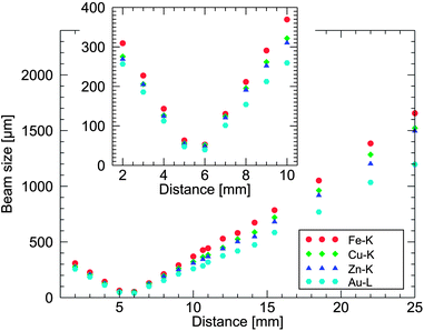
The beam size was measured by scanning brass (Cu/Zn), Fe and Au wires of 25 μm diameter at variable distances from the measuring head. The Full-Width-at-Half-Maximum (FWHM) of the obtained profiles was taken as a measure for the beam size.
Two sets of elemental distribution images of historical paintings are presented below. In Fig. 5 details of a late 15th/early 16th century portable altar are shown. The scan was performed in two hours with a step size of 50 μm and a dwell time of 10 ms per pixel to image an area of 4 by 4.5 cm. The tube settings were 50 kV and 0.6 mA.
Further in Fig. 6 the results obtained from Govert Flinck's Portrait of Dirck Jacobsz. Leeuw are shown. The elemental distribution images of 1300 × 900 pixels were acquired with a dwell time of 10 ms per pixel and a step size of 500 μm. So, nearly the complete surface of the painting (65 × 45 cm2) was scanned in 3.5 hours. The tube settings were 35 kV and 0.5 mA.
The figures of merit in this article were based on least squares fitting with PyMCA.19 The spectra of imaging data were processed by an in-house written Dynamic Analysis (DA)20 routine with DA matrices based on PyMCA least squares fits of the sum spectrum of all pixels.
Figures of merit
The M6 Jetstream was designed to acquire distribution images of elements present at a concentration level of several mass percent with dwell times below one second. The K-line sensitivity plotted in Fig. 2 shows that for a broad range of elements from Ti (Z = 22) to Mo (Z = 42) present at 1 mass percent, more than 1000 cps can be achieved, allowing for acquisition of images with good contrast and low noise with several tens of milliseconds dwell time. Heavy elements such as Au, Hg and Pb can also easily be imaged, as their L-lines fall in the same energy range as the K-lines of the elements mentioned above. | ||
| Fig. 2 K-line sensitivity for the elements Ca (Z = 20) to Sb (Z = 51) of the M6 Jetstream at 50 kV and 0.6 mA. | ||
Calcium is often present in paintings at high concentration levels, e.g. in the commonly employed paint extenders chalk (CaCO3) and gypsum (CaSO4·2H2O), as well as in the black pigment bone black. Thus, its distribution can usually be clearly visualized, even though the scanner's sensitivity for it is lower than for the elements mentioned above (at the level of ca. 400 cps per mass percent).
More problematic is the limited K-line sensitivity for elements heavier than Rh. Especially Cd (Z = 48), Sn (Z = 50) and Sb (Z = 51), the elements associated with the respective yellow pigments such as cadmium yellow, lead tin yellow and Naples yellow feature a fairly lower sensitivity (in the range 200–300 cps per mass percent). In some cases, such as in the study of hidden van Gogh paintings,4 Naples yellow proved to be a crucial pigment. The reason for this low sensitivity is the choice of a polycapillary lens as the beam defining optic which has a lower transmission for the high energy components of the primary radiation.
Although these heavier elements can also be imaged by their L-lines instead of their K-lines, the lower energy of this radiation is easily absorbed by covering layers, so that it does not allow for the visualization of hidden paint layers.
In this framework, it is relevant to mention that the measured sensitivities presented in this work are only valid for elements on the surface of a painting; depending on the composition of the covering paint layers, in practice, a noise free visualization of hidden paint layers may require longer dwell times than those of the surface layers.
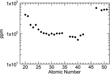
In general, the limits of detection (LOD) shown in Fig. 3 confirm the results obtained by comparing the sensitivity. The LOD for one second for the elements from Ti (Z = 22) to Mo (Z = 42) is around 100 ppm, so that for a scan with a dwell time of 10 milli-seconds the detection limit would still be around 1000 ppm (or 0.1 mass percent) considerably below the demanded level of 1 mass percent. But also here Ca, Cd, Sn and Sb are less detectable than the elements in the range mentioned above.
 | ||
| Fig. 3 K-line limits of detection for the elements Ca (Z = 20) to Sb (Z = 51) of the M6 Jetstream at 50 kV and 0.6 mA. | ||
In Fig. 4 the beam size is plotted as a function of the distance between the painting surface and the measuring head. Beside the distance, the effective beam size also depends on the absorption edge energy of the excited elements due to chromatic aberration of the polycapillary optic. The beam size of the M6 Jetstream can be varied between 40 and 1200 μm for the Au–L lines and 52 and 1650 μm for the Fe–K lines.
 | ||
| Fig. 4 Beam size as a function of the distance between the sample and the measuring head. | ||
This variable beam size allows us to change the beam size of the instrument by adjusting the distance between the measuring head and the paint surface. This allows for easy investigation of smaller regions of special interest in greater detail, after being identified in an overview scan, without a time consuming exchange of the beam defining optic.
While it is possible to image small parts of a painting with a lateral resolution below 100 μm, it is quite challenging to achieve this for the entire surface of a painting. In general, paintings are not flat as the paint itself can have a topography of up to several millimeters in height, depending on the painting style. Also, the support may have a curved or undulated surface making it difficult to keep the distance between the measuring head and the painting within tolerance.
To illustrate the high-resolution imaging capabilities of the instrument, Fig. 5 shows the details of an oil painting which is part of a portable altar dated to the late 15th/early 16th century (unknown artist). This scan was performed with a step size of 50 μm and a dwell time of 10 ms per pixel to image an area of 4 by 4.5 cm. The tube settings were 50 kV and 0.6 mA.
 | ||
| Fig. 5 (Left) Details of a late 15th/early 16th century portable altar triptych showing the face of Christ surrounded by a radiant halo (photograph taken with the M6 camera); (middle and right) high-resolution MA-XRF images of Pb and Au. The images of 900 × 800 pixels were acquired with a step size of 50 μm and a dwell time of 10 ms per pixel in two hours with tube settings of 50 kV and 0.6 mA. | ||
Given the excellent state of the surface paint layers of this painting and the presence of 19th century pigments such as Scheele's green, it is likely that this painting was heavily restored in the recent past. This was confirmed by the investigation with the M6 Jetstream.
In the Pb image of Fig. 5, it can be seen that the head of Jesus was completely repainted, slightly changing its position. The red dots indicate the position of the original, damaged head and shoulder, while the green ones indicate the current position.
It is worth noting that fine details of the face could be resolved and that also the ca. 400 μm wide Au rays can be imaged in a satisfactory manner.
Case study
In order to illustrate the value of MA-XRF imaging in the investigation of historical paintings, we present the following case study on a work by the Amsterdam painter Govert Flinck (1615–1660). Flinck was one of Rembrandt's most successful students and, during his lifetime, even surpassed his former master in success. Fig. 6 shows the portrait Flinck made in 1636 of his nephew Dirck Jacobsz. Leeuw, as well as selected elemental distribution images obtained from the painting. As the earliest of Govert Flinck's signed and dated paintings, it was investigated as part of a broader study of the painting techniques of Rembrandt's students conducted at the Conservation Department of the Rijksmuseum, the Scientific Department of the Cultural Heritage Agency and the Department for Conservation of the University of Amsterdam. Investigations of this painting by X-ray radiography (XRR) and infrared reflectography (IRR) had revealed changes to the composition during its execution (pentimenti) but failed to reveal any details. In order to gain insight into these compositional changes the painting was investigated by MA-XRF. The acquired elemental distribution images provided a most surprising and revealing insight into the social life of the 17th century Low Countries. | ||
| Fig. 6 Govert Flinck, Portrait of Dirck Jacobsz. Leeuw, canvas 64.4 × 47.2 cm, signed and dated 1636, Mennonite Church, Amsterdam on loan to the Rembrandthuis, Amsterdam, the Netherlands. The elemental distribution images (1300 × 900 pixels, 0.5 mm step size, 10 ms dwell time) were acquired in 3.5 hours. The yellow line delimits the area in which the underpainting was executed, while the arrow indicated the position of the sketched branch (see text). | ||
To understand this, we should know that in his early career, Flinck was given many commissions by his prosperous Amsterdam relatives.21,22 Just as Flinck himself, these relatives were members of the Mennonite church, the Christian denomination that is based on the writings of Menno Simons (1496–1561). Mennonites were at the time recognized by their modest old-fashioned costume and the investigated Portrait of Dirck Jacobsz. Leeuw in his sober black attire is often referred to as a typical example of this dress code.
However, it is obvious in the Pb–L image that the costume was originally quite different. At first, it featured a fashionable broad bobbin lace collar and long lace cuffs and a lower hat with a broader brim. Also the position of the legs was different. While in the final painting Leeuw walks towards the observer, in the original portrait he stood with one leg slightly bent; an elegant pose that had just become popular in portraiture.
The Cu-map shows how the probably green copper containing paint used in the landscape precisely follows the contours of the earlier silhouette. The fine details on the areas that were later covered by the final legs and hat show that the landscape was already advanced or even finished before the costume and position were changed.
The Hg distribution image reveals the use of the red pigment vermilion (HgS) used to give a slightly warmer colour in the flesh tones, the tree trunk as well as in the coat. It is also present in the stockings of the original painting, which were bright red as evidenced by small red patches shimmering through the final paint surface. Such coloured stockings were fashionable items worn by the upper class at the time.
The different costumes might at first suggest that Leeuw had been depicted over a portrait of someone else. However, the distribution maps show that no modifications, overpainting or erasure took place in the area of the face, indicating that it was Leeuw who was portrayed from the beginning. This is confirmed by the symbolic object (seemingly an orange) Leeuw holds in his hand, which was present in the earlier version as well.
The change from a highly fashionable to a modest costume suggests that Leeuw's first attire may have caused some debate on Mennonite circles. In the Mennonite congregation not everybody felt comfortable with the strict dress rules. Ministers sometimes struck out at those who dressed (in their opinion) indecently indicating that this must have taken place.23 Leeuw must have been one of those ‘improperly’ dressed. He, at first, ordered a portrait of himself in fashionable clothes but, probably in order to avoid provocation of his fellow-believers, asked Flinck to change it at a later stage. One wonders whether this affair may have played a role in Leeuw's decision to leave the Mennonites. Three years after his portrait was signed by Flinck, Leeuw and his wife joined the more liberal Remonstrant church.
In addition to identifying the pentimenti, the distribution maps provide valuable insight into the painting's state of conservation. It may be tempting to assume that the Mn distribution is the result of sketching or outlining the figure with umber, a brown earth pigment. Yet, the lines seemingly defining the figure can be identified as retouchings due to the presence of Ti in many of the same areas and on the basis of UV-investigation. Ti reveals the use of titanium white (TiO2), a pigment not used before the 1920s; its presence in high concentrations is a sure indicator for restoration treatments in paintings created before the 20th century. In the case of Flinck paintings we know that it was restored in 2005/2006 by Martin Bijl of Bijl Schilderijenrestauratie. In UV light the recent retouchings stand out dark against the fluorescent varnish. Other retouchings can be identified in the Ba distribution images, again indicating the presence of a modern pigment.
Finally, the distribution maps made it possible to visualize some of Flinck's sketching and preparatory paint layers. This is of particular importance since for this aspect in 17th-century paintings, XRR and IRR are often of limited use. In the Fe image we see that the shape of the tree trunk extends beyond the final version (indicated by a yellow line). It was probably laid out as a rough sketch with ochre paint but finally only partly realized. Also in the upper right corner, where the painting today only features the sky, broad indications of tree branches sketched with ochre paint can be seen (indicated with yellow arrows).
While the authenticity of this particular painting was never doubted, the presence of a modified first sketch as well as the presence of pentimenti are often considered as indications that a painting is not a copy but an original first version; thus, for other paintings the visualization of these aspects now made possible by MA-XRF can have great value in discussions of authenticity.
Conclusions
We have presented the first results obtained with the Bruker M6 Jetstream, the first commercial macro-XRF scanner that is based on the experience gained with in-house built scanners of this type.We have presented the technical characteristics and spectrometric figures of merit of the scanner and illustrated the value of elemental distribution images acquired with it in the investigation of historical paintings. As a case study, the investigation of the Portrait of Dirck Jacobsz. Leeuw by Govert Flinck was discussed.
The scanner satisfies the demands made to it: (a) it allows the visualization of many elements at and below the surface of oil paintings with a (shortest) dwell time of 10 ms in (mostly) noise free images; (b) it is transportable in a small van; (c) it allows us to vary the primary beam size between ∼50 μm and 1 mm, although high resolution scans are only possible in areas of limited size and (d) it features an easy to learn, intuitive control software including routines for region-of-interest imaging and principal component analysis of the acquired data.
In its current configuration, employing a polycapillary lens as the beam defining optic, the scanner is lacking sensitivity for the fluorescence lines more energetic than those of Rh (20.2 keV). The difficulties in visualizing heavy elements such as Cd, Sn and Sb by means of their K-lines are especially problematic, as these have proven themselves crucial in, e.g., the study of Vincent van Gogh's paintings and similar works.
Acknowledgements
This research was supported by the Interuniversity Attraction Poles Programme – Belgian Science Policy (IUAP VI/16). The text also presents the results of GOA “XANES meets ELNES” (Research Fund University of Antwerp, Belgium) and from FWO (Brussels, Belgium) projects no. G.0704.08 and G.01769.09. M. Alfeld receives a Ph. D. fellowship of the Research Foundation-Flanders (FWO). We thank J. Langerock for allowing us to examine the portable altar triptych shown in Fig. 5.Notes and references
- K. H. A. Janssens, F. C. V. Adams and A. Rindby, Microscopic X-ray Fluorescence Analysis, Wiley, Chichester, 2000 Search PubMed.
- M. Mantler, M. Schreiner, F. Weber, R. Ebner and F. Mairinger, Adv. X-Ray Anal., 1992, 35, 987–993 CrossRef.
- M. Schreiner, M. Mantler, F. Weber, R. Ebner and F. Mairinger, Adv. X-Ray Anal., 1992, 35, 1157–1163 CrossRef.
- J. Dik, K. Janssens, G. Van der Snickt, L. van der Loeff, K. Rickers and M. Cotte, Anal. Chem., 2008, 80, 6436–6442 CrossRef CAS.
- J. Dik, K. Janssens, M. Alfeld, K. Rickers and E. van de Wetering, in Nouveau regard sur Rembrandt, ed. E. v. d. Wetering, Local World BV, Amsterdam, 2010, pp. xx–xxiv Search PubMed.
- M. Alfeld, K. Janssens, K. Appel, B. Thijsse, J. Blaas and J. Dik, Z. Kunsttechnol. Konserv., 2011, 25, 157–163 Search PubMed.
- L. S. van der Loeff, M. Alfeld, T. Meedendorp, J. Dik, E. Hendriks, G. Van der Snickt, K. Janssens and M. Chavannes, in Van Gogh Studies 4: New Findings, ed. C. Stolwijk and L. van Tilborgh, WBOOKS, Zwolle, 2012, pp. 33–53 Search PubMed.
- M. Alfeld, D. P. Siddons, K. Janssens, J. Dik, A. Woll, R. Kirkham and E. van de Wetering, Appl. Phys. A: Mater. Sci. Process., 2013, 111, 157–164 CrossRef CAS.
- M. Alfeld, G. Van der Snickt, F. Vanmeert, K. Janssens, J. Dik, K. Appel, L. van der Loeff, M. Chavannes, T. Meedendorp and E. Hendriks, Appl. Phys. A: Mater. Sci. Process., 2013, 111, 165–175 CrossRef CAS.
- D. L. Howard, M. D. de Jonge, D. Lau, D. Hay, M. Varcoe-Cocks, C. G. Ryan, R. Kirkham, G. Moorhead, D. Paterson and D. Thurrowgood, Anal. Chem., 2012, 84, 3278–3286 CrossRef CAS.
- M. Alfeld, W. De Nolf, S. Cagno, K. Appel, D. P. Siddons, A. Kuczewski, K. Janssens, J. Dik, K. Trentelman, M. Walton and A. Sartorius, J. Anal. At. Spectrom., 2013, 28, 40–51 RSC.
- H. Bronk, S. Röhrs, A. Bjeoumikhov, N. Langhoff, J. Schmalz, R. Wedell, H.-E. Gorny, A. Herold and U. Waldschläger, Fresenius' J. Anal. Chem., 2001, 371, 307–316 CrossRef CAS.
- F. P. Hocquet, H. C. del Castillo, A. C. Xicotencatl, C. Bourgeois, C. Oger, A. Marchal, M. Clar, S. Rakkaa, E. Micha and D. Strivay, Anal. Bioanal. Chem., 2011, 399, 3109–3116 CrossRef CAS.
- M. Alfeld, K. Janssens, J. Dik, W. de Nolf and G. van der Snickt, J. Anal. At. Spectrom., 2011, 26, 899–909 RSC.
- P. Noble, A. van Loon, M. Alfeld, K. Janssens and J. Dik, Technè, 2012, 35, 36–45 Search PubMed.
- D. Bull, A. Krekeler, M. Alfeld, J. Dik and K. Janssens, Burlington Magazine, 2011, 153, 668–673 Search PubMed.
- M. Haschke, U. Rossek, R. Tagle and U. Waldschläger, Adv. X-Ray Anal., 2012, 55, 286–298 Search PubMed.
- K. P. Jochum, U. Weis, B. Stoll, D. Kuzmin, Q. Yang, I. Raczek, D. E. Jacob, A. Stracke, K. Birbaum, D. A. Frick, D. Günther and J. Enzweiler, Geostand. Geoanal. Res., 2011, 35, 397–429 CrossRef CAS.
- V. A. Solé, E. Papillon, M. Cotte, P. Walter and J. Susini, Spectrochim. Acta, Part B, 2007, 62, 63–68 CrossRef.
- C. G. Ryan, D. N. Jamieson, C. L. Churms and J. V. Pilcher, Nucl. Instrum. Methods Phys. Res., Sect. B, 1995, 104, 157–165 CrossRef CAS.
- S. A. C. Dudok van Heel, Doopsgezinde Bijdragen, 1980, new series 6, pp. 105–123 Search PubMed.
- A. Houbraken, in De groote schouburgh der Nederlantsche konstschilders en schilderessen (3 delen), Amsterdam, 1718–1721, vol. 2, p. 21 Search PubMed.
- M. de Winkel, Fashion and fancy: dress and meaning in Rembrandt's paintings, University Press, Amsterdam, 2006 Search PubMed.
| This journal is © The Royal Society of Chemistry 2013 |
