Stability of diphenylalanine peptide nanotubes in solution†
Karsten Brandt
Andersen
a,
Jaime
Castillo-Leon
a,
Martin
Hedström
b and
Winnie Edith
Svendsen
*a
aDTU - Nanotech, Technical University of Denmark, Lyngby, Denmark. E-mail: winnie.svendsen@nanotech.dtu.dk; Fax: +45 4588 7762; Tel: +45 4525 5731
bDepartment of Biotechnology, Lund University, Lund, Sweden
First published on 3rd December 2010
Abstract
Over the last couple of years, self-organizing nanotubes based on the dipeptide diphenylalanine have received much attention, mainly as possible building blocks for the next generation of biosensors and as drug delivery systems. One of the main reasons for this large interest is that these peptide nanotubes are believed to be very stable both thermally and chemically. Previously, the chemical and thermal stability of self-organizing structures has been investigated after the evaporation of the solvent. However, it was recently discovered that the stability of the structures differed significantly when the tubes were in solution. It has been shown that, in solution, the peptide nanotubes can easily be dissolved in several solvents including water. It is therefore of critical importance that the stability of the nanotubes in solution and not after solvent evaporation be investigated prior to applications in which the nanotube will be submerged in liquid. The present article reports results demonstrating the instability and suggests a possible approach to a stabilization procedure, which drastically improves the stability of the formed structures. The results presented herein provide new information regarding the stability of self-organizing diphenylalanine nanotubes in solution.
1. Introduction
Self-organizing peptide nanotubes can be employed in a large number of intriguing applications ranging from building blocks in the fabrication of new types of biosensors, such as a Biological Field Effect Transistor (BioFET), over drug delivery systems, to the chemical modification of electrodes.1–6Peptide nanotubes based on the self-organization of the dipeptide diphenylalanine (FF) have received much attention as a promising nanostructure. Previously, it has been shown that self-organizing nanotubes based on these particular dipeptides display a high Young's modulus and bending stiffness.7,8 Recently, their electrical and structural properties were investigated in our group.9,10 Furthermore it has been published that they are thermally stable up to 80 °C11,12 and that they are also chemically very stable.13 However, the latter result was obtained based on a study of dried samples of peptide nanotubes, for which reason investigation did not reveal information concerning the chemical stability of the peptide nanotubes when submerged in solution.
This article presents recent results demonstrating that peptide nanotubes can be dissolved in most liquids, including water. In contrast, peptide nanotubes have previously been found to be stable in most solvents. However, the previous data was obtained from the observation of dried droplets of peptide solutions and, therefore, any knowledge of the stability of the nanotubes in a liquid environment was lost. Hence, new investigations of the stability of the peptide nanotubes in solution are required. Within the framework of the present study, tests were performed on the stability of the nanotubes in various solutions ranging from organic solvents to buffer liquids, including Phosphate Buffered Saline (PBS), which is a commonly employed biological buffer and therefore highly relevant for biomedical applications.
Various descriptions of the self-organizing process for the peptide nanotubes exist in literature. The two governing hypotheses include the dipeptide monomers first forming a sheet and then rolling up to form nanotubes,5,6,14 and the dipeptides arranging themselves into hydrophobic-hydrophilic rods which interact to form larger superstructures.15–17 The information gained from the study of the dissolution process can assist in the understanding of the formation process since it is easier to monitor the former rather than the latter.
An interesting application involving the peptide nanotubes is the possibility of reducing silver within the nanotubes to increase their ability to conduct current.5,6 However, the layer of peptides covering the silver nanowire after the reduction process significantly raises the contact resistance between the silver wire and the contact pads. Dry etching clean room processes have been suggested as a possible means for removing the dipeptide nanotubes from the contact area.2 At the end of the paper we will suggest a simpler method for the removal of the peptide nanotubes.
2. Experimental section
2.1. Materials and chemicals
The FF peptide was purchased from Bachem (Cat. no. G-2925, Germany). All other chemicals were purchased from Sigma-Aldrich.The optical microscope images were obtained using an inverted microscope (Olympus IX51) and all the dissolving experiments were conducted in 1.5 ml microwells purchased from Nunc A/S.
2.2. Preparation of the peptide nanotubes
The peptide nanotubes were all formed from a stock solution of FF monomers. The stock solution was prepared by dissolving the FF peptide powder in the alcohol 1,1,1,3,3,3-hexafluoro-2-propanol (HFP) at a concentration of 100 mg/ml. A fresh stock solution was prepared prior to the experiments. In order to form the nanotubes, the stock solution was further diluted in water to a final concentration of 2 mg/ml and aged for a day during which period the nanotubes were formed. The peptide structures were formed in acidic conditions in diluted hydrochloric acid.2.3. Fixation of the nanotubes
An important part of the study was the possibility of observing the nanotubes when placed in a solution. Such a controlled monitoring of the nanotubes in a liquid environment was enabled by fixing them to the substrate before introducing the solvent in question. In these experiments, a 10 μl drop of the peptide solution at a concentration of 2 mg/ml was dried in a microwell under vacuum conditions at room temperature. Control experiments showed that tubes dried under atmospheric pressures showed similar results, see the supplementary material. The microwell containing the dried sample was then placed under an inverted microscope and the peptides were monitored continuously as fresh solution was dispersed on top of the dried nanotubes.2.4. Optical observation of the dissolving process
In these experiments a 10 μl drop of peptide solution (2 mg/ml) was dried as described above. To test the stability of the peptides in various solvents, 1 ml of the solvent was added to the microwell and the peptide structure was monitored using an optical microscope. To verify that the heat generated by the light from the inverted microscope did not influence the obtained results, parallel experiments were performed outside the inverted microscope. These experiments showed equivalent results, and it could thus be concluded that the generated heat in the liquid did not affect the results presented below.2.5. HPLC experiments and mass spectrometry
To confirm observations from the optical microscopy, high pressure liquid chromatography (HPLC) as well as mass spectrometry (MS) analysis was performed on small samples of the fresh solution dispersed on top of the dried nanotubes at different times succeeding the addition of the solvents. If the peptide nanotubes became dissolved, the corresponding peak should increase over time. The solutions analyzed using HPLC were taken during the dissolution experiments as explained above. MS determinations of the peptide nanotubes (PNTs) were performed using turbo ion ® spray MS (QSTAR ®,-i-Q-TOF tandem mass spectrometer, PE Sciex, Toronto, Canada) connected in series to a HPLC system from Perkin Elmer (Boston, USA). The samples (≈10 μl) containing the FF monomers having gradually increased contact times with the solvents studied were serially injected via the LC autosampler. The mass spectrometer was set to positive ion mode with a needle voltage of +5500V and the quadrupole system was set to scan m/z 200–1000 in TOF-MS mode whereas for product ion mode (i.e.MS/MS) a range of m/z 50–1000 was chosen. Analysis of the MS data was performed using the software Analyst ®,QS (PE Sciex, Toronto, Canada).3. Results
The diphenylalanine dipeptide will form two different structures when mixed with water. In the solution both peptide nanotubes and larger microcrystals consisting of these peptide nanotubes will form. In Fig. 1 the two structures formed by this material are illustrated. In this figure also the interaction between the nanotubes and the larger microcrystal structures can be seen, as the nanotubes tend to attach to the sides of the larger microcrystals. In the inset an image of a single peptide nanotube is shown in order to better distinguish its size. | ||
| Fig. 1 SEM image to show the different structures that the dipeptide diphenylalanine is able to form. In this image the larger microcrystals also formed by diphenylalanine are visible along with a pile of peptide nanotubes. The inset shows a single peptide nanotube. The scale bars in both images correspond to 1 μm. | ||
In the initial experiments, the stability of the peptide nanotubes in solution was investigated using solvents in which the nanotubes have previously been shown to be stable.13 The microcrystals were monitored under the microscope immediately after the solvents had been added to the dried samples. In order to better be able to visualize the dissolution process the dissolving of the larger microcrystals was monitored in this part of the investigation. However, later the dissolution of the nanotubes will be followed by the monitoring of the concentration of the dipeptide monomers. Images of the nanotubes were taken at fixed intervals during the dissolution process, and Fig. 2 displays optical images from some of these experiments. As can be seen, the stability of the nanotubes was very different in solution as compared to previously published results. The nanotubes were found to dissolve in most of the tested solvents. Furthermore, the dissolution time of the peptide nanotubes differed drastically between the solutions. Images from long-term monitoring of the peptide tubes are available in the supplementary material. It was found that the peptide nanotubes seemed stable only in acetonitrile and PBS saturated with peptide monomers. Nevertheless, it should be noted that the peptide structures should be stable in any saturated solution; PBS was merely used for illustrative purposes since the peptide tubes dissolved quickly in the nonsaturated solution.
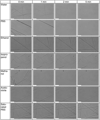 | ||
| Fig. 2 Microscopy images of the dissolving peptide nanotubes in distilled water, PBS (pH 7,4), ethanol, isopropanol, methanol, acetonitrile and a PBS (pH 7,4) solution saturated with peptide monomers. From these images, the nanotubes were found to dissolve in many of the solutions in which it has been claimed that they were stable. All scale bars in the images correspond to 20 μm. | ||
To confirm the results from the optical investigation, HPLC/MS experiments were conducted. If the results presented above were correct and the peptide nanotubes indeed dissolve when submerged in solution, the concentration of peptide monomer should increase over time from zero to a saturated concentration level in the liquid covering the dried nanotubes. Therefore, a way to confirm the results presented above would be to monitor the concentration of the peptide monomers as a function of time. Fig. 3 plots the amplitude of the HPLC peak corresponding to the peptide monomers dissolved in solution as a function of time. From this figure, it was clear that the amplitude of the peak and hence the concentration of the monomers did in fact increase over time and hence that the peptide nanotubes dissolves. By using MS, it was confirmed that the HPLC peak increasing in intensity over time corresponded to the FF monomers. The data from these experiments can be seen in the supplementary material online.
 | ||
| Fig. 3 An example of data from the HPLC experiments, performed to confirm the results presented in Fig. 2. Here the concentration of the FF monomers was monitored in an aqueous solution. The horizontal axis should not be seen as an expression of the dissolution time (but rather as the actual duration of the measurements). However, from the above figure, it is clear that the concentration of the FF peptide monomers increased as the peptides were dissolved in the fresh solution. | ||
In order for the peptide tubes to be successfully incorporated into a variety of sensor systems, it is essential that their stability in solution becomes increased so that they remain intact longer when submerged in the liquid. Thus, it was studied how a change in the formation conditions affected the stability of the formed structures. The results showed that the structures formed in an acidic environment demonstrated an increase in stability over time. However, it should be noted that the exact structural conformation of these is still not known. Fig. 4 presents a number of optical images of the dissolution process of peptide structures formed at pH 3 submerged in PBS. This dissolution process should be compared to standard tubes becoming dissolved in PBS, as presented in Fig. 2. As can be seen from these images, the tubes showed a tendency of being more stable when formed in a more acidic environment. When formed in the most acidic environment, the tubes were found to be stable in the solution for up to several hours. It should be noted that the number of tubes formed in a given solution was strongly dependent on the pH of that particular solution. From the performed experiments, one can see that when formed at a pH between 5 and 6 the density of tubes is highest. In more acidic solutions, much fewer peptide tubes were formed and the formation process was significantly slower. At pH 3, for instance, the peptide solution had to age for several days before the tubes were completely formed.
 | ||
| Fig. 4 Optical images of the dissolution process of peptide structures, formed at pH 3, submerged in PBS. It was clear that the tubes showed a tendency to be more stable when formed in a more acidic environment. All scale bars corresponds to 20 μm. | ||
4. Discussion
The results presented above have revealed new information with regard to the chemical stability of the peptide nanotubes. It was demonstrated that the peptide nanotubes could be dissolved in most solvents including water and PBS. Many of the applications in which nanotubes are planned to play a central role involve their submersion in a solvent. An example of such use could be BioFET or cargo vehicles designed for drug delivery.The dissolution effect observed in the optical microscope was confirmed by HPLC/MS experiments. Here, it was seen that the concentration of FF monomers increased over time in a fresh solution added to the dried peptide structures. The fact that the concentration of the monomers increased continually over time confirmed that the peptide structures became dissolved; otherwise, the concentration of monomers would be constant as a function of time. Through MS analysis, it was determined that it was the concentration of the FF monomers that increased over time.
The above mentioned results suggest that the formation of self-organizing peptide nanotubes was a question of solubility of the peptide monomers in the various solutions. When the concentration of the peptide monomers exceeded the solubility limit of peptide monomers, peptide nanotubes were formed. Naturally, the solubility of the peptide monomers varied with the type of solution. When a fresh solution was added to a dried sample of peptide nanotubes, the latter became dissolved due to the same effect. Initially, the concentration of peptide monomers in the fresh solution was zero, and as long as the concentration of monomers remained below the solubility of peptide monomers, the nanotubes continued to dissolve and eventually disappeared.
The previous section demonstrated that it was possible to drastically increase the stability of the peptide tubes by altering the formation process. This represents a means of obtaining a sufficient stabilization of the peptide tubes in order for them to be useful in a variety of applications. We have shown tubes that remained unaffected by the dissolution process for more than two hours in a PBS solution, whereas tubes formed in the standard approach disappeared after only a couple of minutes, as evidenced in the experiments. This was indeed very promising since a further optimization of the parameters involved in the formation process might result in even more stable tubes.
The time it took to form the peptide tubes varied significantly with pH, as was mentioned above. This might prove useful in future applications since the formation of peptide tubes in distilled water might lead to additional peptide tubes forming in the evaporation phase. This, in turn, could give rise to tubes at unwanted places after a completed manipulation. However, for the tubes formed in the acidic solution, the formation process would be so slow that no new tubes should form in the evaporation phase. Hence, when a tube in solution has been positioned at the place of interest, no formation of new tubes occurs when the solution is evaporated, thus rendering the manipulation of the peptide far more simple. It should be noted that the structural details of the structures formed in the acidic environment is still unknown. Therefore whether they are in fact nanotubes or rather nanowires is a subject for further investigation.
In this work, only the pH of the solution was varied, however many other parameters in the formation process may influence the properties of the tubes. When the peptide monomers are dissolved in distilled water, as described in the standard process for tube formation,6 the pH of the solution actually dropped to around pH 5. It was demonstrated that the tubes formed at this pH were the most unstable (even though the amount of formed tubes was much larger), which indicates that the standard production approach results in the most unstable structures. Therefore, it is strongly suggested that the parameters of the formation process be investigated further and in particular that a new standard fabrication process be developed.
The results presented by Adler-Abramovich et al.13 can also be explained by this description of nanotube formation. These authors mainly studied peptides after solvent evaporation. In this case, the peptide nanotubes will form in the evaporation phase even though they are not present in the solution. This is due to the concentration of the monomers increasing as the solvent evaporates. Our experiments also confirmed that nanotubes reformed in the evaporation phase.
The results presented in this article may explain some of the difficulties with these peptide nanotubes according to prior publications. In the work by Ryu et al.,18 the authors stated that the stability of the peptide nanotubes could not be investigated as a result of the nanotubes detaching from the surface they were placed on when emerged into the solvent. These observations can be explained by the fact that the nanotubes simply dissolved in the different solutions in which they were submerged. The fact that the peptide nanotubes dissolved in nearly all solutions can actually also be used in a wide variety of new and innovative applications. For instance, water could be used instead of the dry etching machines, as suggested previously,2 in the process of achieving electrical contact between the contact pads and the silver wires reduced inside the peptide nanotube. Furthermore, the nanotubes could be employed as a mold in the fabrication of a nanofluidic channel connecting two reservoirs.
5. Conclusion
This article has presented the stability of diphenylalanine peptide nanotubes in solution, which differed significantly from previously published results regarding the stability under dried conditions. In particular, images of the peptide nanotubes dissolving in distilled water, PBS, ethanol, methanol, isopropanol and acetonitrile were shown.It was argued that the dissolution of these peptides could be due to the final solubility of the peptide monomers in the respective solutions. In particular, the nanotubes were found to be stable in a saturated solution, which confirms this hypothesis.
In many potential applications, the peptide nanotubes will be exposed to a solvent. The present investigation is thus of great importance since it proves that the stability of the peptide nanotubes in solution differs from that of the same structure under dry conditions. From this, it is clear that an emphasis should be put on the stabilization of the peptide nanotubes in order to expand the field of possible applications for this material.
The present article has also shown that the stability of the peptide tubes could be increased by simply forming them under, for instance, varying pH conditions of the solution. In particular, it was demonstrated that tubes formed at low pH were more stable than their counterparts formed in the standard fabrication approach used previously. Such a dramatic increase in peptide stability is indeed a promising result. However, room for optimization remains with regard to the formation parameters, which may result in even longer stabilization.
Acknowledgements
The authors would like to thank the European BeNatural project (BeNatural/NMP4-CT-2006-033256) and the Danish Agency for Science Technology and Innovation (FTP 271-08-0968) for financial support and Dr Osvaldo Delgado for the assistance during the HPLC/MS experiments.References
- A. L. Briseno, S. C. B. Mannsfeld, S. A. Jenekhe, Z. Bao and Y. Xia, Mater. Today, 2008, 11, 38–47 CrossRef CAS.
- N. B. Sopher, Z. R. Abrams, M. Reches, E. Gazit and Y. Hanein, J. Micromech. Microeng., 2007, 17, 2360–2365 CrossRef CAS.
- M. Yemini, M. Reches, E. Gazit and J. Rishpon, Anal. Chem., 2005, 77, 5155–5159 CrossRef CAS.
- E. C. Cho, J.-W. Choi, M. Lee and K.-K. Koo, Colloids Surf., A, 2008, 313–314, 95–99 CrossRef.
- M. Reches and E. Gazit, Science, 2003, 300, 625–627 CrossRef CAS.
- O. Carny, D. E. Shalev and E. Gazit, Nano Lett., 2006, 6, 1594–1597 CrossRef CAS.
- N. Kol, L. Adler-Abramovich, D. Barlam, R. Z. Schneck, E. Gazit and I. Rousso, Nano Lett., 2005, 5, 1343–1346 CrossRef CAS.
- L. Niu, X. Chen, S. allen and S. J. B. Tendler, Langmuir, 2007, 23, 7443–7446 CrossRef CAS.
- C. H. Clausen, J. Jensen, J. Castillo, M. Dimaki and W. E. Svendsen, Nano Lett., 2008, 8, 4066–4069 CrossRef CAS.
- J. Castillo, S. Tanzi, M. Dimaki and W. Svendsen, Electrophoresis, 2008, 29, 5026–5032 CrossRef CAS.
- V. L. Sedman, L. Adler-Abramovich, S. Allen, E. Gazit and S. J. B. Tendler, J. Am. Chem. Soc., 2006, 128, 6903–6908 CrossRef CAS.
- V. L. Sedman, S. Allen, X. Chen, C. J. Roberts and S. J. B. Tendler, Langmuir, 2009, 25, 7256–7259 CrossRef CAS.
- L. Adler-Abramovich, M. Reches, V. L. Sedman, S. Allen, S. J. B. Tendler and E. Gazit, Langmuir, 2006, 22, 1313–1320 CrossRef CAS.
- M. Reches and E. Gazit, Nano Lett., 2004, 4, 581–585 CrossRef CAS.
- C. H. Görbitz, Chem.–Eur. J., 2001, 7, 5153–5159 CrossRef CAS.
- C. H. Görbitz, Chem. Commun., 2006, 2332–2334 RSC.
- C. H. Görbitz and F. Rise, J. Pept. Sci., 2008, 14, 210–216 CrossRef CAS.
- J. Ryu and C. B. Park, Biotechnol. Bioeng., 2010, 105, 221–230 CrossRef CAS.
Footnote |
| † Electronic supplementary information (ESI) available: Experimental mass spectroscopy data and overview of dissolution in solution in which the peptides dissolves slowly. See DOI: 10.1039/c0nr00734j |
| This journal is © The Royal Society of Chemistry 2011 |