Research highlights
Šeila
Selimović
ab and
Ali
Khademhosseini
*abcd
aCenter for Biomedical Engineering, Department of Medicine, Brigham and Women's Hospital, Harvard Medical School, Cambridge, Massachusetts 02139, U. S. A. E-mail: alik@rics.bwh.harvard.edu
bHarvard-MIT Division of Health Sciences and Technology, Massachusetts Institute of Technology, Cambridge, Massachusetts 02139, U. S. A.
cWyss Institute for Biologically Inspired Engineering, Harvard University, Boston, Massachusetts 02115, U. S. A.
dWorld Premier International – Advanced Institute for Materials Research (WPI-AIMR), Tohoku University, Sendai 980-8577, Japan
First published on 30th September 2011
High-throughput nanoparticle measurement
Nanoscale objects, ranging from viruses to synthetic molecules, are widely used in industries ranging from drug development to cosmetics.1 Despite the widespread utility for these particles, however, there are relatively few methods available that allow for high-throughput sensing and analysis of nanoparticles without the addition of labeling dyes. The integration of microfluidic technology and electrical components may provide a potential solution to this long-standing problem.Cleland and colleagues have taken a step to address this issue by developing a microscale electrofluidic platform for analyzing nanoparticles.2 In particular, they introduced a device based on fluid flow in a channel of varying resistances. The device was fabricated from poly(dimethylsiloxane) using standard molding techniques, and the electric components were patterned using optical lithography. The nanoparticles suspended in an aqueous solution were propelled into the microfluidic channel by an external pressure source and flowed from a high voltage to a low voltage electrode, with potentials in the order of ±1–3 V. As the particles passed through a nanofilter or fluidic resistor and reached a high-resistance channel termed “nanoconstriction”, the ionic electrical current generated by the electrodes in the solution was modified. As a consequence, the potential difference between the solution and a directly connected sensing electrode also changed and was recorded. Small nanoparticles caused a smaller potential difference than larger ones. Effectively, the device was designed to function as a voltage divider in a fluidic environment.
The device allowed for a high detection rate, more than 104 times higher than standard sensing methods such as dynamic light scattering (DLS), and was capable of measuring particles as small as 50 nm. Pressure was determined to be the driving force in particle transport, rather than electroosmotic and kinetic mechanisms. By controlling the fluid pressure, the particles’ transit time was minimized to 2–3 μs per particle, enabling high-throughput analysis of the suspension. In the case of 117 nm polystyrene particles, the pressure required for transport across the nanoconstriction was between 0.1 and 2 psi. At higher pressures and thus shorter transit times the signal-to-noise ratio remained high, down to a minimum detectable transit time of 750 ns. This was equivalent to measuring 500![[thin space (1/6-em)]](https://www.rsc.org/images/entities/char_2009.gif) 000 nanoparticles per second.
000 nanoparticles per second.
The device was also capable of detecting heterogeneous nanoparticle populations, e.g. with average particle sizes of 51 nm, 79 nm, and 117 nm. The 117 nm particle signal from the previous experiments was used for calibration. DLS measurements of the same sample, however, were almost indistinguishable from those of a homogenous 117 nm population. Additionally, the present method was also shown to offer concentration measurements of the various sample constituents. For example, in an detection test of a bacteriophage suspension mixed with large nanoparticles, there were few recorded voltage peaks associated with the 117 nm particles, but many more signaling the transit of the bacteriophages (∼55 nm). As expected, the measured bacteriophage size also depended on the ionic strength of the solution. Namely, when the phage was suspended in a 1 M NaCl solution, the average size was determined to be 10 nm larger. This was likely due to the difference in capsid size in the different media. Interestingly, in both cases the presence of phage dimers was also observed, as 81 nm particles.
The high-throughput and high sensitivity of this simple device make it particularly useful for the detection of rare particles in liquids as different as blood and waste water. In either case, the present mechanism requires much less time for particle sensing than standard methods. Potential areas of remaining concern for real-world application include the ability analyze other features of the nanoparticles such as charge, biospecificity, composition and shape. In addition, it is useful to detect larger objects in field samples as well as fabricate robust, and ideally portable, units that can be used for desired applications.
Microfluidic barcoding
Quantum dots are semiconductor particles whose size and shape can be used to control the particles' electronic properties. This characteristic is exploited in quantum dot applications such as labelling of molecules. Given the diverse range of excitation and emission wavelenghts of quantum dots, as well as their photostability, they are advantageous as a tool for studies of large numbers of samples.3 To enable such studies, Weitz and colleagues have now developed a method for generating droplets and particles labeled with quantum dots in a capillary system.In their paper, Zhao et al.4 describe a simple glass capillary setup for formulation of oil/water/oil emulsions. These double emulsions were formed by co-flowing a lipid inner liquid containing trimethylolpropane triacrylate (ETPTA), quantum dots and colloidal silica nanoparticles together with an aqueous middle liquid of poly(ethylene glycol) diacrylate (PEG-DA). When the two phases encountered an outer lipid phase—hexadecane—they were sheared off into oily droplets encased by an aqueous shell. This shell prevented leakage of the quantum dots into the outer oily phase. Both the middle and the outer phases contained surfactants for droplet stabilization. The droplet size was controlled by the applied flow rates and was in the order of 10 to 100 μm. In all droplets, the distribution of coding particles was uniform as shown by the similar fluorescence intensities. After emulsification, the droplets were crosslinked via light to yield solid particles with a protective shell. Neither the shell nor the particle core suffered damage during subsequent washing, drying and resuspension steps, even when the suspending fluid was aqueous. Additionally, the shell was permeable to water and certain biomolecules from the suspension, but the quantum dots could not diffuse out of the core, as they were strongly bound inside it.
The main advantage of using quantum dots in this manner for barcoding is in their optical properties. Quantum dots of different sizes emit light at different wavelengths, as shown by different photoluminescence spectra, even when excited by the same wavelength. The authors exploited this property to form solid particles with distinct cores. In one case, two cores were generated, one of which was filled with quantum dots, and the other with magnetic nanoparticles (Fig. 1). By turning the flow on and off selectively in the inner and middle liquid phases, particle cores with two distinct halves could also be formed, often referred to as Janus particles.
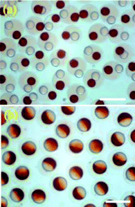 | ||
| Fig. 1 Solid spheres with two cores: one was labelled with quantum dots (white cores), the other with magnetic nanoparticles (red) (a). Barcoded Janus particles surrounded by a transparent shell (b). Both types of particles were formed in a double emulsion capillary system. Scale bars are 200 μm. Figure adapted and reprinted with permission from Yuanjin Zhao et al., Microfluidic Generation of Multifunctional Quantum Dot Barcode Particles, J. Am. Chem. Soc., 2011, 133(23), 8790–8793. Copyright 2011 American Chemical Society. | ||
The ability to produce particles with functionalized or multiple cores has appeal for multiplexing and directed flow applications. Here, quantum dots of different sizes could be used for barcoding particles, and the presence of magnetic nanoparticles in a distinct region of the spheres could be utilized for sorting. Both of these approaches would be useful in bioassays or drug screening studies.
Thiol-ene soft lithography
Soft lithography is a popular and wide-spread method of fabricating microfluidic devices. Benefits of soft lithography include the ability to prepare many replicates from a single master, use of affordable polymers such as poly(dimethylsiloxane) and high feature fidelity.5 However, a key limitation of soft lithography is the difficulty in generating high-aspect ratio (height:width) features when most common resins are used. In addition, acrylic resins have a relatively high polymerization shrinkage rate, which often results in stress fractures and misshapen features, which reduce the quality of the device. Bowman and colleagues have recently introduced a thiol-ene based light-sensitive resin, which promises to overcome these limitations.
Thiol-ene chemistry involves the addition of a sulfur atom via a double or triple bond. As part of this process radicals are formed and transferred, requiring smaller initiator amounts. Delayed gelation is another consequence of the thiol-ene reaction. Here, the resin remains liquid until a very high degree of polymerization has been reached, reducing polymerization-related fracture stresses, resulting in cleaner features.
Ashley et al.6 exploited these characteristics of thiol-ene reactions to fabricate high-aspect ratio features (Fig. 2) using the soft lithographic method of CLiPP or contact liquid photolithographic polymerization. The thiol-ene monomers were poured onto a siloxane-treated glass substrate and exposed to 365 nm wavelength light under a transparency mask. After the photopolymerization the un-crosslinked thiol-ene was washed away, and the features remained attached to the glass.
 | ||
| Fig. 2 High-aspect features fabricated via thiol-ene soft lithography: raised cylinders (a) and recessed columns (b), both with a 4.2 aspect ratio. Figure reprinted with permission from the Royal Society of Chemistry from Ashley et al.6 | ||
The optimal feature quality at an aspect ratio of 8 was found when the inhibitor:initiator ratio was 1:1 or 1.5:1, with a curing time of 15 s. This was observed for both recessed and raised structures, although the latter had a slightly higher quality coefficient. Exposure times longer than 15 s affected the feature shapes adversely, as did initiator concentrations above 0.05 wt%. In both these cases the curing process extended beyond the regions exposed to the UV source through the transparency mask, and more material than desired was crosslinked.
Once the optimal curing conditions for the thiol-ene were determined, its fabrication properties were compared to an acrylate resin (urethane diacrylate and TEGDA mixed with Irgacure 184 and acrylic acid). For aspect ratios up to 10 the thiol-ene resin outperformed the acrylic compound. The curing time was one to two orders of magnitude shorter (few to 50 s compared to several minutes) and the feature fidelity was much improved. In contrast, the quality of the acrylic features deteriorated above an aspect ratio of 3.
The ability to fabricate high-aspect ratio structures in microfluidic devices opens up a range of design innovations, e.g. in filtering or cantilever applications. Beyond that, the use of thiol-ene could be advantageous in microscale studies of biological materials, such as cells. Since the optimized manufacturing process requires minimal amounts of cytotoxic photoinitiator, thiol-ene could potentially be utilized in bioengineering applications.
References
- R. J. Aitken,
et al., Manufacture and use of nanomaterials: current status in the UK and global trends, Occup. Med., 2006, 56(5), 300–306 CrossRef CAS
.
- J.-L. Fraikin,
et al., A high-throughput label-free nanoparticle analyser, Nat. Nanotechnol., 2011, 6(5), 308–313 CrossRef CAS
- W. C. W. Chan,
et al., Luminescent quantum dots for multiplexed biological detection and imaging, Curr. Opin. Biotechnol., 2002, 13(1), 40–46 CrossRef CAS
- Y. Zhao,
et al., Microfluidic Generation of Multifunctional Quantum Dot Barcode Particles, J. Am. Chem. Soc., 2011, 133(23), 8790–8793 CrossRef CAS
- Y. Xia and G. M. Whitesides, Soft Lithography, Annu. Rev. Mater. Sci., 1998, 28, 153–184 CrossRef CAS
- J. F. Ashley,
et al., Soft-lithography fabrication of microfluidic features using thiol-ene formulations, Lab Chip, 2011, 11, 2772–2778 RSC
| This journal is © The Royal Society of Chemistry 2011 |