Efficacy of surface sampling methods for different types of beryllium compounds
A.
Dufresne
*a,
T.
Mocanu
b,
S.
Viau
d,
G.
Perrault
c and
C.
Dion
d
aDepartment of Environmental and Occupational Health, Faculty of Medicine, Université de Montréal, Pavillon Marguerite-d'Youville, 2375 Chemin de la Côte-Ste-Catherine, Montréal, Québec H3T 1A8, Canada
bMcGill University Environmental Health and Safety, 3610 McTavish, Room 426, Montréal, Québec H3A 1Y2, Canada
cConsultation en R&D et Expertise en SST, 3285 de Bercy, Laval, Québec H7E 1V7, Canada
dInstitut de recherche Robert-Sauvé en santé et en sécurité du travail, 505 ouest de Maisonneuve, Montréal, Québec H3A 3C2, Canada
First published on 28th October 2010
Abstract
The objective of the research work was to evaluate the efficiency of three different sampling methods (Ghost Wipe™, micro-vacuum, and ChemTest®) in the recovery of Be dust by assessing: (1) four Be compounds (beryllium acetate, beryllium chloride, beryllium oxide and beryllium aluminium), (2) three different surfaces (polystyrene, glass and aluminium) and (3) inter-operator variation. The three sampling methods were also tested on site in a laboratory of a dental school for validation purposes. The Ghost Wipe™ method showed recovery ranging from 43.3% to 85.8% for all four Be compounds and for all three quantities of Be spiked on Petri dishes, while recovery with the micro-vacuum method ranged from 0.1% to 12.4%. On polystyrene dishes with 0.4 µg Be, the recovery ranged from 48.3% to 81.7%, with an average recovery of 59.4% for Operator 1 and 68.4% for Operator 2. The ChemTest® wipe method with beryllium acetate, beryllium chloride, and AlBeMet® showed analogous results that are in line with the manufacturer's manual, but collection of beryllium oxide was negative. In the dental laboratory, Ghost Wipe™ samplings showed better recovery than the micro-vacuum method. The ratios between the recovered quantities of Be in each location where the Ghost Wipe™ was tested differed substantially, ranging from 1.45 to 64. In the dental laboratory, a faint blue color indicating the presence of Be was observed on the ChemTest® wipes used in two locations out of six. In summary, the Ghost Wipe™ method was more efficient than micro-vacuuming in collecting the Be dust from smooth, non-porous surfaces such as Petri dishes by a factor of approximately 18. The results obtained on site in a dental laboratory also showed better recovery with Ghost Wipes™. However, the ratio of Be recovered by Ghost Wipes™ versus micro-vacuuming was much lower for surfaces where a large amount of dust was present. Wet wiping is preferred over micro-vacuuming for beryllium forms, but this conclusion probably applies to the ultra-low particulate loading levels (0.4 micrograms or less) which was tested in this study.
Environmental impactWhen occupational site contamination by beryllium is present multiple exposure pathways are likely. Containment followed by cleaning is the logic steps to prevent workers' exposure and environmental contamination pollution. As such, a proper sampling and analysis method is prerequisite to ascertain the presence of the contaminant. In addition, thorough evaluation of airborne or surface contamination will help in choosing the appropriate cleaning method to prevent its migration in the outside environment and protect the individuals. This general procedure may be extrapolated to investigate other toxic pollutants that may become airborne and settle on surfaces. |
Introduction
The manner in which the contaminant adheres to a surface can vary from surface to surface and is dependent on the adhesive forces that retain the particles. Characteristics of the surface such as porosity, permeability, smoothness or roughness are determinants that will affect the sampling of particles. If particles are deposited in surface cracks, they might not be removed by wiping. Nevertheless, even the smoothest, most highly polished surface contains microscopic fissures from which it is difficult to dislodge all of the contamination.1Accurate sampling of particles on surfaces depends on the selection of appropriate wetting agents for the types of residues and surfaces. Several types of sampling methods have been employed in surface contamination studies for collecting dust from different surfaces such as in passive sampling, vacuum sampling and wipe sampling.2 The most common surface sampling method used for chemicals is wipe sampling and the most common technique is hand pressure to move, filter, or wipe in a “Z” or “S” motion.1 Researchers have generally focused on the physical and chemical properties of dust samples collected by these methods. Decades ago, Royster and Fish et al.3 found that a number of factors can influence the efficiency and variability of manual wipe sampling and they are: (1) concentration and particle size distribution of the surface contaminant, (2) surface roughness and porosity, (3) area of surface sampled and wiping pressure applied, (4) the operator (surveyor) effect, and (5) others.
Some surfaces are more difficult to evaluate by surface wipe sampling, and thus the interest in using the vacuum sampling technique. This technique was mainly studied during its use for residential samples, especially on carpets in lead-contaminated homes.1,4–7 Creek et al. discussed the possibility of using the “micro-vacuum” technique for surface sampling of Be particles and concluded that the method, as described in ASTM D7144, should be improved in order to obtain a better recovery efficiency.8 The vacuum method has become an important aspect of surface sampling, particularly for collecting residential samples.1 In a review focused on beryllium sampling evaluation, various standardized techniques relevant for surface contamination were identified. “Wet” or “dry” filters/wipes were mostly used for smooth surfaces, while the micro-vacuum method was required for carpets or rough, porous, uneven or fragile surfaces.9
Beryllium may be an integral part of the surface such as in Be–Cu alloy plates, which contain 2% Be or less, or powdered Be material adhering to the surface by means of weak electrostatic forces or gravity. It has been shown in recent articles that the transfer of beryllium compounds from a cleaned device made of Be–Cu is possible at any time due to the continuous oxidation that occurs on a freshly cleaned surface.10 On the other hand, loose particles of beryllium compounds that had adhered to different types of structures such as unpainted metal, wood frames, painted metal, concrete, painted concrete and Plexiglas™ on which decontamination was attempted were reduced to below the clearance reference value, except on concrete floors.11,12
If the objective of surface sampling is to detect the presence of beryllium or to measure whether the beryllium load is above 0.2 µg per 100 cm2 (the US Department of Energy reference, 10 CFR 850,13 for release of beryllium-contaminated equipment to a non-beryllium area), colorimetry may be an inexpensive alternative direct-reading method to detect the level of beryllium in relation to the regulatory limit.14 However, the efficacy of this method in comparison to wiping and suction methods is not known.
The objective of the present research work is to evaluate the efficiency of three different sampling methods (Ghost Wipes™, micro-vacuuming, and the ChemTest®) in the recovery of Be dust by assessing: (1) four Be compounds (beryllium acetate, beryllium chloride, beryllium oxide and beryllium aluminium), (2) three different surfaces of Petri dishes (polystyrene, glass and aluminium), and (3) inter-operator variation. The three sampling methods were also tested on site, in a dental technique teaching college, for validation purposes.
Material and methods
Tested surfaces
“Fisherbrand” sterile polystyrene Petri dishes (surface of approximately 62 cm2), “Fisherbrand” aluminium dishes (surface of approximately 42 cm2) and “Pyrex” glass dishes (surface of approximately 68 cm2) from Fisher (Montreal, Qc) were used as the manipulation surfaces on which the various Be solutions or suspensions were spiked.Tested beryllium compounds
Four different Be compounds were chosen for manipulation. In order to obtain the required mass of Be that was spiked on the Petri dishes, the solutions/suspensions were prepared as explained below.Nitric acid 1% (HNO3) (Fisher, Montreal) was used to dilute the solution of soluble beryllium acetate and to prepare the solution of soluble beryllium chloride. The suspensions of insoluble compounds (beryllium oxide and AlBeMet®) were prepared using isopropanol (Fisher, Montreal), which was chosen based on NIOSH method 750015 which requires the use of this solvent for the preparation of suspensions of silica, an insoluble material.
Beryllium acetate (Be4O(O2CCH3)6)
A 10![[thin space (1/6-em)]](https://www.rsc.org/images/entities/char_2009.gif) 000 mg L−1 solution of beryllium acetate (SPEX CertiPrep®Group) (Metuchen, NJ) was repeatedly diluted with nitric acid 1% (HNO3). The final solution, with a concentration of 100 µg L−1, contained 10 µg (0.01 mg) Be. Proper volumes of this solution were spiked on the Petri dishes to obtain approximately 0.1 µg, 0.2 µg and 0.4 µg Be.
000 mg L−1 solution of beryllium acetate (SPEX CertiPrep®Group) (Metuchen, NJ) was repeatedly diluted with nitric acid 1% (HNO3). The final solution, with a concentration of 100 µg L−1, contained 10 µg (0.01 mg) Be. Proper volumes of this solution were spiked on the Petri dishes to obtain approximately 0.1 µg, 0.2 µg and 0.4 µg Be.
Beryllium chloride (BeCl2)
In preparing the beryllium chloride solution, a beryllium chloride to Be conversion factor of 0.11 was taken into consideration, based on the ratio between the atomic weight of Be (9 g mol−1) and the molar mass of beryllium chloride (79.9 g mol−1). Beryllium chloride powder (Fluka, Buchs, Switzerland) was diluted in water. This solution was spiked on the Petri dishes in order to obtain approximately 0.1 µg, 0.2 µg and 0.4 µg Be.Beryllium oxide (BeO)
The conversion factor for beryllium oxide to Be is 0.36, based on the ratio between the atomic weight of Be (9 g mol−1) and the molar mass of beryllium oxide (25 g mol−1). Beryllium oxide powder (Fluka, Buchs, Switzerland) was mixed with isopropanol to obtain a suspension that was spiked on the Petri dishes to obtain approximately 0.1 µg, 0.2 µg and 0.4 µg Be.Beryllium aluminium alloy (AlBeMet®)
Beryllium aluminium alloy (AlBeMet®) has a mass percentage of 62% Be and 38% aluminium according to the manufacturer. The powder (Brush Wellman, Cleveland, OH) was mixed with isopropanol to obtain a suspension that was spiked on the Petri dishes to obtain approximately 0.1 µg, 0.2 µg and 0.4 µg Be.Sampling methods
The three sampling methods used in the present study were the Ghost Wipe™, micro-vacuuming with a sampling pump and a cassette, and the beryllium ChemTest® wipe.Ghost Wipe™
The Ghost Wipe™ is a sampling product manufactured by Environmental Express (Mt. Pleasant, South Carolina) that is made of a resistant material used to collect metal dusts from various surfaces. The surface area of the wipe is 225 cm2 (15 cm × 15 cm) and is previously humidified with deionized water. After manipulation, a sampling cup is used to store the wipes until analysis. Because of their strength, these wipes are the ones used most by industrial hygienists.16The wipe sampling was carried out in accordance with ASTM D6966.17 Initially, wiping was done in a horizontal direction (following an “S” pattern) and the wipe was folded so that the collected dust remained inside the wipe. Subsequently, the same maneuver was applied vertically and the wipe was folded once more. At the end, wiping was done on the inner perimeter of the Petri dishes, and the wipe was folded and stored in a container and sent for analysis.
Micro-vacuum sampling equipment
The metal dust was collected in a 37 mm leak-free filter cassette manufactured by SKC Inc. (Concept Controls Inc., Montreal). At the inlet, a short (5.5 cm) micro-vacuum tubing cut at 45° was connected to the filter cassette; at the outlet, the filter cassette was connected by a longer vacuum tubing to the pump used to suck in the dust. The pump flow of 2.5 L min−1 required by the American Society for Testing and Materials (ASTM) D7144 standard18 was increased to 16 L min−1 based on the United States Department of Housing and Urban Development (US HUD)19 as used by Farfel et al.6 in a study that compared the efficiency of wipes and micro-vacuum sampling methods.The end of the short vacuum tubing cut at 45° was passed over the surface of the Petri dish previously spiked with Be compounds. Analogous to the previous sampling method, the sampling was carried out using an “S” pattern, first in the horizontal and then in the vertical direction. Finally, the inner perimeter of the Petri dish was sampled.
Beryllium ChemTest® wipe
The Be ChemTest® kit is manufactured by Microteq, LLC (Virginia Beach, VA) for Nextteq, LLC (Tampa, FL).20 The ChemTest® wipe is a semi-quantitative method that is reported by the manufacturer to identify the presence of 0.2 µg Be or more when the wipe develops a blue color. The minimum detectable limit of 0.2 µg for the ChemTest® corresponds to the maximum value per 100 cm2, established by the Department of Energy (DOE) for working areas where workers no longer use Be or for equipment required to be removed from Be working areas.13 The ChemTest® kit consists of special wipes for collecting the dust from contaminated surfaces, three different developing solutions, and additional equipment for processing (wetting solution, developing plates, and sample bags). According to the manufacturer, this method uses the colorimetric technique to characterize whether or not Be is present in an amount above 0.2 µg on the surface analyzed. The different shades of blue color show increasing quantities of Be as shown in Fig. 1.20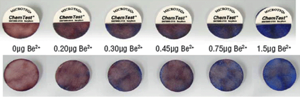 | ||
| Fig. 1 Manufacturer's results obtained with the ChemTest® wipe for different quantities of beryllium. Beryllium ChemTest® Kit—Validation Report.© Microteq, LLC (2005). | ||
The Petri dish surface spiked with Be solution was wiped with the ChemTest® wipe that was previously humidified with deionized water. Treatments with three different developing solutions were used to determine whether the amount of Be present was 0.2 µg or more by a change of the color of the wipe from pink to blue. The first solution contained 10% sulfuric acid in water, while the second solution contained ammonium buffer with a pH between 10 and 11. Interference with other metals (Al3+, Ba2+, Ca2+, Cd2+, Co2+, Cu2+, Cr3+, Cr6+, Fe2+, Fe3+, Hg2+, Pb2+, and Zn2+) could occur in the analysis process. The third developing solution, which contained the complexing reagent in water, was used to confirm the presence of Be.20
Sampling tests done in the laboratory
The four Be compounds were spiked using an electronic pipette (VMR, Montreal) on the Petri dishes' surfaces in three different quantities: 0.1, 0.2 and 0.4 µg Be. Three dishes were prepared for each compound and each Be quantity. Three different sampling methods were analyzed: Ghost Wipes™, micro-vacuuming and the ChemTest® wipe. In order to qualify the polystyrene Petri dishes, the experiments with the highest amount (0.4 µg Be) were applied to two other types of Petri dishes, i.e., glass and aluminium.In order to allow the Be compounds to reach a dust state, the manipulation always took place 72 hours after the preparation of the dishes. During this period, the Be solutions/suspensions dried out by evaporation in calm air at room temperature.
For each solution, a total of 27 polystyrene dishes were spiked with Be solutions/suspensions. For each sampling method, eight blank samples (two for each solution) were analyzed. Petri dishes that were not spiked with Be solutions/suspensions were placed in the hood along with the other dishes. These experiments were carried out by a single operator (Operator 1).
For the other two types of dishes, glass and aluminium, an amount of 0.4 µg Be was spiked. Six dishes were prepared for each of them and were manipulated by Operators 1 and 2 for individual inter-comparison. For these experiments, the Ghost Wipe™ method was used because it showed a higher efficiency of recovery when compared with the micro-vacuum technique. Table 1 and Table 2 show the factors selected and the number of experiments carried out in this study.
| Beryllium concentrations | Beryllium solutions | Sampling methods | |||
|---|---|---|---|---|---|
| Be acetate | Be chloride | Be oxide | ALBeMet® | ||
| a These experiments coincide with those marked in Table 2. | |||||
| 0.1 µg | 3 | 3 | 3 | 3 | ChemTest™ |
| 0.1 µg | 3 | 3 | 3 | 3 | Ghost Wipe® |
| 0.1 µg | 3 | 3 | 3 | 3 | Micro-vacuum |
| 0.2 µg | 3 | 3 | 3 | 3 | ChemTest™ |
| 0.2 µg | 3 | 3 | 3 | 3 | Ghost Wipe® |
| 0.2 µg | 3 | 3 | 3 | 3 | Micro-vacuum |
| 0.4 µg | 3 | 3 | 3 | 3 | ChemTest™ |
| 0.4 µg | 3 | 3 | 3 | 3 | Ghost Wipe®a |
| 0.4 µg | 3 | 3 | 3 | 3 | Micro-vacuum |
| 2 | 2 | 2 | 2 | ChemTest™ | |
| Blank | 2 | 2 | 2 | 2 | Ghost Wipe® |
| 2 | 2 | 2 | 2 | Micro-vacuum | |
| Type of Petri dishes | Beryllium solutions | Operators | |||
|---|---|---|---|---|---|
| Be acetate | Be chloride | Be oxide | AlBeMet® | ||
| a These experiments coincide with those marked in Table 1. | |||||
| Polystyrene | 3 | 3 | 3 | 3 | Operator 1a |
| Polystyrene | 3 | 3 | 3 | 3 | Operator 2 |
| Glass | 3 | 3 | 3 | 3 | Operator 1 |
| Glass | 3 | 3 | 3 | 3 | Operator 2 |
| Aluminium | 3 | 3 | 3 | 3 | Operator 1 |
| Aluminium | 3 | 3 | 3 | 3 | Operator 2 |
Sampling tests done in the dental laboratory
A sampling campaign was carried out in the dental laboratory of a collegial school where Be alloys were used. The samples were collected from a 100 cm2 surface using a standardized square template. Six locations with different surface conditions were chosen for the three sampling methods used in this study: (1) location 1, on fluorescent light 1 (smooth, painted metallic surface, and very dusty); location 2, on fluorescent light 2 (small porous, painted metallic surface, and slightly dusty); location 3, on the duct of the ventilation system (smooth, painted metallic surface, and very dusty); location 4, on a student's desk (smooth, painted surface, and not dusty); location 5, on the bottom edge of the window (windowsill) (old tinted, wooden surface, and not dusty); and location 6, on the ceiling, above the ventilation system (uneven, painted surface, and not dusty). The ChemTest® wipe could not be used at location 6 due to the uneven surface.Analytical methods
Ghost Wipe™ and filter cassette and content in tubing
The filter cassette used during micro-vacuum sampling and the wipes were analyzed by using inductively coupled plasma mass spectrometry (ICP-MS). The tubing connected to the filter cassette was not removed in order to allow recovery of the possible dust that remained on the inner side of the tubing. During storage, the end of the tubing was sealed using Parafilm M® laboratory film. The moistened wipes were stored in small containers.The samples were digested and then analyzed using an ICP-MS Perkin Elmer Model Elan 6000 DRC II (Perkin Elmer, Toronto) according to IRSST Method 359.21 The limit of detection for filter cassettes was 0.0005 µg and for wipes was 0.05 µg. Half of the limit of detection was reported whenever the result was below the limit of detection.22
In order to verify the accuracy of the amount of Be that was spiked onto Petri dish surfaces, 1 mL of each Be solution/suspension was put into three different beakers and was directly analyzed using the same ICP-MS method. The average quantity obtained from these three assays was considered as the real amount of Be that was spiked onto the Petri dish surfaces. For the 2 mL and 4 mL spiked on the Petri dishes, the concentration of Be was inferred by multiplying by 2 and 4 the true concentration obtained with 1 mL. The estimated amount of 0.1 µg Be was confirmed for beryllium acetate and beryllium chloride, while for beryllium oxide, 0.09 µg Be was found from direct analysis. The recovery amount for AlBeMet® was 0.03 µg Be. Due to the low result obtained with AlBeMet®, a new suspension was prepared under the same conditions, and a quantity of 0.041 µg was obtained. The first suspension was analyzed again after several weeks and a quantity of 0.037 µg was then found. The results, while reproducible, show the difficulty in obtaining a suspension of specific concentration for this compound. Therefore, the calculations for AlBeMet® were done using the amount of 0.1 µg Be in 1 mL of suspension.
ChemTest® wipe
The ChemTest® wipes gave the final result immediately after the treatment with the third developing solution. A blue color on the wipe meant the presence of at least 0.2 µg Be.20Data treatment
Since the tests carried out in this study were done to evaluate the efficiency of different sampling methods in the recovery of pre-determined amounts of beryllium spiked on Petri dishes, the laboratory results are reported as mass of beryllium and are not assessed in relation to the area of the Petri dish surfaces. On site, the samples were collected on 100 cm2 surfaces and the results are reported as mass of beryllium recovered per 100 cm2.Statistical analysis
Means, standard deviations, coefficient of variation, and the difference between the results that were obtained by the two operators and analyzed with a paired t-test procedure were computed by using Minitab™ 15 statistical software.Results
Surfaces tested with the Ghost Wipe™ and micro-vacuum sampling techniques
Table 3 shows the average percentage recovery obtained for the four different Be compounds and the three different target amounts of Be spiked on Petri dishes after applying Ghost Wipe™ and micro-vacuum sampling methods. These experiments were carried out by a single person (Operator 1) using only polystyrene Petri dishes. The Ghost Wipe™ method showed better recovery (ranging from 43% to 86%) for all four Be compounds and for all three quantities of Be spiked on dishes compared to the micro-vacuum method (recovery ranging from 0.1% to 12%). The coefficient of variation obtained with the Ghost Wipe™ method varied from 4% to 46%, while the micro-vacuum method had a coefficient of variation ranging from 19% to 126%.| Micro-vacuum | Ghost Wipe™ | |||||||||||
|---|---|---|---|---|---|---|---|---|---|---|---|---|
| 0.1 µg Be | 0.2 µg Be | 0.4 µg Be | 0.1 µg Be | 0.2 µg Be | 0.4 µg Be | |||||||
| Recovery (SD) | COV (%) | Recovery (SD) | COV (%) | Recovery (SD) | COV (%) | Recovery (SD) | COV (%) | Recovery (SD) | COV (%) | Recovery (SD) | COV (%) | |
| a SD = Standard Deviation and COV = Coefficient of Variation. The italicized columns are reported in Table 4. | ||||||||||||
| Be acetate | 1.9 (2.4) | 126 | 4.5 (3.5) | 79 | 0.1 (0.06) | 48 | 73 (4.5) | 6.2 | 86 (6.5) | 7.6 | 73 (9.1) | 12 |
| Be chloride | 2.1 (1.1) | 54 | 3.0 (2.5) | 83 | 3.0 (1.9) | 65 | 73 (5.8) | 7.9 | 57 (2.9) | 5.1 | 48 (3.8) | 7.9 |
| Be oxide | 2.5 (0.8) | 31 | 1.5 (0.6) | 37 | 1.8 (0.3) | 19 | 82 (6.4) | 7.9 | 63 (12) | 18 | 67 (2.8) | 4.2 |
| AlBeMet® | 4.8 (3.7) | 76 | 3.9 (2.8) | 72 | 12 (16) | 125 | 52 (24) | 46 | 43 (10) | 24 | 49 (8.0) | 16 |
Table 4 shows the results obtained for the same Be compounds spiked on the different Petri dishes (polystyrene, glass and aluminium), while the experiments were carried out by two persons (Operator 1 and Operator 2). On polystyrene dishes, the recovery ranged from 48% to 82%, with an average recovery of 59% for Operator 1 and 68% for Operator 2. On glass dishes, the recovery ranged from 14% to 77%, with an average recovery of 70% for Operator 1 and 56% for Operator 2. On aluminium dishes, the recovery ranged from 45% to 97%, with an average recovery of 79% for Operator 1 and 74% for Operator 2. The recovery ratio between the two operators varied from 0.6 to 4.2.
| Polystyrene dish | Glass dish | Aluminium dish | |||||||||||||
|---|---|---|---|---|---|---|---|---|---|---|---|---|---|---|---|
| Operator 1 | Operator 2 | Recovery ratio (Op1/Op2) | Operator 1 | Operator 2 | Recovery ratio (Op1/Op2) | Operator 1 | Operator 2 | Recovery ratio (Op1/Op2) | |||||||
| Recovery (SD) | COV (%) | Recovery (SD) | COV (%) | Recovery (SD) | COV (%) | Recovery (SD) | COV (%) | Recovery (SD) | COV (%) | Recovery (SD) | COV (%) | ||||
| a SD = Standard Deviation and COV = Coefficient of Variation. | |||||||||||||||
| Be acetate | 73 (9.1) | 12 | 82 (7.2) | 8.8 | 0.9 | 74 (3.8) | 5.1 | 75 (11) | 15 | 1.0 | 96 (1.4) | 1.5 | 96 (1.4) | 1.5 | 1.0 |
| Be chloride | 48 (3.8) | 7.9 | 78 (5.8) | 7.4 | 0.6 | 60 (16) | 26 | 14 (14) | 97 | 4.2 | 97 (1.4) | 1.5 | 97 (1.4) | 1.5 | 1.0 |
| Be oxide | 67 (2.8) | 4.2 | 62 (4.3) | 6.8 | 1.1 | 71 (1.6) | 2.2 | 77 (3.2) | 4.2 | 0.9 | 80 (5.8) | 7.3 | 56 (7.3) | 13 | 1.4 |
| Be AlMet | 49 (8.0) | 16 | 52 (17) | 32 | 1.0 | 74 (22) | 29 | 59 (17) | 28 | 1.3 | 45 (2.5) | 5.6 | 46 (8.8) | 19 | 1.0 |
| Average recovery (%) | 59 | 68 | 70 | 56 | 79 | 74 | |||||||||
An obvious difference was observed between the two operators for beryllium chloride solution spiked on polystyrene (Operator 1) and glass dishes (Operator 2). The four tests that showed differences between the two operators (recovery ratios of 0.6, 4.2, 1.3 and 1.4 in Table 4) were analyzed by a paired t-test. Beryllium oxide on aluminium dishes and AlBeMet® on glass dishes showed no statistically significant difference (beryllium oxide on aluminium dishes: 95% CI, p-value = 0.083 and AlBeMet® on glass dishes: 95% CI, p-value = 0.414). However, a statistically significant difference between the operators' results was observed for beryllium chloride spiked on polystyrene dishes (95% CI, p-value = 0.002) and on glass dishes (95% CI, p-value = 0.021). These tests were repeated, after training of the operators, and this second set of results is shown in Table 5. Notice that Operator 1 only repeated the test on polystyrene dishes and Operator 2 only repeated the test on glass dishes. Collection was improved in the repeat of both tests.
| Polystyrene dish | Glass dish | |||||||
|---|---|---|---|---|---|---|---|---|
| Operator 1 | Operator 2 | Operator 1 | Operator 2 | |||||
| Recovery (%) (SD) | COV (%) | Recovery (%) (SD) | COV (%) | Recovery (%) (SD) | COV (%) | Recovery (%) (SD) | COV (%) | |
| a SD = Standard Deviation and COV = Coefficient of Variation. | ||||||||
| First set | 48 (3.8) | 7.9 | 78 (5.8) | 7.4 | 60 (16) | 26 | 14 (14) | 97 |
| Second set | 68 (4.3) | 6.4 | 84 (5.8) | 6.9 | ||||
Surfaces tested with ChemTest®
The results obtained with the ChemTest® colorimetric method are not quantifiable but can be visually analyzed. This method was applied to four different Be compounds and three different target amounts of Be spiked on polystyrene Petri dishes. The outcome of these experiments is shown in Fig. 2. Beryllium acetate, beryllium chloride, and AlBeMet® showed analogous results that are in line with the manufacturer's manual, but beryllium oxide was negative. The results are summarized as follows: (1) blue coloration for dishes spiked with 0.2 and 0.4 µg Be, with a darker shade for 0.4 µg; (2) little or no coloration for dishes spiked with 0.1 µg Be, (3) lack of coloration for dishes not spiked with Be compounds (blank samples), and (4) beryllium oxide did not respond to the ChemTest® wipe under the conditions recommended by the manufacturer. | ||
| Fig. 2 ChemTest® wipe coloration from three different quantities of four beryllium compounds spiked on polystyrene Petri dishes. | ||
Surfaces tested in the dental laboratory of a school building
| Sampling method | Location 1a | Location 2b | Location 3c | Location 4d | Location 5e | Location 6f |
|---|---|---|---|---|---|---|
| a Location 1: on a fluorescent light (smooth, painted metallic surface, and very dusty). b Location 2: on a fluorescent light (small porous, painted metallic surface, and slightly dusty). c Location 3: on the duct of the ventilation system (smooth, painted metallic surface, and very dusty). d Location 4: on a student's desk (smooth, painted surface, and not dusty). e Location 5: on the bottom edge of the window (windowsill) (old tinted, wooden surface, and not dusty). f Location 6: on the ceiling, above the ventilation system (uneven, painted surface, and not dusty). | ||||||
| Quantity of beryllium recovered (µg per 100 cm2) | ||||||
| Ghost Wipe™ | 0.32 | 26 | 44 | 0.03 | 0.16 | 0.06 |
| Micro-vacuum | 0.23 | 6.7 | 38 | 0.001 | 0.003 | 0.004 |
| Ratio Ghost Wipe™/micro-vacuum technique | ||||||
| 1.5 | 3.9 | 1.2 | 31 | 64 | 17 | |
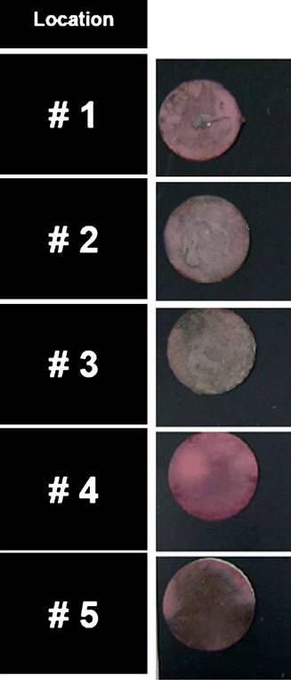 | ||
| Fig. 3 ChemTest® wipe coloration obtained in the dental laboratory. Location 1—on fluorescent light 1; Location 2—on fluorescent light 2; Location 3—on the duct of the ventilation system; Location 4—on a student's desk; and Location 5—on the bottom edge of the window (windowsill). | ||
Discussion
The rationale of sampling the working surface is to determine the level of contamination, which can help to estimate the amount of contaminants that could penetrate the skin. As such, surface sampling is essentially performed with wipe and suction techniques. A major determinant in surface sampling is certainly the physical nature of the surface, such as its smoothness or roughness. It was intuitively felt that a smooth surface should be a starting point for exploring the performance of both techniques. Hence, Petri dishes spiked with three different chemicals containing Be were used to compare the efficiency of the Ghost Wipe™, micro-vacuum, and ChemTest® wipe sampling methods.Ghost Wipe™ and micro-vacuum sampling methods
Ghost Wipe™ and micro-vacuum methods were applied to estimate the efficiency of recovery of the dust of different Be compounds spiked in different quantities on polystyrene Petri dishes with smooth and non-porous surfaces. The Ghost Wipe™ was more efficient than micro-vacuuming in the recovery of the Be compounds used in this study which was limited to the ultra-low particulate loading levels (0.4 micrograms or less). These results are in line with the results of other studies that have compared “wet” and “dry” methods when applied on smooth surfaces to recover lead dust.5,6 In their study, Farfel et al.6 showed that the geometric mean lead dust estimates in wipe sampling exceeded those for vacuum samples by a factor of 3.9 and 5.7 for the floor and sills respectively.At present, the major discrepancy observed between the calculated mass of Be (0.1 µg) for AlBeMet® and the average amount recovered by the direct analytical method (0.03 µg) cannot be explained, owing to the fact that the maximum recovery with the Ghost Wipe™ was 0.074 µg, a mass that was twice the direct analytical value (see the Analytical methods section). It is hypothesized that either the stock suspension was heterogeneously dispersed or, after the suspension dried out, the small particles of Be may have been accidentally lost from the vessel, due to the fact that the Be concentration was analyzed approximately one week after the preparation of the Petri dishes. The second hypothesis is less likely since the Petri dishes were covered.
Table 3 shows the average percentage recoveries calculated from three experiments carried out for each group of tests, the standard deviation, and the coefficient of variation reported as a percentage. The micro-vacuum method showed an average coefficient of variation (68%) higher than the Ghost Wipe™ method (14%). Besides the fact that the recovery was much lower than for the Ghost Wipe™ method (an average of 3.5% versus 64%), micro-vacuuming also gave a higher coefficient of variation, showing a large range of recovery within a group of tests. Although there are limited data in the present study, it can be concluded that Ghost Wipes provide better recovery at the specific loading levels (0.4 micrograms or less) than micro-vacuum. However, the micro-vacuum method is a better one when sampling quantities of 5–25 milligrams of particulate as it was shown in a recent article.23
The Ghost Wipe™ method showed better recovery by the two operators for Be solutions of acetate and chloride (approximately 96%) than for Be suspensions of oxide and AlBeMet® (ranging from 45% to 80%) spiked on aluminium dishes as well as for all Be compounds spiked on polystyrene (ranging from 48% to 82%) and glass dishes (ranging from 14% to 75%), as shown in Table 4. No reliable justification was found for this variation, and further studies need to investigate whether the solubility of the metal surface or reference material may facilitate the recovery. Nevertheless, one has to remember that tenths of one microgram of dust are extremely small amounts manipulated, and a slight loss (by adsorption) on the wall of a vessel will drastically affect the recovery results.
A higher coefficient of variation was observed for AlBeMet® sampled with the Ghost Wipe™ method compared to the other three Be compounds (Table 4 and the columns corresponding to Ghost Wipe™ in Table 3). This observation is also corroborated by the lower than expected amount of Be found in the AlBeMet® suspension during the direct analysis (see the Analytical methods section). The two observations might suggest that this alloy did not allow consistent dispersion of the particulate in suspension. AlBeMet® also showed the lowest ratio between recoveries obtained with the Ghost Wipe™ and micro-vacuum methods (6.9) compared with the other Be compounds (approximately 36 for beryllium acetate and beryllium chloride, and 22 for beryllium oxide), suggesting a lower adherence potential of Be particles on the surfaces, which led to better results with the micro-vacuum method.
The Ghost Wipe™ and micro-vacuum methods were also applied on site, at six locations in a dental laboratory with different types of surfaces. The Ghost Wipe™ technique gave a better recovery, even though the micro-vacuum method showed analogous recovery for three locations (locations 1 to 3, as can be seen in Table 6), where a large amount of loose dust was present. This is in line with previous work that showed that the micro-vacuum performed well with heavy load recoveries (5–25 milligrams).23 The different concentrations of Be recovered by the two methods (Ghost Wipe™/micro-vacuum) might be explained by the surface conditions (painted, smooth, small or non-porous surfaces for locations 1 to 4, wooden surface for location 5, and painted, uneven surface for location 6).
ChemTest® wipe method
This semi-quantitative analysis is based on colorimetry, which allows visual identification of the presence of an amount equal to or higher than 0.2 µg Be as reported by the manufacturer. Fig. 1 shows diverse colorations obtained by the manufacturer for different quantities of Be.20As can be seen in Fig. 2, three of the Be compounds used in this study (beryllium acetate, beryllium chloride and AlBeMet®) had positive responses to ChemTest® wipe sampling. It was observed that the blue color was fairly regularly spread over the ChemTest® surface in the case of Be solutions (acetate and chloride), while for the AlBeMet® suspension, the blue color was visible in the form of distinct spots. An explanation for this is that beryllium acetate and beryllium chloride form ions easily dispersed in solution, while the AlBeMet® suspension does not. However, when they were spiked with 0.1 µg Be (below the detection limit as reported by the manufacturer) the ChemTest® wipes showed a faint sign of presence, a definite presence, or a lack of Be. The irregular blue color developed at the level of 0.1 µg was caused by the non-uniform residue of soluble beryllium on the dish surface. The blank samples showed an absence of color.
The beryllium oxide suspension did not show positive results in both sets of experiments (Fig. 2). A hypothesis is that the beryllium oxide molecule, which has a very strong bond between the Be and oxygen atoms could not be ionized by either solution, the one containing 10% sulfuric acid and the one containing the ammonium buffer (pH = 10–11). It was shown that aggressive digestion procedures gave more complete recovery of beryllium from BeO.24 This observation is important due to the fact that beryllium oxide is one of the main chemical forms to which workers can be exposed in occupational settings.25
On site, in the dental laboratory, the large amount of settled dust on tested surfaces most likely prevented the chemical reaction of Be in contact with the ChemTest® wipes (Fig. 3). Instead, the Ghost Wipe™ analysis showed an amount higher than 0.2 µg Be at locations 1, 2 and 3. Location 4 (student's desk) showed a negative result with the ChemTest® wipe and this was also confirmed by the Ghost Wipe™ technique. Overall, our results show that ChemTest® wipes are affected by surface conditions.
Inter-operator variation
In order to evaluate the reproducibility of the tests, two operators carried out the Ghost Wipe™ experiments at the maximum amount of Be (0.4 µg) spiked on three different Petri dishes (polystyrene, glass and aluminium). The results were considered comparable when the recovery ratio between the two operators (Table 4) varied from 0.9 to 1.1 (±10%). The four tests that showed differences between the two operators (recovery ratio of 0.6, 4.2, 1.3 and 1.4) were analyzed by a paired t-test. Beryllium oxide on aluminium dishes and AlBeMet® on glass dishes showed no statistically significant difference (beryllium oxide on aluminium dishes: 95% CI, p-value = 0.083 and AlBeMet® on glass dishes: 95% CI, p-value = 0.414). A statistically significant difference between the operators' results was observed for beryllium chloride spiked on polystyrene dishes (95% CI, p-value = 0.002) and on glass dishes (95% CI, p-value = 0.021). The tests with the lowest recovery were repeated (Operator 1 on polystyrene dishes and Operator 2 on glass dishes in Table 4) and this time no statistically significant difference was observed (polystyrene dishes: 95% CI, p-value = 0.204 and glass dishes: 95% CI, p-value = 0.172) (Table 5) which may indicate that sampling training is probably an important determinant for obtaining consistency between operators.Inter-operator reproducibility showed good results for the twelve tests performed, although the difference between the two sets of results obtained by the same operator (Table 5) may be an indication of human error that occurred as a result of performing two different techniques (pressure, wiping the whole surface, folding of wipes, etc.) during the sampling procedure.
Limitations of the study
A number of limitations of this study were identified:(1) Laboratory experiments were carried out on smooth, non-porous surfaces that are less likely to be present on site, so the conclusions of this study should be applied only to similar surfaces.
(2) Adherence of Be particles on dishes is likely, which could have affected the collection efficiency.
(3) It is not known to what extent the use of different solvents during the preparation of the solutions/suspensions (watervs.isopropanol) introduced errors in the Be concentration.
(4) The calculated amount of Be in the AlBeMe® suspension and the homogeneity of the suspension were not confirmed by direct analysis and therefore the true concentration is possibly underestimated.
Conclusion
This study evaluated the efficiency of three different surface sampling methods (Ghost Wipe™, micro-vacuum and ChemTest®) in the recovery of Be dust, by comparing four Be compounds (beryllium acetate, beryllium chloride, beryllium oxide and beryllium aluminium) and three different surfaces (polystyrene, glass and aluminium). Inter-operator reproducibility was also assessed. The efficiency of the three sampling methods was tested at a working site.In the present experimental procedures, the Ghost Wipe™ was more efficient than micro-vacuuming in collecting the Be dust from smooth, non-porous surfaces by a factor of approximately 18. This conclusion probably applies to the ultra-low particulate loading levels (0.4 micrograms or less) which was tested in this study.
The results obtained on site, in a dental technique teaching college, also showed better recovery with the Ghost Wipe™ method; however, the ratio of Be recovered by Ghost Wipe™ versus micro-vacuum sampling was lower for surfaces where a large amount of dust was present. This is in line with previous work that showed that the micro-vacuum performed well with heavy load recoveries (5–25 milligrams).23
The ChemTest®, a colorimetric method, showed comparable results with those reported by the manufacturer for three Be compounds; however, beryllium oxide did not respond to this test. It is concluded that the newly released ChemTest® needs to improve its efficiency since the surface conditions and type of Be could affect the result. The Ghost Wipe™ is recommended for dust sampling on smooth surfaces. Inter-operator variation was not found to be significant in this study. Nevertheless, human errors may occur during manipulation, and instruction and training of the operators are critical.
Funding
Institut de recherche Robert-Sauvé en santé et sécurité du travail (G206745 IRSST 099-303); Évaluation des paramètres de surveillance environnementale des travailleurs exposés au béryllium and (G213800 IRSST 099-302); Guide de nettoyage et de décontamination des lieux de travail où il y a présence de béryllium.Notes and references
- S. A. Ness, Surface Sampling for Chemicals, in Surface and Dermal Monitoring for Toxic Exposures, Van Nostrand Reinhold, New York, 1994, ch. 7, pp. 163–207 Search PubMed.
- E. Wang and G. G. Rhoads, Appl. Occup. Environ. Hyg., 1995, 10(2), 111–118.
- G. W. Royster, J. R. Birney and R. Fish, Technique for Assessing “Removable” Surface Contamination, in Surface Contamination Proceedings of a Symposium Held at Gatlinburg, ed. B. R. Fish, Pergamon Press, Oxford, 1967, pp. 201–207 Search PubMed.
- Z. Bai, L.-M. Yiin., D. Q. Rich, J. L. Adgate, P. J. Ashley, P. J. Lioy, G. G. Rhoads and J. Zhang, Am. Ind. Hyg. Assoc. J., 2003, 64, 528–532 CAS.
- S. J. Reynolds, L. Etre, P. S. Thorne, P. Whitten, M. Selim and W. J. Popendorf, Am. Ind. Hyg. Assoc. J., 1997, 58, 439–446 CrossRef CAS.
- M. R. Farfel, P. S. Lees, C. A. Rohde, B. S. Lim, D. Bannon and J. J. Chisolm, Environ. Res., 1994, 65, 291–301 CrossRef CAS.
- B. P. Lanphear, M. Emond, D. E. Jacobs, M. Weitzman, M. Tanner, N. L. Winter, B. Yakir and S. Eberly, Environ. Res., 1995, 68, 114–123 CrossRef CAS.
- K. L. Creek, G. Whitney and K. Ashley, J. Environ. Monit., 2006, 8, 612–618 RSC.
- K. Ashley, M. J. Brisson, S. D. Jahn, J. ASTM Int., 2005, 2, 1–12, http://www.astm.org/DIGITAL_LIBRARY/JOURNALS/JAI/TOC/292005.htm Search PubMed.
- A. Dufresne, V. Turcotte, H. Golshahi, S. Viau, G. Perrault and C. Dion, Ann. Occup. Hyg., 2009, 53, 353–362 CrossRef CAS.
- A. Dufresne, C. Dion, S. Viau and G Perrault, Ann. Occup. Hyg., 2009, 53, 669–675 CrossRef CAS.
- S. Viau, C. Dion, G. Perrault, A. Dufresne, V. Turcotte, H. Golshahi, B. Campbell, T. Mocanu, A. Ouellet, P. J. Desormeaux, Studies and Research Projects/Report R-614, IRSST, Montréal, 2009, 74 p., http://www.irsst.qc.ca/fr/_publicationirsst_100470.html Search PubMed.
- 10 CFR 850, United States Department of Energy, Office of Environment, Safety and Health Department of Energy (US DOE), Fed. Regist., Chronic beryllium disease prevention program, Final Rule, 64(235), 68854–68914, 1999, http://www.hss.energy.gov/healthsafety/wshp/be/docs/berule.pdf Search PubMed.
- T. P. Taylor and N. N. Sauer, J. Hazard. Mater., 2002, B93, 271–283 CrossRef.
- Silica, Crystalline, by XRD (filter redeposition), NIOSH Method 7500, Issue 4, Manual of Analytical Methods, 4th edn, DDHS, Cincinnati, OH, 1994, http://www.cdc.gov/niosh/nmam/pdfs/7500.pdf Search PubMed.
- S. K. Dufay and M. Archuleta, J. Environ. Monit., 2006, 8, 630–633 RSC.
- ASTM International, American Society for Testing and Materials D6966, Standard Practice for Collection of Settled Dust Samples Using Wipe Sampling Methods for Subsequent Determination of Metals, American Society for Testing and Materials, West Conshohocken, PA, 2005, vol. 11.03, http://www.astm.org/Standards/D6966.htm Search PubMed.
- ASTM International, American Society for Testing and Materials, D7144, Standard Practice for Collection of Surface by Micro-vacuum Sampling for Subsequent Metals Determination, Annual Book of ASTM Standards, ASTM International, West Conshohocken, PA, 2005, http://www.astm.org/Standards/D7144.htm Search PubMed.
- United States Department of Housing and Urban Development (HUD), Comprehensive and Workable Plan for the Abatement of Lead-Based Paint in Privately Owned Housing: A Report to Congress, 1990, http://biotech.law.lsu.edu/cases/research/HUD-RptCongress-LeadBasedPaint.pdf.
- Beryllium ChemTest® Kit—ValidationReport.© Microteq, LLC, 2005, http://www.nextteq.com/docs/Manuals/ChemTest%20Manuals/ChemTest%20Beryllium%20Validation%20Report/ChemTest%20Beryllium%20Validation%20Report%20Rev%20C%20905.pdf, accessed June 22, 2008 Search PubMed.
- Institut de recherche Robert-Sauvé en santé et en sécurité du travail (IRSST), Method 359-en, Determination of beryllium [7440-41-7] in workplace air, Montréal, Canada, 2008, 18 p., http://www.irsst.qc.ca/files/documents/PubIRSST/M-359-en.pdf.
- R. W. Hornung and L. D. Reed, Appl. Occup. Environ. Hyg., 1990, 5, 46–51 CAS.
- K. Ashley, G. T. Applegate, T. J. Wise, J. E. Fernback and M. J. Goldcamp, J. Occup. Environ. Hyg., 2007, 4, 215–223 CrossRef CAS.
- A. B. Stefaniak, M. D. Hoover, G. A. Day, A. A. Ekechukwu, G. E. Withney, C. A. Brink, R. C. Scripsik, J. ASTM Int., 2005, 2, 15, www.astm.org Search PubMed.
- S. S. Tinkle, J. M. Antonini, B. A. Rich, J. R. Roberts, R. Salmen, K. DePree and E. J. Adkins, Environ. Health Perspect., 2003, 111, 1202–1208 CAS.
| This journal is © The Royal Society of Chemistry 2011 |