Electron induced dissociation (EID) tandem mass spectrometry of octaethylporphyrin and its iron(III) complex†
Malgorzata A.
Kaczorowska
and
Helen J.
Cooper
*
School of Biosciences, University of Birmingham, Birmingham B15 2TT, UK. E-mail: h.j.cooper@bham.ac.uk; Fax: +44 (0)121 414 5925; Tel: +44 (0)121 414 7527
First published on 17th September 2010
Abstract
EID tandem mass spectrometry of singly-charged electrosprayed ions of octaethylporphyrin (OEP) and its iron(III) complex results in ionisation to give doubly-charged precursor and fragment ions. Singly-charged fragments are also observed. EID fragmentation differs significantly to that observed in electron ionisation mass spectrometry.
Electron-based tandem mass spectrometry (MS/MS) methods for the elucidation of molecular structure, such as electron capture dissociation,1 have become increasingly popular because they provide complementary information to that obtained from traditional MS/MS techniques. Most of these electron-based methods require that the precursor ions be multiply-charged. Initial studies2 suggested that electron-based MS/MS of singly-charged ions produced fragments similar to collision-induced dissociation (CID), however electron induced dissociation (EID) in which precursor ions are bombarded with high energy (>10 eV) electrons is emerging as a useful orthogonal tool for structural elucidation of such species.3 EID has been applied to both biomolecules3–5 and inorganic species.6–8 We have applied EID to the analysis of (metallo)-porphyrin ions generated by electrospray ionisation. On irradiation with ∼24 eV electrons, the ions readily undergo tandem ionisation and characteristic fragmentation. The EID fragmentation is significantly different to that observed in electron ionisation (EI) MS.
Metallo-porphyrins, the ‘pigments of life’,9 comprise a tetrapyrrolic unit, which has planar geometry as a result of electronic delocalization and which serves as a ligand for metal ions. The metal ion is located in the axial position and the system has square pyramidal geometry. The structure and properties of these species depend also on the side groups attached to the porphyrin ring and anionic ligation of the metal ion.10 Several mass spectrometry methods have been applied to the analysis of porphyrins and their metal complexes.11–18MS/MS, in the form of CID, particularly low energy (eV) CID, provides19,20 very limited structural information on OEP and MeOEPs.
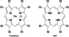
Here we describe the EID behaviour of singly-charged ions of octaethylporphyrin (OEP) and its iron(III) complex, Scheme 1. These species constitute models for biologically and geologically important porphyrins.21–23 We compare the EID behaviour with the fragmentation observed in electron ionization (EI) mass spectrometry.2,24 Singly-charged ions of OEP and Fe(OEP) were generated by electrospray of chloroform/methanol/formic acid solutions (30![[thin space (1/6-em)]](https://www.rsc.org/images/entities/char_2009.gif) ∶
∶![[thin space (1/6-em)]](https://www.rsc.org/images/entities/char_2009.gif) 70
70![[thin space (1/6-em)]](https://www.rsc.org/images/entities/char_2009.gif) ∶
∶![[thin space (1/6-em)]](https://www.rsc.org/images/entities/char_2009.gif) 2, v/v) of OEP and Fe(OEP)Cl (10 μM) and subjected to EID by use of a Thermo Finnigan LTQ FT mass spectrometer as described previously.8 Precursor ions were bombarded with electrons for 70 ms at 25% energy (corresponding to a cathode potential of − 23.79 V). Electron ionisation mass spectra of these species were recorded on a VG Zabspec mass spectrometer.
2, v/v) of OEP and Fe(OEP)Cl (10 μM) and subjected to EID by use of a Thermo Finnigan LTQ FT mass spectrometer as described previously.8 Precursor ions were bombarded with electrons for 70 ms at 25% energy (corresponding to a cathode potential of − 23.79 V). Electron ionisation mass spectra of these species were recorded on a VG Zabspec mass spectrometer.
 | ||
| Scheme 1 Structure of Fe(OEP)Cl and OEP. | ||
The electrospray ionization FT-ICR mass spectrum of Fe(OEP)Cl solution revealed two peaks; the dominant corresponding to [Fe(OEP)]+ (m/zmeas 588.2905, m/zcalc 588.2915), and the minor corresponding to the radical molecular cation [Fe(OEP)Cl]+˙ (m/zmeas 623.2615, m/zcalc 623.2604).
The electrosprayed [Fe(OEP)]+ ions fragment in EID as shown in Fig. 1a. Two distinct groups of fragments are observed. The first, in m/z range ≈ 410 to 588, comprises abundant singly-charged ions and the second, in m/z range ≈ 210 and 290, less abundant doubly-charged ions. For comparison, the electron ionisation mass spectrum of Fe(OEP)Cl is shown in Fig. 1b. Note that the two processes, EID and EI, are fundamentally different: the first involves tandem mass spectrometry, i.e., controlled fragmentation of a selected precursor ion, whereas the second involves single stage mass spectrometry and reveals in-source unimolecular decomposition. The dominant peaks in the EI spectrum correspond to singly-charged [Fe(OEP)]+ ions at m/z 588.33 and singly-charged [Fe(OEP)Cl]+˙ ions at m/z 623.30. Singly- and doubly-charged fragments, of comparative abundance, are also observed. It has been shown previously that EI of porphyrins, particularly metallo-porphyrins, yields unusually high abundances of doubly-charged molecule and fragment ions as a result of the stability of the highly aromatic macrocyclic ring system.25,26 The singly-charged EI fragments arise through cleavage of the bond β to the porphyrin nucleus, with loss of up to eight methyl groups, whereas the doubly-charged fragments arise via both α- and β-cleavage and corresponding loss of ethyl and methyl groups.26 At first glance, the EID product ion spectrum and the EI mass spectrum appear to be qualitatively similar. Both contain regions of singly- and doubly-charged fragments. However, closer inspection reveals the fragmentation to be markedly different. Whereas the singly-charged region of the EI mass spectrum reveals loss of a maximum of eight methyl groups, the singly-charged region of the EID MS/MS spectrum reveals loss of up to eleven substituents. Losses of both methyl and ethyl radicals are observed. The most abundant peaks at m/z 544.2313 and m/z 500.1685 can be assigned to losses of even numbers of radicals from the parent, i.e., loss of one methyl and one ethyl radical, and two methyl and two ethyl radicals respectively. Fragmentation via α- and β-cleavage has also been observed in collision-induced dissociation (CID) experiments,20 however the particular stability of the peaks corresponding to loss of (˙C2H5 + ˙CH3)n, where n = 1, 2, is unprecedented (Scheme 2). Clearly, losses of even numbers of radicals from the singly-charged precursor result in formation of stable even-electron fragments. Nevertheless, in the case of the metallo-porphyrin loss of (˙C2H5 + ˙CH3) is favoured over loss of (˙CH3 + ˙CH3).
![(a) EID mass spectrum of [FeOEP]+ ions. Inset: expanded m/z region showing doubly-charged fragment ions. * denotes even-electron fragments. (b) EI mass spectrum of [FeOEP]Cl. Inset: expanded m/z region showing singly- (top) and doubly-charged (bottom) fragment ions. (c) EID mass spectrum of [OEP+H]+. * denotes even-electron fragments. (d) EI mass spectrum of OEP. Inset: expanded m/z region showing singly- (top) and doubly-charged (bottom) fragment ions. Double-headed arrows indicate differences in molecular composition.](/image/article/2011/CC/c0cc02198a/c0cc02198a-f1.gif) | ||
| Fig. 1 (a) EID mass spectrum of [FeOEP]+ ions. Inset: expanded m/z region showing doubly-charged fragment ions. * denotes even-electron fragments. (b) EI mass spectrum of [FeOEP]Cl. Inset: expanded m/z region showing singly- (top) and doubly-charged (bottom) fragment ions. (c) EID mass spectrum of [OEP+H]+. * denotes even-electron fragments. (d) EI mass spectrum of OEP. Inset: expanded m/z region showing singly- (top) and doubly-charged (bottom) fragment ions. Double-headed arrows indicate differences in molecular composition. | ||
EID of Fe(OEP) resulted in the formation of doubly-charged fragments, that is, electron ionisation dissociation4 occurred. This is rarely observed but is perhaps not surprising for these species. As mentioned above, EI of OEP and its metal complexes produces doubly-charged species of a much greater abundance than typically observed in EI25,26 as a consequence of aromaticity and the presence of nitrogen. Those same characteristics should also favour ionisation as a result of EID. The electron could either be ejected from the metal ion or the porphyrin ligand. Ejection of the electron from the metal would result in a change in iron oxidation state from Fe(III) to Fe(IV). We have shown previously that it is possible to access oxidation states not readily available in solution-phase by gas-phase electron capture dissociation.27 Nevertheless, the fourth ionisation energy of Fe is 54.8 eV, whereas the first ionisation energy of OEP is 6.24 eV. The energy of electrons here was ∼24 eV. The most abundant peak in the doubly-charged region can be assigned to loss of a single ethyl radical from doubly-charged [Fe(OEP)]2+. The [Fe(OEP)]2+ ion is an odd-electron species and the most abundant fragments are those that derive from the loss of odd numbers of radicals, i.e., stable even-electron species.
The EID product ion spectrum of protonated base porphyrin [OEP + H]+, shown in Fig. 1c, shows some similarities to the EID product ion spectrum of Fe(OEP). Two distinct groups of fragment ions are observed, one in the high m/z region comprising singly-charged ions and one in the low m/z region containing very low abundance doubly-charged ions. As for Fe(OEP), the most abundant fragment, m/z 491.3192 is the result of loss of (˙C2H5 + ˙CH3), see Scheme 2. However the second most abundant fragment, m/z 461.2720, arises via loss of (˙CH3 + ˙CH3). The fragment is an even-electron species but it is not clear why the presence of Fe(III) facilitates multiple α-cleavages. Unlike Fe(OEP), EID of [OEP + H]+ results in loss of methane, in addition to loss of the methyl radical. (Loss of methane has also been observed in the high energy CID of protonated OEP.)20 Losses of up to thirteen substituents are observed in the singly-charged fragments revealing the occurrence of both α- and β-cleavages. The doubly-charged region of the EID MS/MS spectrum of the base porphyrin differs significantly to that of its iron(III) complex. Only the doubly-charged precursor, [OEP + H]2+ at m/z 267.6952, and the fragment resulting from the loss of one methyl radical, [OEP − CH3 + H]2+ at m/z 260.1784, are observed. Again, electron ionisation dissociation has occurred but appears to be a far less favourable process. This might suggest that in the case of the Fe(OEP), further ionisation of Fe occurs to produce the doubly-charged species. Alternatively, we postulate that the presence of the metal ion lowers the ionisation energy of the porphyrin, thus promoting tandem ionisation. In either case, the doubly-charged precursor is an odd-electron species which fragments via loss of a single methyl radical to give an even-electron species. (That is in contrast to the Fe(OEP) results, further suggesting that α-cleavage is favoured in the presence of the metal ion).
For comparison, the EI mass spectrum of the base porphyrin is shown in Fig. 1d. The EID MS/MS spectrum and the EI mass spectrum appear similar but on closer inspection are significantly different. The base peak in the EI mass spectrum of OEP corresponds to singly-charged [OEP − 4H]+˙ ions, m/z 530.93, which is formed as a result of loss of four hydrogen atoms followed by rearrangement of the porphyrin macrocycle. The minor peak at m/z 534.97 can be assigned to singly-charged molecular ions of OEP. The EI mass spectrum is also characterized by regions of singly- and doubly-charged fragment ions. The singly-charged fragments result from the loss of up to eight methyl groups (β-cleavages) in contrast to the combinations of α- and β- cleavages observed in EID. Nine doubly-charged fragments are observed in contrast to the sole doubly-charged fragment observed following EID.
In conclusion, electron induced dissociation tandem mass spectrometry of electrosprayed octaethylporphyrin and its Fe(III) complex results in characteristic fragmentation which is unlike that observed in electron ionisation and collision-induced dissociation.19,20,26 The formation of singly-charged even-electron species via loss of one ethyl and one methyl group is favoured. EID of Fe(OEP) results in abundant doubly-charged fragment ions. Doubly-charged fragments are also formed, although less readily, following EID of OEP. Tandem ionisation following irradiation of ions with these fairly low energy electrons (∼24 eV) is rare. For doubly-charged tandem ionised precursors, fragmentation proceeds via loss of a single radical group, either ethyl for Fe(OEP) or methyl for OEP, resulting in even-electron fragments. The presence of the metal ion appears to facilitate α-cleavage, with loss of ethyl group(s) observed in both singly- and doubly-charged fragments.
Notes and references
- R. A. Zubarev, N. L. Kelleher and F. W. McLafferty, J. Am. Chem. Soc., 1998, 120, 3265–3266 CrossRef CAS.
- R. B. Cody and B. S. Freiser, Anal. Chem., 1979, 51, 547–551 CrossRef CAS.
- H. Lioe and R. A. J. O'Hair, Anal. Bioanal. Chem., 2007, 389, 1429–1437 CrossRef CAS.
- Y. M. E. Fung, C. M. Adams and R. A. Zubarev, J. Am. Chem. Soc., 2009, 131, 9977–9985 CrossRef CAS.
- H. J. Yoo, H. C. Liu and K. Hakansson, Anal. Chem., 2007, 79, 7858–7866 CrossRef CAS.
- G. N. Khairallah, R. A. J. O'Hair and M. I. Bruce, Dalton Trans., 2006, 3699–3707 RSC.
- L. Feketeova and R. A. J. O'Hair, Rapid Commun. Mass Spectrom., 2009, 23, 60–64 CrossRef CAS.
- M. A. Kaczorowska and H. J. Cooper, J. Am. Soc. Mass Spectrom., 2010, 21, 1398–1403 CrossRef CAS.
- A. R. Battersby, Pure Appl. Chem., 1993, 65, 1113–1122 CrossRef CAS.
- J. L. Hoard, G. H. Cohen and M. D. Glick, J. Am. Chem. Soc., 1967, 89, 1992–1996 CrossRef CAS.
- J. M. E. Quirke, Mass spectrometry of porphyrins and metalloporphyrins, in The Porphyrin Handbook, ed. K. M. Kadish, K. M. Smith and R. Guilard, Elsevier Science, Oxford, 2000, vol. 7, p. 371 Search PubMed.
- G. J. Van Berkel, G. L. Glish, S. A. McLuckey and A. A. Tuinman, Anal. Chem., 1990, 62, 786 CrossRef CAS.
- M. J. Dale, K. F. Costello, A. C. Jones and P. R. R. Langridge-Smith, J. Mass Spectrom., 1996, 31, 590 CrossRef CAS.
- F. M. Rubino, S. Banfi, G. Pozzi and S. Quici, J. Am. Soc. Mass Spectrom., 1993, 4, 249–254 CrossRef CAS.
- S. Feil, M. Winkler, P. Sulzer, S. Ptasinska, S. Denifl, F. Zappa, B. Krautler, T. D. Mark and P. Scheier, Int. J. Mass Spectrom., 2006, 255–256, 232–238 Search PubMed.
- G. J. Van Berkel, S. A. McLuckey and G. L. Glish, Anal. Chem., 1991, 63, 1098 CrossRef CAS.
- V. E. Vandell and P. A. Limbach, J. Mass Spectrom., 1998, 33 Search PubMed.
- R. S. Brown and C. L. Wilkins, Anal. Chem., 1986, 58, 3196–3199 CrossRef CAS.
- T. Gozet, L. Huynh and D. K. Bohme, Int. J. Mass Spectrom., 2009, 279, 113–118 Search PubMed.
- M. R. Domingues, O. V. Nemirovskiy, M. Graco, O. S. Marques, M. Graca Neves, J. A. S. Cavaleiro, A. J Ferrer-Correia and M. L. Gross, J. Am. Soc. Mass Spectrom., 1998, 9, 767–774 CrossRef CAS.
- C. K. Chang, K. M. Barkigia, K. L. Hanson and J. Fajer, J. Am. Chem. Soc., 1986, 108, 1352–1354 CrossRef CAS.
- G. J. Shaw, J. M. E. Quirke and G. J. Eglinton, J. Chem. Soc., Perkin Trans. 1, 1978, 1655–1659 RSC.
- B. D. Beato, R. A. Yost and J. M. E. Quirke, Chem. Geol., 1991, 185–192 CrossRef CAS.
- R. A. Zubarev, Mass Spectrom. Rev., 2003, 22, 57–77 CrossRef CAS.
- A. H. Jackson, G. W. Kenner, K. M. Smith, R. T. Aplin, H. Budzikiewicz and C. Djerassi, Tetrahedron, 1965, 21, 2913 CrossRef CAS.
- B. D. Beato, R. A. Yost and J. M. E. Quirke, Org. Mass Spectrom., 1989, 24, 875 CrossRef CAS.
- M. A. Kaczorowska and H. J. Cooper, J. Am. Soc. Mass Spectrom., 2010, 21, 300–309 CrossRef CAS.
Footnote |
| † This article is part of the ‘Emerging Investigators’ themed issue for ChemComm. |
| This journal is © The Royal Society of Chemistry 2011 |

![Loss of methyl and ethyl radicals on EID of [Fe(OEP)]+.](/image/article/2011/CC/c0cc02198a/c0cc02198a-s2.gif)