Direction control of photomechanical bending of a photochromic molecular fiber†
Hideyuki
Nakano
*
Department of Applied Chemistry, Faculty of Engineering, Osaka University, Yamadaoka, Suita, Osaka 565-0871, Japan. E-mail: nakano@chem.eng.osaka-u.ac.jp; Fax: +81-6-6879-7365; Tel: +81-6-6879-7367
First published on 9th February 2010
Abstract
A photochromic molecular fiber was developed and the photomechanical bending motions of the fiber were demonstrated. The bending direction of the fiber was found to be controllable by altering the polarization direction of the irradiated light without changing the position of the light source and the wavelength of the light.
Photomechanical effects observed for photochromic materials have attracted a great deal of attention from both viewpoints of fundamental science and practical applications such as light-driven actuators. Films and fibers of azobenzene-based liquid-crystalline polymers have been reported to exhibit bending motions by light irradiation.1–4 With regard to materials composed of low molecular-weight compounds, needle- and plate-shaped microcrystals of diarylethene, anthrathene, and azobenzene derivatives have been reported to exhibit reversible bending motions by photoirradiation.5–7 The bending directions of such materials were suggested to depend on the kinds of molecules and their alignments in the material,2,5 therefore, it is difficult to change the bending direction without the change of the position of the light source and the wavelength of the light. In the present study, a novel fiber composed of a low molecular-weight photochromic compound, referred to as a “photochromic molecular fiber”, has been developed and the direction of the photomechanical bending of the fiber was found to be controllable by altering the polarization direction of the irradiated light.
The low molecular-weight photochromic compound using in the present study was 4-[bis(9,9-dimethylfluoren-2-yl)amino]azobenzene (BFlAB: the chemical structure was shown in Fig. 1). We have already reported the synthesis and properties of BFlAB, which readily formed an amorphous glass with a glass-transition temperature of 97 °C and exhibited photochromism as amorphous film as well as in solution.8,9
 | ||
| Fig. 1 Two-dimensional X-ray diffraction pattern of BFlAB fiber. The arrow indicates the direction of the fiber axis. | ||
A photochromic molecular fiber of BFlAB was fabricated as follows. The powder of BFlAB was heated above the melting point (167 °C) on a glass substrate. After that, the molten sample was slightly cooled and kept at the appropriate temperature just under the melting point. A part of the resulting viscous supercooled liquid sample was anchored with a pair of tweezers and then pulled it up to readily form a fiber with a diameter in the range of 10 to 100 μm. In some cases, the fiber reached ca. 40 cm in length. No significant anisotropy was observed by polarizing optical microscopy, indicating the resulting fiber was amorphous. In contrast to the melt-spun fibers in which the polymer chains often align parallel to the drawing direction, two-dimensional X-ray diffraction pattern of the BFlAB fiber, monitored by means of a Rigaku RAXIS-RAPID Imaging Plate diffractometer with graphite-monochromated Cu-Kα (1.54186 Å) radiation, showed only an isotropic halo as shown in Fig. 1, indicating no significant alignment of the molecules in the fiber. The result may be due to no polymer chain existing in the material.
Experimental set-up for photomechanical bending of the photochromic molecular fiber was schematically illustrated in Fig. 2. The fiber with a diameter of ca. 15 μm and with a proper length was put on an edge of a glass substrate. Then a part of the sample fiber was irradiated (length of the irradiated area: ca. 2 mm) with a linearly polarized laser beam (488 nm: CYAN-488-100NH-W, Spectra Physics) with an intensity of ca. 400 mWcm−2 and with a polarization direction either parallel (H-polarized light) or perpendicular (V-polarized light) to the fiber axis through an ND filter, a wave plate, and a polarizer at room temperature (ca. 20 °C). Bending behaviour of the fiber was monitored using a Stereoscopic Microscope (Nikon) attached with a CCD camera (VB-7010, Keyence) through a cut-off filter (Y-51, AGC Techno Glass Co. Ltd). The bending directions of the fiber opposite and toward the light source were defined here as a positive and a negative directions, respectively, as shown in Fig. 2.
 | ||
| Fig. 2 Schematic experimental set-up for photoirradiation of BFlAB fiber. | ||
When irradiated with the H-polarized light, the sample fiber was found to bend rapidly in the positive direction. When the irradiation was stopped, the reverse motion in the negative direction gradually took place. The light-on (1 s) and light-off (3 s) cycles caused the repetition of bending and reverse motions as shown in Fig. 3a and Movie 1, ESI.† Such motions were almost similar to those observed when the V-polarized light was used. However, these repetition behaviors of bending and reverse motions were not completely reversible (Fig. 3b and 3c), and interestingly, the tip positions gradually shifted in the negative direction with increasing the number of the on-off cycles when the V-polarized light was used (Fig. 3c), whereas the positions shifted in the positive direction when the H-polarized light was used (Fig. 3b).
 | ||
| Fig. 3 Photomechanical bending motion of the fiber. (a) Photographs of the fiber upon the on-off cycles of the H-polarized light. Scale bar: 1 mm. (b) Change in the tip positions of the fiber upon the on-off cycles of the H-polarized light. (c) Change in the tip positions of the fiber upon the on-off cycles of the V-polarized light. | ||
Then, the photomechanical bending motion upon continuous irradiation for a longer period was investigated. When the H-polarized light was used, the fiber rapidly bent in the positive direction just after starting the irradiation (from Fig. 4a-i to 4a-ii). And then, further bending in the positive direction gradually took place (Movie 2, ESI†). The structure of the fiber after 10 min irradiation was shown in Fig. 4a-iii. After the irradiation was stopped at this time, the tip position was slightly shifted in the negative direction (Fig. 4a-iv) and then the resulting bending structure of the fiber remained in the dark at room temperature.
 | ||
| Fig. 4 Bending motion of the fiber upon continuous irradiation. (a) Photographs of the fiber when the H-polarized light was used. (b) Photographs of the fiber when the V-polarized light was used. i) Before irradiation. ii) After irradiation for 2 s. iii) After irradiation for 10 min. iv) After stopping the irradiation. Scale bar: 1 mm. | ||
When the V-polarized light was used, initial bending motion in the positive direction (from Fig. 4b-i to 4b-ii) was almost similar to that observed when the H-polarized light was used. However, the subsequent continuous irradiation for a longer period induced the mechanical motion in the negative direction (from Fig. 4b-ii to 4b-iii and Movie 3, ESI†). When the irradiation was stopped, the tip position was further shifted in the negative direction and then the resulting bending structure (Fig. 4b-iv) remained in the dark. In addition, the reverse motion in the positive direction for the resulting fiber could be induced by irradiation with the H-polarized light. Likewise, the reverse motion in the negative direction for the fiber bent in the positive direction by irradiation with the H-polarized light could be induced by irradiation with the V-polarized light. Thus, the fiber could be bent in both directions easily by altering the polarization direction of the irradiated light without changing the position of the light source and the wavelength of the light.
The bending behaviors observed in the present study suggested that the bending motions of the fiber upon photoirradiation include two modes, one being a “reversible mode” concerned with the reversible motions as observed mainly for the on–off cycles in the shorter periods and the other an “irreversible mode” concerned with the motions as observed during continuous irradiation for a longer period. Regarding the reversible mode, the bending behaviors can be explained as follows. The diameter of the fiber was considerably larger than the expected penetration depth of the irradiated light (∼100 nm) due to a large extinction coefficient of BFlAB at 488 nm, suggesting that the photochemical reaction of the trans-isomers to the cis-isomers took place only near the irradiated surface of the fiber. Since the photoisomerization reactions of the trans-isomers to the cis-isomers of azobenzene derivatives need excess free volume,9,10 the reaction induced the volume expansion only near the irradiated side of the fiber, resulting in bending in the direction opposite the light source, i.e. the positive direction. When the irradiation stopped, the photo-generated cis-isomers recovered to the trans-isomers, resulting in the reverse motion of the fiber. Volume expansion caused by increase in temperature near the irradiated surface due to absorption of the irradiated light, namely photo-thermal effect, was also presumable to induce the bending regarding the reversible mode. Although the temperature at the irradiated area and the relationship between the degree of bending and the content of cis-isomers have not been clear, the fact that the rate of recovering motion of the bent fiber was faster than the reaction rate of cis–trans thermal isomerization of BFlAB as amorphous film at room temperature (in the order of 10−3 min−1)9 implied that the photo-thermal effect was the main factor for bending regarding the reversible mode. Because of optically isotropic nature of the fiber, it is understandable that the bending motion regarding the reversible mode was independent of the polarization direction of the irradiated light.
In contrast to the reversible mode, the bending motion observed upon the continuous irradiation for a longer period, i.e. the irreversible mode, depended on the polarization direction as described above. It has been reported that the interference irradiation of the amorphous films of azobenzene-based polymers11–17 and amorphous molecular materials8,18–22 including BFlAB with coherent laser beams induced mass transport to inscribe surface relief gratings and those behaviors strongly depended upon the polarization direction of the laser beams. It was suggested that the mass transport was induced in the direction from the bright area to the dark area parallel to the polarization direction of the laser beam.15 Although the mechanism of the photoinduced mass transport depending on the polarization direction of the irradiated laser beam has not been clear yet,13–16 such anisotropic mass transport upon polarized-light irradiation can explain the photomechanical bending motions regarding the irreversible mode as follows. When the fiber was irradiated with the H-polarized light, the mass transport was induced near the surface in the direction from the middle of the irradiated area toward both ends of the fiber, being parallel to the fiber axis as indicated by white arrows in Fig. 5a. Such mass transport induced bending moments as indicated by black arrows in the figure, resulting in bending the fiber in the direction opposite the light source, i.e. the positive direction. On the other hand, the V-polarized light induced the mass transport in the direction from the irradiated side toward the back side of the fiber, being perpendicular to the fiber axis, which induced the bending moments with opposite directions to those when the H-polarized light was used (Fig. 5b). As a result, the fiber bent in the direction toward the light source, i.e. the negative direction. Since the mass transport was thermally irreversible, the resulting bent structure of the fiber remained in the dark.
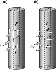 | ||
| Fig. 5 Schematic illustration for plausible mechanism of the bending motions regarding the irreversible mode. (a) When the H-polarized light was used. (b) When the V-polarized light was used. White and black arrows indicate directions of photoinduced mass transport and resulting bending moment, respectively. | ||
In order to gain further information about the mechanism of the photomechanical bending of the fiber, the change in the diameter of the fiber at the irradiated site was investigated. In order to avoid the change in the diameter due to bending motion, both ends of the sample fiber were fixed at the glass substrates and then the change in the diameter of the fiber upon irradiation was monitored by using an Optiphot X2 (Nikon) microscope. It was found that the diameter of the fiber viewed from the back of the irradiated surface increased drastically by irradiation with the V-polarized light (ca. 400 mWcm−2) for 20 min (Fig. 6a). In addition, the diameter of the fiber viewed from the side of the irradiated surface was found to decrease by the irradiation (Fig. 6b). These results suggested that the mass transport was induced by irradiation with the V-polarized light as indicated by the white arrows in the Fig. 5b. Due to the curved surface of the fiber, the gradient of the light intensity at the surface of the fiber in the direction perpendicular to the fiber axis was thought to be large enough so that such observable mass transport could be induced. On the other hand, no significant change in diameter at the irradiated site of the fiber was observed by irradiation with the H-polarized light, maybe because the change in the diameter was too small to detect due to relatively small intensity gradient in the direction parallel to the axis.
 | ||
| Fig. 6 Change in diameter of the BFlAB fiber by irradiation with the V-polarized light for 20 min. (a) Photographs of the fiber at the irradiated area viewed from the back of the irradiated surface (i) before and (ii) after irradiation. (b) Photographs of the fiber at the irradiated area viewed from the side of the irradiated surface (i) before and (ii) after irradiation. Scale bar: 10 μm. Note that the sample fiber shown in (a) was not identical to that shown in (b). | ||
Since anisotropic orientation of azobenzene moieties by linearly polarized light, namely Weigert effect,23 has been known for azobenzene-based materials,24,25 the Weigert effect might play a role for the bending motion of the BFlAB fiber observed in the present study. However, the fact that the anisotropic orientation of BFlAB molecules could not be induced so much by irradiation of the BFlAB amorphous film with linearly polarized light26 suggested that the Weigert effect did not so much contribute to the present photomechanical motions.
In conclusion, a photochromic molecular fiber was developed using BFlAB and the photomechanical bending motions of the resulting fiber were demonstrated. The bending direction of the fiber was found to be controllable by altering the polarization direction of the irradiated light without changing the position of the light source and the wavelength of the light. The bending motions were suggested to be related with the photoinduced mass transport taking place near the irradiated surface of the fiber.
Acknowledgements
This work was partly supported by a Grant-in-Aid for Science Research in Priority Areas “New Frontiers in Photochromism (No. 471)” from the Ministry of Education, Culture, Sports, Science and Technology (MEXT), Japan.Notes and references
- Y. Yu, M. Nakano and T. Ikeda, Nature, 2003, 425, 145 CrossRef CAS.
- M. Kondo, Y. Yu and T. Ikeda, Angew. Chem., Int. Ed., 2006, 45, 1378 CrossRef CAS.
- H. J. Choi, K.-U. Jeong, L.-C. Chien and M.-H. Lee, J. Mater. Chem., 2009, 19, 7124 RSC.
- C. L. van Oosten, C. W. M. Bastiaansen and D. J. Broer, Nat. Mater., 2009, 8, 677 CrossRef CAS.
- S. Kobatake, S. Takami, H. Muto, T. Ishikawa and M. Irie, Nature, 2007, 446, 778 CrossRef CAS.
- R. O. Al-Kaysi, A. M. Muller and C. J. Bardeen, J. Am. Chem. Soc., 2006, 128, 15938 CrossRef CAS.
- H. Koshima, N. Ojima and H. Uchimoto, J. Am. Chem. Soc., 2009, 131, 6890 CrossRef CAS.
- H. Nakano, T. Takahashi, T. Kadota and Y. Shirota, Adv. Mater., 2002, 14, 1157 CrossRef CAS.
- T. Tanino, S. Yoshikawa, T. Ujike, D. Nagahama, K. Moriwaki, T. Takahashi, Y. Kotani, H. Nakano and Y. Shirota, J. Mater. Chem., 2007, 17, 4953 RSC.
- T. Naito, K. Horie and I. Mita, Polymer, 1993, 34, 4140 CrossRef CAS.
- P. Rochon, E. Batalla and A. Natansohn, Appl. Phys. Lett., 1995, 66, 136 CrossRef CAS.
- D. Y. Kim, S. K. Tripathy, L. Li and J. Kumar, Appl. Phys. Lett., 1995, 66, 116.
- C. Barret, A. Natansohn and P. Rochon, J. Phys. Chem., 1996, 100, 8836 CrossRef CAS.
- P. Lefin, C. Fiorini and J.-M. Nunzi, Pure Appl. Opt., 1998, 7, 71 CrossRef CAS.
- N. K. Viswanathan, D. Y. Kim, S. Bian, J. Williams, W. Liu and L. Li, J. Mater. Chem., 1999, 9, 1941 RSC.
- A. Natansohn and P. Rochon, Chem. Rev., 2002, 102, 4139 CrossRef CAS.
- H. Ando, T. Tanino, H. Nakano and Y. Shirota, Mater. Chem. Phys., 2009, 113, 376 CrossRef CAS.
- H. Nakano, T. Tanino, T. Takahashi, H. Ando and Y. Shirota, J. Mater. Chem., 2008, 18, 242 RSC.
- Y. Shirota, H. Utsumi, T. Ujike, S. Yoshikawa, K. Moriwaki, D. Nagahama and H. Nakano, Opt. Mater., 2003, 21, 249 CrossRef CAS.
- H. Ueda, T. Tanino, H. Ando, H. Nakano and Y. Shirota, Chem. Lett., 2004, 33, 1152 CrossRef CAS.
- T. Takahashi, T. Tanino, H. Ando, H. Nakano and Y. Shirota, Mol. Cryst. Liq. Cryst., 2005, 430, 9 CrossRef CAS.
- H. Nakano, T. Takahashi, T. Tanino and Y. Shirota, Dyes Pigm., 2009, 84, 102.
- F. Weigert, Naturwissenschaften, 1921, 29, 583.
- K. Ichimura, Chem. Rev., 2000, 100, 1847 CrossRef CAS.
- T. Ikeda, J. Mater. Chem., 2003, 13, 2037 RSC.
- T. Tanino, T. Takahashi, H. Nakano and Y. Shirota, Mol. Cryst. Liq. Cryst., 2005, 430, 193 CrossRef CAS.
Footnote |
| † Electronic supplementary information (ESI) available: Movies 1–3. See DOI: 10.1039/b924718a |
| This journal is © The Royal Society of Chemistry 2010 |