Indole-containing pharmaceuticals: targets, pharmacological activities, and SAR studies

Wei Zeng a, Chi Han a, Sarah Mohammed a, Shanshan Li a, Yixuan Song b, Fengxia Sun *c and Yunfei Du *a
aSchool of Pharmaceutical Science and Technology, Tianjin University, Tianjin 300072, China. E-mail: duyunfeier@tju.edu.cn
bDepartment of Chemical Engineering & Biotechnology, University of Cambridge, CB2 3RA Cambridge, UK
cResearch Center for Chemical Safety & Security and Verification Technology & College of Chemical and Pharmaceutical Engineering, Hebei University of Science and Technology, Shijiazhuang 050018, China. E-mail: fxsun001@163.com

Received 1st December 2023 , Accepted 29th January 2024

First published on 30th January 2024


Abstract

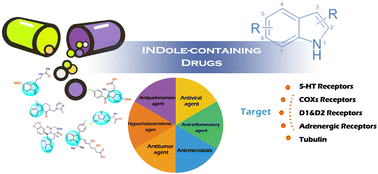
Indole is a prestigious heterocyclic skeleton widely found in both naturally-occurring and biologically-active compounds. Pharmaceutical agents containing an indole skeleton in their framework possess a wide range of pharmacological properties, including antiviral, antitumor, analgesic, and other therapeutic activities, and many indole-containing drugs have been proven to have excellent pharmacokinetic and pharmacological effects. Over the past few decades, the FDA has approved over 40 indole-containing drugs for the treatment of various clinical conditions, and the development of indole-related drugs has attracted significant attention from medicinal chemists. This review aims to provide an overview of all the approved drugs that contain an indole nucleus, focusing on their targets, pharmacological activities, and SAR studies.


1. Introduction

Structurally, indole is an aromatic heterocyclic molecule with a double-ring structure made up of a benzene ring and a nitrogenous pyrrole ring. The nitrogen atom in indole donates its lone pair of electrons to form a conjugated system, which stabilizes the indole structure. Additionally, this lone pair of electrons causes the indole compound to exhibit weak alkalinity (pKa = 16.2 and pKb = 17.6). Furthermore, due to the relatively high electron density in the pyrrole ring and the conjugation effect of the nitrogen atom, indole can undergo an electrophilic substitution reaction at 3-position in its pyrrole ring (Fig. 1a).
image file: d3md00677h-f1.tif
Fig. 1 (a) Structure of indole and (b) its multiple clinical applications.

Indole is an important privileged heterocyclic motif in medicinal chemistry and life sciences. Many endogenous substances, including tryptamine, serotonin, and melatonin, contain an indole skeleton in their structures.1 Furthermore, over 70 synthetic medications containing indole are globally available in the market and have been proven to exhibit excellent medical treatment outcomes (Fig. 1b). Some of the ailments that these indole-containing medications are advertised to treat include diabetes, cancer, leukemia, hepatitis C, psychosexual dysfunction, and other clinical problems. Since 2015, the US Food and Drug Administration (FDA) has specifically approved 14 indole-containing drugs, three of which were approved in 2021 (Fig. 2).


image file: d3md00677h-f2.tif
Fig. 2 Number of indole-containing drugs approved by the FDA in recent years.

Several review papers2,3 focusing on the biological activities of synthesized and naturally-occurring indole compounds have been documented. However, to date, few reports have focused on the indole-containing pharmaceuticals that are used clinically. Thus, in this review, we summarize all the marketed indole-containing drugs, highlighting their target receptors, pharmacological activities, and respective structure–activity relationship (SAR) studies. It should be noted that the pharmaceutical agents that are still under development, peptide derivatives whose main functions are related to their amino acid sequence, veterinary drugs, and herbicides are not included. The indole-containing pharmaceuticals discussed in this review are categorized based on their therapeutic areas with an aim to highlight the importance of the indole scaffold in quantitative structure–activity relationship (QSAR) analysis.

2. Indole-containing drugs targeting 5-HT receptors

Dihydroergotamine (DHE, 1), with an indole scaffold in its chemical structure, is an ergot alkaloid employed in the treatment of migraines and cluster headaches (Fig. 3a).4 This medication is commonly administered via the subcutaneous and intramuscular routes with 93% plasma protein bound, and its apparent steady-state volume of distribution is approximately 800 L. Four dihydroergotamine mesylate metabolites have been identified in human plasma following oral administration. The major metabolite, i.e., 8′-β-hydroxydihydroergotamine, exhibits affinity equivalent to its parent for adrenergic and 5-HT receptors and demonstrates equivalent potency in several venoconstrictor activity models both in vivo and in vitro. The decline in plasma dihydroergotamine after intramuscular or intravenous administration is multi-exponential with a terminal half-life of about 9 h. Its agonistic activity on 5-HT1b receptors in the smooth muscle of the cranial vasculature may reduce the blood vessel dilation caused by the release of CGRP during migraine attacks, thus providing relief.5,6 Specifically, ability of the ventroposteromedial thalamus to transmit nociceptive signals to the trigeminal sensory neurons is inhibited by the antagonist activity of dihydroergotamine (1) on 5-HT1b and 5-HT1d receptors. The agonist activity of the trigeminal nucleus caudalis on 5-HT1f, coupled with its additional action on 5-HT1b and 5-HT1d receptors reduces afferent signaling to the trigeminal sensory neurons and aids in the development of central sensitization. Finally, vasoactive neuropeptide release is inhibited by the activity at the 5-HT1d receptors on trigeminal nerve terminals.7 This is how dihydroergotamine (1) mechanistically reduces sensitivity to the pain of a migraine attack. The indole part of the drug structure is a significant element contributing to the total drug structure (Fig. 3b). This pharmaceutical agent can bind to the active pocket stably due to the existence of an H-bond between the N–H of indole and Ser212.8
image file: d3md00677h-f3.tif
Fig. 3 (a) Structure of dihydroergotamine (1) and ondansetron (2). (b) Key-target interactions of dihydroergotamine (1) with its target (left) and the key-target interactions of ondansetron (2) with its target (right).

Having been first developed in the 1980s by GlaxoSmithKline and approved by the US FDA in January 1991, ondansetron (2), a serotonin 5-HT3 receptor antagonist, is used for preventing nausea and vomiting during cancer treatment and after surgery (Fig. 3a). Ondansetron (2) can be well absorbed from the gastrointestinal tract and undergoes some first-pass metabolism. Its mean bioavailability in healthy subjects, following administration of a single 8 mg tablet, is approximately 56%. This drug is extensively metabolized in humans, where its primary metabolic pathway is hydroxylation on the indole ring, followed by subsequent glucuronide or sulfate conjugation. Besides, ondansetron (2) has a mean elimination half-life of 3.1 h to 6.2 h based on the age of the patient. It has been proven that cytotoxic chemotherapy and radiotherapy cause the enterochromaffin cells of the small intestine to produce serotonin (5HT), which is thought to cause the vomiting reflex by activating 5HT3 receptors on vagal afferents.9,10 Ondansetron (2) may block the initiation process of the vomiting reflux. The central release of serotonin from the chemoreceptor trigger zone in the area of postrema, which is found on the floor of the fourth ventricle, may also result from the activation of vagal afferents. Thus, the antiemetic effect of ondansetron is likely brought on by the specific inhibition of 5-HT3 receptors on the neurons found in the peripheral, central, or maybe both nervous systems. Mechanistically, ondansetron (2), which has an N-alkylimidazolium moiety, interacts cationically with Trp63,11,12 considerably increasing the affinity of this drug to the target (Fig. 3b).

For the treatment of irritable bowel syndrome constipation (IBS-C), tegaserod (3) was initially given the green light by the US FDA in 2002 (Fig. 4). The absolute bioavailability of this drug when administered to fasting subjects is approximately 10% with a volume of distribution at steady state of 368 ± 223 L. The main metabolite of tegaserod (3) exhibited poor affinity for 5-HT4 receptors in in vitro trials. The plasma clearance of this medication is 77 ± 15 L h−1 with an estimated terminal half-life of 11 ± 5 h following intravenous dosing.


image file: d3md00677h-f4.tif
Fig. 4 Structure of tegaserod (3).

Tegaserod was withdrawn from the market in the US in 2007 because of worries that it would cause patients have risky cardiovascular events. Subsequently, it was given approval to resume use in April of 2019. Tegaserod (3) is thought to exert its effect by facilitating activities related to the activation and antagonism of the 5-HT(4) and 5-HT(2B) receptors, such as stimulating peristaltic gastrointestinal reflexes and intestinal secretion, inhibiting visceral sensitivity, and enhancing basal motor function. Tegaserod (3) is considered to be an agonist of the 5-HT(4) receptor and an antagonist of the 5-HT(2B) receptor at clinically relevant levels,12 and among its actions, it normalizes gastrointestinal tract motility that has been compromised.13,15 However, more details about the interaction between the medication and the target are not available.

Dolasetron (4) is used in moderately emetogenic cancer chemotherapy and for the treatment of postoperative nausea and vomiting (Fig. 5). Oral dolasetron is well absorbed and rapidly metabolized to the most clinically relevant species, namely, hydrodolasetron. Hydrodolasetron is widely distributed in the body with a mean apparent volume of distribution of 5.8 L kg−1 in adults. This medication has an elimination half-life of 5.5 h to 11.0 h according to the disease stage. It is a highly effective and selective antagonist of the serotonin 5-HT3 receptor. The main active metabolite of this drug, hydrodolasetron, is quickly formed in vivo, which appears to be substantially responsible for its pharmacological effect. The antiemetic effects of this drug are produced by inhibiting the 5-HT3 receptors in the GI tract and the medullary chemoreceptor zone, which are centrally and peripherally located, respectively. Through direct inhibition of serotonin activity in the area of postrema and the chemoreceptor trigger zone, as well as indirect inhibition at the level of the area postrema, this inhibition of 5-HT3 receptors in turn inhibits the visceral afferent stimulation of the vomiting center. There are no available structure-relationship results, but it was reported that the indole scaffold has a strong hydrophobic effect and electrostatic interaction with the residues of the active pocket, which is the same as ondansetron (2).14


image file: d3md00677h-f5.tif
Fig. 5 Structure of dolasetron (4).

Tropisetron (5) is an indole-containing pharmaceutical agent that has antiemetic properties (Fig. 6a). Oral tropisetron has a bioavailability of around 60% and is rapidly absorbed into the plasma following its administration. This drug is mainly metabolized by hydroxylation with subsequent sulfation and glucuronidation of the hydroxylated metabolites. Besides, it has a plasma half-life of around 6 h. As a selective serotonin receptor antagonist, tropisetron (5) inhibits the activity of serotonin at the 5HT3 receptors, suppressing nausea and vomiting induced by chemotherapy and radiotherapy. The indole ring of tropisetron (5) is adjacent to R92, which was found to interact with the indazole ring of granisetron previously19 and can interact with this moiety through a cation-π interaction (Fig. 6b).


image file: d3md00677h-f6.tif
Fig. 6 (a) Structure of tropisetron (5) and vilazodone (6). (b) Key target interactions of tropisetron (5) with its target (left) and key target interactions of vilazodone (6) with its target (right).

On January 21, 2011, the FDA approved the drug vilazodone (6), which binds to the 5-hydroxytryptamine (5-HT) transporter and 5-HT(1A) receptors with great affinity and selectivity (Fig. 6a).20 The pharmacokinetics of this drug is due to the parent drug, which is dose dependent (5–80 mg). The steady state should be achieved in about 3 days. Elimination of vilazodone is primarily by hepatic pre-systemic metabolism with a terminal half-life of approximately 25 h. At the steady-state, after daily dosing of 40 mg under fed conditions, its mean Cmax value is about 156 ng mL−1 and its mean AUC (0–24 h) value is 1645 ng h mL−1. Vilazodone (6) functions as a partial agonist of 5HT-1A receptors and specifically inhibits serotonin reuptake in the central nervous system.21 It has been reported that vilazodone (6) may also be less likely to cause weight gain and sexual problems.22 However, although there is a correlation between these effects and antidepressive activity, the precise mechanism by which these effects lead to its antidepressant effects is unknown. Tyr579, Pro561, Ser559, Pro561 and Pro561 of the target generate significant hydrophobic contacts with the indole on vilazodone (6), as demonstrated by the SAR,23 while also facilitating improved binding of the complete molecule to the drug pocket (Fig. 6b).24

Tritan medications (5-HT receptor agonists) are more effective and tolerable than other medications for treating acute migraine attacks. This pharmacological class exhibits the benefits of quick action, prolonged use, and few adverse effects. Sumatriptan (7), zolmitriptan (8), naratriptan (9), rizatriptan (10), almotriptan (11), frovatriptan (12), eletriptan (13), and oxitriptan (14) are representative examples of the triptan medications that have been commercialized abroad. Additionally, triptans hold the largest market share for antimigraine medications (Fig. 7).


image file: d3md00677h-f7.tif
Fig. 7 Structure of triptans.

Triptans are thought to function in three different ways. The pharmacokinetics of triptans greatly vary due to the different characteristics of individuals but can generally be explained by the effect of P-glycoprotein efflux transporters, triptan-metabolizing enzymes (CYP450 and MAO), and drug bioavailability. The primary mechanism involves the 5-HT1B/1D receptors found in the trigeminocervical complex (TCC) and other brain regions that are responsible for modulating nociceptive nerve signaling in the central nervous system. Triptans can also increase vasoconstriction by directly expanding the cranial blood arteries through 5-HT1B-mediated mechanisms and suppressing CGRP release through 5-HT1D-mediated mechanisms. Triptans also work as 5-HT1F agonists, although they are typically only discussed regarding how they affect 5-HT1B/1D receptors. However, although the TCC is also home to this 5-HT subtype, the cerebral vasculature does not contain a significant amount of it. Current descriptions of the importance of triptan-mediated 5-HT1F activation are not available. Triptans may have some influence on CSD-mediated symptoms, given that CSD that starts in the ipsilateral parietal region may exercise its effects in a way that depends on 5-HT1B/1D receptor activation.25

Alosetron (15) is a pharmaceutical agent applied for the treatment of irritable bowel syndrome in women (Fig. 8a). Alosetron is rapidly absorbed after oral administration with a mean absolute bioavailability of approximately 50% to 60%. This medication has a volume of distribution of approximately 65 L to 95 L with 82% plasma protein bound. However, the function of its metabolites is still unclear. The terminal elimination half-life of alosetron is approximately 1.5 h with a plasma clearance of approximately 600 mL min−1. This pharmaceutical agent is an antagonist of 5-HT3, which can influence the serotonin-sensitive gastrointestinal (GI) processes. Nevertheless, alosetron (15) can generate several adverse effects, and thus the FDA published a supplemental drug application and restricted its use.26 In the binding pocket, Trp156 (loop B), Tyr207 (loop C), Trp63 (loop D) and Tyr126 (loop E) of the target are likely to be involved in polar interactions with this medication, which enhance its binding affinity27 (Fig. 8b).


image file: d3md00677h-f8.tif
Fig. 8 (a) Structure of alosetron (15). (b) Key target interaction of alosetron (15) with its target.

Methysergide (16) is an ergot derivative, which can treat migraines and other vascular headaches efficiently. The systemic availability of methysergide is about 13%, which is probably due to the high level of its pre-systemic metabolism to methylergometrine. Besides, it has a mean elimination half-life of about 1 h following oral administration. This drug can be synthesized using lysergic acid by adding a butanolamide group and methyl group (Fig. 9a). Methysergide (16) functions by antagonizing the effects of serotonin in blood vessels and gastrointestinal smooth muscle.28 The binding mode reveals that Gly221 and Thr140 interact with the indole scaffold of methysergide (16) by H-bonding, and therefore this medication can bind to the 5-HT receptor and function as an antagonist27 (Fig. 9b).


image file: d3md00677h-f9.tif
Fig. 9 (a) Structure of methysergide (16). (b) Key target interaction of methysergide (16) with its target.

Dihydroergocornine (17) is a pharmaceutical agent derived from a dihydrogenated ergot compound (Fig. 10). However, its pharmacokinetic property is not available. Its function is similar to that of ergocornine.29 This medication can bind not only 5-HT receptors but also dopamine receptors. Upon binding to 5-HT receptors, it works by decreasing the mean arterial pressure and lowering the cerebral blood flow, cerebral vascular resistance and oxygen uptake in the brain.30


image file: d3md00677h-f10.tif
Fig. 10 Structure of dihydroergocornine (17).

3. Indole-containing drugs targeting COX receptors

Indomethacin (18) is a member of the class of indole-3-acetic acids whose functional groups include p-chlorobenzoyl, methyl, and methoxy groups. It is a nonsteroidal anti-inflammatory medication (NSAID) with anti-inflammatory, analgesic, and antipyretic characteristics (Fig. 11a). Indomethacin is well absorbed and reaches peak plasma concentrations at 2 h. Furthermore, its bioavailability is approximately 100%. The high lipid solubility of indomethacin allows it to cross the blood–brain barrier easily. Indomethacin is metabolized through enterohepatic circulation and demethylation and deacylation. Besides, this medication has a mean elimination half-life of 7 h. It was first developed in 1963 and licensed for use in the US by the Food and Drug Administration in 1965.31
image file: d3md00677h-f11.tif
Fig. 11 (a) Structure of indomethacin (18). (b) Key target interaction of indomethacin (18) with its target.

The cyclooxygenase (COX) enzyme is known as a prostaglandin G/H synthase, and indomethacin is a non-specific and reversible cyclooxygenase inhibitor. It binds to the active site of this enzyme and inhibits its interaction with its original substrate, i.e., arachidonic acid. Diminished prostaglandin synthesis is responsible for the analgesic, antipyretic, and anti-inflammatory benefits of indomethacin as well as its side effects. Its hypothalamic actions, which lead to increased peripheral blood flow, vasodilation, and subsequent heat dissipation, may cause antipyretic effects. The interaction between this medication and its target is a classic case revealing the mechanism of many well-known pharmaceutical agents such as tadalafil and morphine. Indomethacin (18) has low binding, given that it can only attach to its receptor in a hydrophobic manner. This demonstrates that indomethacin may occupy the pocket of the receptor without being able to create enough bonds with the various amino acid residues found in the active site. In this case, the function of indole is to interact with Lys69 and strengthen the bond between the medication and the receptor (Fig. 11b).32

Acemetacin (19) is another strong nonsteroidal anti-inflammatory medicine derived from indol-3-acetic acid (Fig. 12).35 It is mainly metabolized by esterolytic cleavage to indomethacin. Besides, this medication can be strongly and almost completely bound to plasma proteins. The half-life of this drug is about 4.5 h. Also, 40% of acemetacin is excreted by the kidneys and 60% in the feces. This pharmaceutical agent is a non-selective inhibitor of COX, which can mediate the synthesis of pro-inflammatory mediators such as prostaglandin E2 and F2 made from fatty acids and kept in cell membranes.33 Mechanistically, acemetacin (19) can be converted to its main metabolite, i.e., indomethacin, which can inhibit the activity of COX and reduce cerebral blood flow. Besides, this medication has the ability to inhibit polymorphonuclear leukocyte motility by modifying the nitric oxide pathway.34


image file: d3md00677h-f12.tif
Fig. 12 Structure of acemetacin (19).

Etodolac (20), as an NSAID, exhibits anti-inflammatory, analgesic, and antipyretic effects (Fig. 13a). Etodolac is well absorbed and has a relative bioavailability of 100% following oral administration of 200 mg capsules. Its mean peak plasma concentrations (Cmax) range from approximately 14 ± 4 to 37 ± 9 μg mL−1. The mean apparent volume of distribution (Vd/F) of etodolac is approximately 390 mL kg−1. Besides, it is bound to plasma protein with high affinity. Etodolac (20) is mainly metabolized in the liver by the cytochrome P450 system. The mean oral clearance of etodolac following oral dosing is 49 (±16) mL h−1 kg−1. It is used to manage acute pain and treat rheumatoid arthritis and osteoarthritis. The anti-inflammatory effects of etodolac, similar to other NSAIDs, are caused by its suppression the COX enzyme. Consequently, the production of peripheral prostaglandins that play a role in mediating inflammation is reduced. By attaching to the top region of the active site of the COX enzyme, etodolac (20) blocks arachidonic acid from entering it. Formerly believed to be a non-selective COX inhibitor, etodolac (20) is now considered 5–50-times more selective for COX-2 than COX-1. The hypothalamus may be affected by its central action to cause antipyresis, causing increased cutaneous blood flow, peripheral dilation, and subsequent heat loss.36 The SAR demonstrates that the water molecule located in the active pocket mediates the formation of a hydrogen bond between the nitrogen atom of etodolac and the nitrogen atom of the side-chain of Arg256 (Fig. 13b).36


image file: d3md00677h-f13.tif
Fig. 13 (a) Structure of etodolac (20) and carprofen (21). (b) Key target interaction of etodolac (20) with its target (left) and carprofen (21) with its target (right).

Carprofen (21), another NSAID utilized by veterinarians as a supportive therapy for the alleviation of arthritic symptoms in older dogs, was previously used in human medicine from 1985 to 1995 (Fig. 13a). Carprofen can be rapidly and nearly absorbed (more than 90% bioavailable) via oral administration. This medication achieves its peak plasma concentration within 1 to 3 h. Besides, its mean terminal half-life is approximately 8 h. Similar to other NSAIDs, it is thought that the mechanism of action of carprofen (21) involves suppressing cyclooxygenase activity. Mammals have been identified to possess two distinct cyclooxygenases. The prostaglandins produced by the constitutive cyclooxygenase COX-1 are essential for healthy renal and gastrointestinal function. The cyclooxygenase COX-2, which is induced, produces prostaglandins that are involved in inflammation. Although COX-2 inhibition has anti-inflammatory properties, COX-1 inhibition is thought to be linked to gastrointestinal and renal toxicity. Carprofen showed specific suppression of COX-2 over COX-1 in an in vitro investigation utilizing canine cell cultures. According to the SAR, the water molecules establish H-bonds with the carbazole nitrogen,37 stabilizing the binding of the whole molecule to the drug pocket (Fig. 13b).

4. Indole-containing drugs targeting D1 & D2 receptors

4.1. Drugs to treat Parkinson's disease

Cabergoline (22), a dopamine receptor agonist, is used for the treatment of hyperprolactinemic disorders and Parkinsonian syndrome (Fig. 14a). This medication reaches mean peak plasma levels of 30 to 70 picograms (pg) mL−1 within 2 to 3 h. Cabergoline is 40% to 42% bound to human plasma proteins in a concentration-independent manner and extensively distributed in tissues. It is metabolized predominately via the hydrolysis of the acylurea bond or the urea moiety with minimal effect on the cytochrome P450 system. The nonrenal and renal clearances for cabergoline (22) are about 3.2 L min−1 and 0.08 L min−1, respectively. It is both a prolactin inhibitor and a long-acting dopamine agonist. Cabergoline (22) has strong dopamine D2 receptor agonist properties.
image file: d3md00677h-f14.tif
Fig. 14 (a) Structure of cabergoline (22). (b) Structure and activity of cabergoline and analogs in 5-HT binding.

Instead of adenylyl cyclase inhibition, dopamine-stimulated growth hormone release from the pituitary gland is mediated by a decrease in intracellular calcium influx through voltage-gated calcium channels. In people with movement problems, stimulation of dopamine D2 receptors in the nigrostriatal pathway improves coordinated muscular action. Cabergoline (22) is a long-acting dopamine receptor agonist with a strong affinity for D2 receptors (21). According to receptor-binding tests, cabergoline (22) has a low affinity for the 5-HT1 and 5-HT2 serotonin receptors as well as the dopamine D1, 1, and 2 adrenergic receptors. By comparing the 5-HT binding activity of cabergoline with that of its analogs (Fig. 14b), it was determined that the existence of indole rings is necessary for its pharmacological activity because indole-methylation lessens functional agonism at the 5-HT2B receptor.38

Lisuride (23) is a product of ergot that stimulates dopamine D2 receptors. Its bioavailability is 14% via oral administration. Besides, this drug is extensively distributed with a volume of distribution of 1.73 L kg−1 and clearance of 13.30 mL min−1 kg−1. Also, its mean elimination half-life is about 1.8 h. Lisuride (23) may function as an agonist at some serotonin receptors and an antagonist at dopamine D1 receptors (Fig. 15a). It has been proven that lisuride (23) reduces the prolactin levels and can stop migraines in modest dosages.39 Pharmacologically, lisuride (23) is a Parkinson's medication that shares chemical similarities with dopaminergic ergoline Parkinson's medications. Lisuride (23) has many targets, binding not only the 5-HT(1A) and 5-HT(2A/2C) receptors, but also the dopamine receptors. When it binds to dopamine receptors, this medication functions as an agonist.39 According to the SAR, the hydrogen link between Ser194 and the nitrogen of the indole ring was discovered,40 strengthening the drug-receptor binding force (Fig. 15b).


image file: d3md00677h-f15.tif
Fig. 15 (a) Structure of lisuride (23). (b) Key target interaction of lisuride with its target.

Due to the possibility of cardiac valvulopathy, a long-acting dopamine agonist, i.e., pergolide (24) has been infrequently used to treat Parkinson's disease. The dopamine D2 receptor agonist bromocriptine (25) is also used to treat early Parkinson's syndrome41 and other prolactin-related diseases, as well as galactorrhea brought on by hyperprolactinemia (Fig. 16a). The bioavailability of pergolide (24) is about 55% following oral administration. It is approximately 90% bound to plasma proteins. The metabolites of pergolide (24), i.e., pergolide sulfoxide and pergolide sulfone, still have the same effects as the parent drug. Also, the drug is mainly excreted by the kidneys. Alternatively, the time taken for bromocriptine (25) to reach peak plasma concentrations (465 ± 226 pg mL−1) and its elimination half-life are, 2.5 h ± 2 and 4.85 h, respectively. The Cmax and AUC of bromocriptine (25) at the steady state are 628 ± 375 pg mL−1 and 2377 ± 1186 pg h mL−1, respectively. Bromocriptine is 90–96% bound to serum albumin and is mainly metabolized via first-pass metabolism.


image file: d3md00677h-f16.tif
Fig. 16 (a) Structure of pergolide (24) and bromocriptine (25). (b) Key target interaction of pergolide (24) with its target (left) and bromocriptine (25) with its target (right).

In contrast to adenylyl cyclase inhibition, which is how these two medicines work, growth hormone release from the pituitary gland, stimulated by dopamine, is caused by a corresponding reduction in intracellular calcium influx via voltage-gated calcium channels. In individuals with movement problems, stimulation of the dopamine D2 receptors in the nigrostriatal pathway improves their coordinated muscular action. Meanwhile, the drug–target interactions for both indoles and Ser194 are significantly influenced by their hydrogen bond interactions (Fig. 16b).41

4.2. Drugs to treat uterine atony

Methylergometrine (26), an ergot alkaloid, has been clinically used to prevent and treat postpartum and post-abortion hemorrhage (Fig. 17). Following oral administration, it is completely absorbed. The absolute oral bioavailability of this drug is approximately 40% because of its first-pass metabolism. Its volume of distribution is about 2.4 L kg−1. Methylergometrine (26) has a half-life of approximately 2.5 h and is mainly metabolized by the cytochrome-P 450 system. This medication binds to the dopamine D1 receptor and works as an antagonist, increasing the tone, pace, and amplitude of rhythmic contractions in the smooth muscle of the uterus. Consequently, it causes a quick and long-lasting tetanic uterotonic impact, shortening the third stage of labor and minimizing blood loss.42 However, the SAR between methylergometrine (26) and the D1 receptor has not been reported in detail.
image file: d3md00677h-f17.tif
Fig. 17 Structure of methylergometrine (26).

4.3. Drugs to treat mental disease

One of the more recent antipsychotic drugs is sertindole (27), a neuroleptic that was initially launched in 1996 in several European nations (Fig. 18a). Sertindole is slowly absorbed by the gastrointestinal tract with the bioavailability of 75% following oral administration. This medication readily distributes in tissues with a large volume distribution of approximately 20 L kg−1. Also, it is over 99% bound with plasma protein. Sertindole (27) is mainly metabolized by liver and has a mean terminal half-life of 53–102 h. However, it was withdrawn in 1998 due to suspected cardiac adverse effects, and subsequently approved again in 2001 after the CPMP reviewed the evidence submitted and reassessed the benefit/risk profile of sertindole-containing medicinal products. The Danish pharmaceutical company H. Lundbeck is the creator of sertindole (27), which is an anti-schizophrenia drug and a phenylindole derivative. Clinical trials have verified that sertindole (27) is efficacious at low levels of dopamine D2 occupancy, and preclinical investigations suggest that it operates selectively on limbic and cortical dopaminergic neurons.43–45 A crucial component of sertindole (27) is the indole framework, which is firmly positioned between Tyr652, Ala653, and two copies of Phe656, as seen in Fig. 18b. Based on the SAR studies, one can generally comprehend why indole is crucial in the functionality of sertindole, including changes in its affinity.41
image file: d3md00677h-f18.tif
Fig. 18 (a) Structure of sertindole (27). (b) Key target interaction of sertindole (27) with its target.

5. Indole-containing drugs targeting adrenergic receptor

5.1. Drugs to treat nervous system disease

On September 3 1982,46,47 the FDA approved pindolol (28) (Fig. 19a), which is a non-selective beta-adrenoceptor antagonist used for treating atrial fibrillation, edema, hypertension, and ventricular tachycardias. This medication is highly and rapidly absorbed with a bioavailability of over 95% and reaches its peak plasma concentration within 1 h. The volume of distribution of pindolol (28) is about 2 L kg−1. Pindolol (28) undergoes extensive metabolism in humans. In humans, 35–40% of pindolol is excreted unchanged in the urine and 60–65% is metabolized primarily to hydroxy-metabolites, which are excreted as glucuronides and ethereal sulfates. Also, it has a mean elimination half-life of about 3–4 h. Pindolol (28) reduces the heart rate and blood pressure by inhibiting beta-1 adrenergic receptors in the heart.48 It also suppresses the release of renin, which prevents the production of angiotensin II and aldosterone by inhibiting beta-1 receptors in the juxtaglomerular apparatus.49 Vasoconstriction is inhibited by decreased angiotensin II, while water retention is prevented by decreased aldosterone. Similar mechanisms are used by beta-2 adrenoceptors in peripheral blood arteries and the kidneys to activate cAMP-dependent kinase A and promote smooth muscle contractility. Thus, vasodilation results from smooth muscle relaxation caused by beta-2 adrenoceptors. It was predicted that pindolol (28) interacts hydrophobically with Ala363, Tyr365, Val365, and Leu513 and π-stacking with Phe392.50 The environment has an opportunity to create hydrogen bonds as a result of the nitrogen atoms in indole, which improves the interactions between this medication and the receptor (Fig. 19b).
image file: d3md00677h-f19.tif
Fig. 19 (a) Structure of pindolol (28) and carvedilol (29). (b) Key target interaction of pindolol (28) with its target (left) and carvedilol (29) with its target (right).

Carvedilol (29), a non-selective beta-adrenergic antagonist approved by the FDA on September 14 1995, is used to treat mild to severe chronic heart failure, hypertension, and left ventricular dysfunction after myocardial infarction in patients who are clinically stable (Fig. 19a). Carvedilol (29) is rapidly and extensively absorbed following oral administration, with absolute bioavailability of approximately 25% to 35% due to its significant degree of first-pass metabolism. The apparent mean terminal elimination half-life of this medication is generally in the range of 7 to 10 h following its oral administration. The plasma concentrations achieved are proportional to the oral dose administered. This drug is metabolized primarily by aromatic ring oxidation and glucuronidation. Carvedilol (29) prevents tachycardia brought on by exercise by blocking beta adrenoceptors.51 Its effects on alpha-1 adrenergic receptors cause the smooth muscle in the vasculature to relax, lowering peripheral vascular resistance and blood pressure. Antioxidant action and calcium channel blockage are also visible at larger doses. Low-density lipoproteins cannot be oxidized or absorbed into the coronary circulation due to the antioxidant action of carvedilol.52 The nitrogen on the indole interacts with Ser203 to produce hydrogen bonds, which enable carvedilol (29) to attach to the target more firmly (Fig. 19b).53

Nicergoline (30) is a semisynthetic ergot derivative, which can be applied for the treatment of cognitive function disorders. This pharmaceutical agent plays a role as a selective alpha-1A adrenergic receptor inhibitor for clinical use (Fig. 20). It is administered orally and rapidly and almost completely absorbed in the gut and rapidly hydrolyzed to an alcohol derivative, i.e., 1-methyl-10 alpha-methoxy-9,10-dihydrolysergol (MMDL), which is further N-demethylated to form 10 alpha-methoxy-9,10-dihydrolysergol (MDL). Nicergoline (30) inhibits the postsynaptic alpha(1)-adrenoceptors on vascular smooth muscle, and thus the circulating and locally released catecholamines cannot play a role as a vasoconstrictor, leading to peripheral vasodilation. Thus, this mechanism can enhance the circulation in the brain, resulting in an increase in the transmission of neurotransmitters.54


image file: d3md00677h-f20.tif
Fig. 20 Structure of nicergoline (30).

Dihydroergocristine (31) is another semisynthetic ergot derivative, which can slow down the mental decay in Alzheimer's disease (Fig. 21). The peak plasma concentration of this medication is about 5.63 μg L−1 and its half-life is about 3.5 h. This medication functions as an antagonist of beta and alpha adrenergic receptors. Its inhibition mechanism results in a vasoregulation effect, which can protect the brain.55


image file: d3md00677h-f21.tif
Fig. 21 Structure of dihydroergocristine (31).

5.2. Drugs to treat reproductive system disease

Ergometrine (32), an ergot alkaloid, is used to manage both postpartum and post-abortion hemorrhage in women with a condition known as uterine atony (Fig. 22). Ergometrine has a rapid onset of action following intravenous injection. Besides, it is mainly metabolized by the liver with a mean elimination half-life of 2 h. However, the distribution of ergometrine (32) is unclear.
image file: d3md00677h-f22.tif
Fig. 22 Structure of ergometrine (32).

This drug directly activates the uterine muscle, causing contractions to become stronger and more frequent. Similar to other ergot alkaloids, ergometrine (32) causes arterial vasoconstriction by stimulating serotonin and alpha-adrenergic receptors, while inhibiting the release of endothelial-derived relaxation factors.56

Yohimbine (33) is an alpha 2-adrenergic pre-synaptic blocker (Fig. 23a). This drug shows a comparatively rapid absorption and its plasma level reaches the peak after 45–60 min of ingestion. It is primarily metabolized in the liver, where it undergoes oxidation to its pharmacologically active metabolite 11-hydroxy-yohimbine. Besides, yohimbine (33) has a mean elimination half-life of less than 1 h. Its use in impotence has not yet been thoroughly explained in terms of its underlying mechanism. However, yohimbine may improve erectile function by blocking central alpha 2-adrenergic receptors, which results in an increase in sympathetic drive as a result of an increase in norepinephrine release and the rate of activity of brain noradrenergic cells. As revealed by its SAR graph (Fig. 23b), Val132, Cys135 and Ser218 of alpha 2-adrenergic receptor interact with this medication by H-bonding.57 This binding mode explains why yohimbine (33) has high affinity with the alpha 2-adrenergic receptor.


image file: d3md00677h-f23.tif
Fig. 23 (a) Structure of yohimbine (33). (b) Key target interaction of yohimbine (33) with its target.

6. Indole-containing drugs targeting tubulin

Vinblastine (34) is an isolated vinca alkaloid, which has been approved as anti-tumor medication for the treatment of breast cancer, testicular cancer, neuroblastoma, Hodgkin's and non-Hodgkins lymphoma, mycosis fungoides, histiocytosis, and Kaposi's sarcoma (Fig. 24a).
image file: d3md00677h-f24.tif
Fig. 24 (a) Structure of vinblastine (34) and vinorelbine (35). (b) Key target interaction of vinblastine (34) with its target (left) and vinorelbine (35) with its target (right).

Pharmacokinetic studies in patients with cancer have shown a triphasic serum decay pattern following the rapid intravenous injection of vinblastine. Its initial, middle, and terminal half-lives are 3.7 min, 1.6 h and 24.8 h, respectively. The volume of the central compartment is 70% of the body weight, probably reflecting the very rapid tissue binding to the formed elements of the blood. This medication is mainly metabolized by the CYP450 system and excreted by the biliary system. The primary mechanism of this medication is thought to be the inhibition of mitosis at the metaphase based on the fact that it interacts with the tubulin. This mechanism can lead to the crystallization of microtubules, mitotic arrest, and cell death.58 In the binding mode, vinblastine (34) is stabilized in the active pocket because of its huge amounts of polar interaction with Asn329, Asp179, Thr223 and Pro175 (Fig. 24b).59

Vinorelbine (35) is another vinca alkaloid that has been approved by FDA as anti-mitotic chemotherapy medication for the treatment of metastatic non-small cell lung carcinoma. This pharmaceutical agent can influence the chromosomal segregation, and then inhibit mitosis by binding with tubulin (Fig. 24a).60 Following intravenous administration, the concentration of this drug in plasma decays in a triphasic manner. The initial rapid decline primarily represents the distribution of the drug in the peripheral compartments, followed by its metabolism and excretion during subsequent phases. The prolonged terminal phase is due to the relatively slow efflux of vinorelbine from the peripheral compartments. Its terminal-phase half-life averages 27.7 to 43.6 h and its mean plasma clearance is in the range of 0.97 to 1.26 L h−1 kg−1. Its steady-state volume of distribution (Vss) values range from 25.4 to 40.1 L kg−1. It is also metabolized in the liver and excreted by the biliary system. According to its SAR graph, vinorelbine (35) inhibits tubulin because it can bind with it tightly with assistance of the polar interaction between vinorelbine (35) and Asn329, Thr221, and Asp179 (Fig. 24b).61

Vindesine (36) is a derivative of vinblastine (34), which is applied for the treatment of various types of cancer in chemotherapy (Fig. 25). The pharmacokinetic property of this drug is similar to vinblastine (34). Besides, its mechanism is the same as that of vinblastine (34); however, vindesine is characterized by the specific inhibition of the S phase during mitosis.62


image file: d3md00677h-f25.tif
Fig. 25 Structure of vindesine (36) and vinflunine (37).

Vinflunine (37) is a third-generation vinca alkaloid derivative used for the treatment of advanced or metastatic transitional cell cancer (Fig. 25).63 Vinflunine displays a linear pharmacokinetic profile in the range of administered doses and large terminal volume of distribution (about 35 L kg−1). The metabolites of vinflunine (37) are mostly generated by cytochrome P450 3A4. Also, it is mainly excreted by the fecal pathway with a mean terminal half-life of 40 h and total blood clearance of 40 L h−1. This drug also inhibits tubulin to prevent its polymerization, resulting in the inhibition of mitosis and cell death. Vinflunine (37) shows a higher efficiency of anti-tumor activity than vinblastine (34) and vinorelbine (35) in a study on murine tumor and human tumor xenografts.64

7. Indole-containing drugs targeting less classical receptors

7.1. Drugs with well-defined structure–activity relationship

A selective phosphodiesterase-5 (PDE5) inhibitor called tadalafil (38) is used to treat benign prostatic hypertrophy (BPH), pulmonary arterial hypertension, and erectile dysfunction (ED). This medication has a longer half-time than other phosphodiesterase-5 (PDE5) inhibitors (Fig. 26a). After single oral-dose administration, the maximum observed plasma concentration (Cmax) of tadalafil (38) is achieved between 30 min and 6 h (median time of 2 h). It is distributed in tissues with a mean apparent volume of distribution following oral administration of approximately 63 L. Besides, it is predominantly metabolized by CYP3A4 to a catechol metabolite and excreted with the mean oral clearance of 2.5 L h−1 and the mean terminal half-life of 17.5 h. By promoting sexual stimulation-dependent smooth muscle relaxation in the penis, tadalafil (38) has a therapeutic impact on erectile dysfunction (ED).65,66 In pulmonary arterial hypertension (PAH), endothelial dysfunction leads to several mechanisms that increase blood pressure in the pulmonary arteries. Pharmacologically, tadalafil (38) can treat ED and PAH by inhibiting phosphodiesterase-5 (PDE5) and increasing NO-cGMP signaling, which can relax smooth muscle.67 The nitrogen of the indole scaffold and Gln817 interact by hydrogen bonding,68 allowing the drug molecule to be strongly attached to the target pocket (Fig. 26b).
image file: d3md00677h-f26.tif
Fig. 26 (a) Structure of tadalafil (38) and melatonin (39). (b) Key target interaction of tadalafil (38) with its target (left) and melatonin (39) with its target (right).

Melatonin (39), an over-the-counter supplement, is an endogenous hormone produced by the pineal gland, which controls the biological clock. L-Tryptophan, an important amino acid, is a precursor in the production of melatonin (39) (Fig. 26a). The absorption and bioavailability of melatonin varies widely due to its various dosage forms. This drug has a mean elimination half-life of 1–2 h and is mainly metabolized by the cytochrome P450 system. Administering this drug exogenously improves sleep onset latency and regulates circadian rhythms. Also, it is beneficial for the regulation of mood, memory, dreaming, immune activity and reproduction.69,70 The circadian disorder may be treated by targeting MT1 and MT2 receptors. Thr191, Phe192 and Met120 can form hydrogen bonds with the amide bond of MT and nitrogen of the indole scaffold (Fig. 26b).71

Alectinib (40), a second-generation oral medication licensed under rapid approval in 2015, can selectively inhibit the activity of anaplastic lymphoma kinase (ALK) (Fig. 27a). It reaches the maximum concentrations at 4 h following twice daily administration under fed conditions in patients. Also, this medication has an apparent volume of distribution of 4016 L and apparent clearance of 81.9 L h−1. Alectinib (40) is metabolized by CYP3A4 and excreted mainly by the fecal pathway with a mean half-life of 33 h. It is intended as chemotherapy in patients who have progressed from or could not tolerable crizotinib. It is used primarily to treat non-small cell lung cancer (NSCLC), which expresses the fusion protein ALK-EML4 (echinoderm microtubule-associated protein-like 4). ALK inhibition reduces the viability of tumor cells by preventing the signal pathways of STAT3 and AKT.72 According to its SAR graph, the carbonyl oxygen of alectinib (red color) forms a crucial hydrogen bond with the backbone NH of Met1199 in the hinge region. Moreover, other hydrogen bonds are also formed with the NH group of the indole structure and the nitrile group (Fig. 27b).73


image file: d3md00677h-f27.tif
Fig. 27 (a) Structure of alectinib (40). (b) Key target interaction of alectinib (40) with its target.

Tezacaftor (41) belongs to the group of medications known as cystic fibrosis transmembrane conductance regulator (CFTR) potentiators used to treat cystic fibrosis. It was approved by the FDA for the treatment of the cystic fibrosis combined with ivacaftor (Fig. 28a). This drug reaches its peak plasma concentration within 2 to 4 h. Also, it has a mean apparent volume of distribution of about 82.0 L. Tezacaftor (41) is mainly metabolized by CYP3A4/5 and excreted via the fecal pathway with a mean effective half-life of about 25.1 h. The cystic fibrosis transmembrane regulator (CFTR) protein ordinarily facilitates the transit of charged ions across the cell membrane. The mutation of F508del in the CFTR gene results in the deletion of a single amino acid at position 508, which compromises the function of the CFTR channel and causes mucus discharge to thicken. Tezacaftor and other CFTR correctors can repair the cystic fibrosis transmembrane regulator (CFTR) protein misprocessing caused by the mutation74 by adjusting the location of the CFTR protein on the cell surface to the proper position, modulating the formation of the ion channel, and enhancing water and salt transport across the cell membrane. As indicated by the SAR graph, tezacaftor forms an H-bond with Arg74 to enhance the molecular interaction (Fig. 28b).75


image file: d3md00677h-f28.tif
Fig. 28 (a) Structure of tezacaftor (41). (b) Key target interaction of tezacaftor (41) with its target.

On February 23, 2015, the FDA approved the oral deacetylase (DAC) inhibitor panobinostat (42) for the treatment of multiple myeloma (Fig. 29a). The absolute oral bioavailability of this drug is approximately 21% and its peak concentration is observed within 2 h following oral administration. Panobinostat (42) is approximately 90% bound to human plasma proteins in vitro, which is independent of its concentration. It is metabolized via various pathways and mainly excreted by the fecal pathway with an oral clearance (CL/F) and terminal elimination half-life (t1/2) of approximately 160 L h−1 and 37 h. Panobinostat (42), an efficient DAC inhibitor on the market, functions as a non-selective histone deacetylase inhibitor (pan-HDAC inhibitor).76 Deacetylase can catalyze the acetylation mechanisms of over 1750 proteins in the body, which are related to necessary biological progresses such as DNA replication and transcription. It is thought that the regulation of gene expression and the suppression of protein metabolism are the main anti-tumor functions of panobinostat (42). The indole ring can create a stable framework for the SAR by H-bond interactions with Asn645 (Fig. 29b).77


image file: d3md00677h-f29.tif
Fig. 29 (a) Structure of panobinostat (42). (b) Key target interaction of panobinostat (42) with its target.

Bazedoxifene (43) is a synthetic selective modulator of estrogen receptors used alone or combined with estrogens to treat mild to severe vasomotor symptoms associated with menopause and osteoporosis (Fig. 30a).78,79 The Tmax for this drug is 2.5 ± 2.1 h, and the area under the curve (AUC) is 71 ± 34 ng h mL−1. This medication has an absolute bioavailability of about 6%. The volume of distribution for bazedoxifene (43) is 14.7 ± 3.9 L kg−1. It undergoes extensive metabolism primarily through glucuronidation by uridine diphosphate (UDP) and is mainly excreted with a half-life of around 30 h. Bazedoxifene, a component of the combination medication DUAVEE, was approved by the FDA in late 2013 for the prevention (not the treatment) of postmenopausal osteoporosis. Depending on the cell and tissue type, as well as the target genes, bazedoxifene (43) can either be an oestrogen-receptor agonist or antagonist. This drug can lower biochemical markers of bone turnover and bone resorption, resulting in an increase in the bone mineral density (BMD) and lowering the risk of fractures. Regarding its anti-cancer property, bazedoxifene (43) can inhibit the STAT3, PI3K/AKT, and MAPK signaling pathways and induce apoptosis.80 According to the SAR of bazedoxifene (43) targeting the IL-6/GP130/STAT3 cancer signaling pathway (Fig. 30b),81 the indole and seven-membered ring azepanyl of bazedoxifene almost overlap with the native Trp157 and Leu57 residues of IL-6, respectively, which reveals its high affinity to target this receptor, resulting in a high medication effect.


image file: d3md00677h-f30.tif
Fig. 30 (a) Structure of bazedoxifene (43). (b) Key target interaction of bazedoxifene (43) with its target.

Osimertinib (44), invented by AstraZeneca Pharmaceuticals (Fig. 31a),81 is an orally administered medication belonging to the third-generation tyrosine kinase inhibitor (TKI) of the epidermal growth factor receptor EGFR. The median time to Cmax of osimertinib is 6 h. Besides, its mean volume of distribution at steady-state (Vss/F) is 918 L. The plasma protein binding of osimertinib (44) is 95%. It is mainly metabolized by CYP3A via oxidation and has a mean half-life of 48 h and oral clearance (CL/F) of 14.3 L h−1.


image file: d3md00677h-f31.tif
Fig. 31 (a) Structure of osimertinib (44). (b) Key target interaction of osimertinib (44) with its target.

This medication can be applied to treat metastatic non-small cell lung cancer (NSCLC) when T790M mutation occurs. Osimertinib (44) is an EGFR tyrosine kinase inhibitor (TKI) that binds to specific mutant EGFR isoforms (T790M, L858R, and exon 19 deletion prevailing in non-small cell lung cancer (NSCLC) tumors after treatment with first-line EGFR-TKIs). Osimertinib (44) targets the gate-keeper T790M mutation, increasing the ATP binding activity to EGFR. Additionally, it has been demonstrated that osimertinib (44) spares wild-type EGFR during treatment, lowering non-specific binding and limiting toxicity.82,83 Osimertinib (44) has a 200-fold greater affinity in vitro for EGFR molecules with the L858R/T790M mutation than for wild-type EGFR.84,85 The core of anilinopyrimidine of osimertinib (44) forms two H-bonding interactions with the backbone of Met790 and the indole scaffold of osimertinib (44) forms van der Waals interactions with various hydrophobic side chains of the amino acid residues in the active pocket (Fig. 31b).86

Rucaparib (45) received accelerated approval from the FDA in December 2016 after being designated as a “Breakthrough Therapy” in April 2015. Subsequently, in May 2018, the European Commission authorized the use of this medication (Fig. 32a). The mean absolute bioavailability of rucaparib immediate-release tablet is 36% with a range of 30% to 45% and it has a steady-state volume of distribution of 113 L to 262 L. It has a low metabolic turnover rate and metabolized primarily by CYP2D6. Besides the mean terminal T1/2 of this drug is 17 to 19 h and its clearance ranges from 13.9 to 18.4 L h−1. Rucaparib (45) can be utilized to treat patients suffering recurrent epithelial ovarian, fallopian tube, or primary peritoneal cancer with a BRCA deletion mutation and having a partial or complete response to platinum-based chemotherapy.87 Mechanistically, rucaparib (45) inhibits the activity of the poly(ADP-ribose) polymerases. The inhibition of enzymes stops the DNA repair process and generates toxic PARP-DNA complexes. Although other DNA repair procedures, such as error-prone nonhomologous end joining (NHEJ) or alternative end-joining pathways, can be started, which may result in mutations or chromosomal changes,88 apoptosis and cell death in cancer cells can be brought on by further DNA damage.89 According to the SAR graph of rucaparib (45), it can bind PARP with high affinity because of its significant H-bonds with Ser904 and Gly863 (Fig. 32b).


image file: d3md00677h-f32.tif
Fig. 32 (a) Structure of rucaparib (45) and its (b) key target interaction.

Arbidol (46) is an indole-based, hydrophobic, dual-acting direct antiviral/host-targeting agent used for the treatment and prevention of influenza and other respiratory infections (Fig. 33a).90 It is rapidly absorbed following oral administration, with an estimated Tmax in the range of 0.65–1.8 h. Its Cmax has been estimated to be 415–467 ng mL−1, which appears to increase linearly with dose. Arbidol (46) is highly metabolized in the body, primarily in the hepatic and intestinal microsomes and it has a half-life of 17–21 h via oral administration. Besides, this drug has a clearance of 99 ± 34 L h−1.


image file: d3md00677h-f33.tif
Fig. 33 (a) Structure of arbidol (46). (b) Key target interaction of arbidol (46) with its target.

This drug can work against a variety of enveloped and non-enveloped RNA and DNA viruses including Ebola virus,91 herpes simplex,92 and hepatitis B and C viruses93 through multiple pathways, inhibiting the life cycle of the virus. Arbidol (46) is currently being investigated as a potential treatment and prophylactic agent for COVID-19 caused by SARS-CoV2 infections in combination with both currently available and investigational HIV therapies.94–96 It is a hydrophobic molecule capable of forming aromatic stacking interactions with certain amino acid residues, which contributes to its ability to directly act against viruses. Its antiviral activity may also be due to its interactions with the aromatic residues within the viral glycoproteins involved in fusion and cellular recognition,97,98 the plasma membrane to interfere with clathrin-mediated exocytosis and intracellular trafficking,99 or directly with the viral lipid envelope itself. As indicated by its SAR graph, the binding pocket of the HA protomers with arbidol is a hydrophobic cavity at the interface of the HA protomers in the upper region of the stem. This interaction creates an extensive network of noncovalent interactions, which stabilize the prefusion conformation of HA to prevent its conformational rearrangement, and ultimately inhibit membrane fusion (Fig. 33b).100–102

Fluvastatin (47) is typically an effective cholesterol-lowering medication, which is also an indole derivative (Fig. 34a). The bioavailability of the fluvastatin capsule is 24%, while that of its extended-release tablet is 29%. Fluvastatin (47) is 98% bound to plasma proteins and its volume of distribution (Vd) is estimated to be 0.35 L kg−1. It is mainly metabolized by hydroxylation in the liver and excreted by the fecal pathway with a half-life of 3 h. This medication functions as an inhibitor of 3-hydroxy-3-methylglutaryl-coenzyme A reductase (HMGR). Taking this medication 20 mg per day results in a reduction from baseline in serum levels of low density lipoprotein (LDL)-cholesterol (19 to 31%). According to its SAR graph, several polar interactions are formed between fluvastatin (47) and residues that are located in the cis loop (Ser684, Asp690, Lys691 and Lys692), which is the characteristic active binding pocket (Fig. 34b).103


image file: d3md00677h-f34.tif
Fig. 34 (a) Structure of fluvastatin (47). (b) Key target interaction of fluvastatin (47) with its target.

Delavirdine (48) is a HIV-1 reverse transcriptase inhibitor containing an indole scaffold. It functions by binding HIV-1 transcriptase to inhibit the DNA polymerase function of this enzyme (Fig. 35a). Delavirdine is rapidly absorbed following oral administration, with its peak plasma concentrations occurring at approximately 1 h. It is 98% bound to plasma proteins, primarily albumin. Delavirdine is extensively converted to several inactive metabolites by CYP3A. The parent plasma half-life of delavirdine increases with dose, where its mean half-life following 400 mg 3 times daily is 5.8 h, with a range of 2 to 11 h. Besides, this medication can enhance the immunological response and generate enduring reduction in plasma viral loads.104 According to its SAR graph, the medication is surprisingly stabilized due to the hydrogen bond between Lys103 and nitrogen of the indole scaffold (Fig. 35b).105,106


image file: d3md00677h-f35.tif
Fig. 35 (a) Structure of delavirdine (48). (b) Key target interaction of delavirdine (48) with its target.

Midostaurin (49) is an anti-cancer pharmaceutical agent that has been applied for treating acute myeloid leukemia (AML) with FLT3 mutation by regulating cell survival and proliferation (Fig. 36a). The time to its maximum concentrations (Tmax) varies between 1 to 3 h post dose in the fasted state. It has an estimated geometric mean volume of distribution of 95.2 L. Midostaurin (49) is mainly metabolized by CYP3A4 and excreted by the fecal pathway with a half-life of 21 h. This medication is efficient for increasing the overall survival rate of patients through adjunct therapy with chemotherapy agents. Midostaurin (49) can inhibit multiple types of tyrosine kinase. Mechanistically, this medication inhibits the FLT3 receptor and stops related cascade signaling pathways, resulting in the apoptosis of leukemia cells and mast cells expressing target receptor.107 The structure–activity relationship graph (Fig. 36b) of midostaurin (49) revealed that it can block the ATP-binding pocket by forming H-bonds with Glu239 and Leu241 existing in the active pocket of tyrosine phosphorylation-regulated kinase 1A.108


image file: d3md00677h-f36.tif
Fig. 36 (a) Structure of midostaurin (49). (b) Key target interaction of midostaurin (49) with its target.

Zafirlukast (50) is a synthetic indole derivative for the treatment of asthma (Fig. 37a). It reaches the peak plasma concentrations within 3 h after oral administration and is more than 99% bound to plasma proteins. Zafirlukast is extensively metabolized by CYP2C9 to hydroxylated metabolites. The apparent oral clearance of zafirlukast (50) is approximately 20 L h−1. Besides, it contains a mean terminal half-life of zafirlukast of 10 h. This medication can work as a leukotriene receptor antagonist, inhibiting the action of cysteinyl leukotrienes. This inhibition can induce remission airway edema, smooth muscle constriction, and inflammation caused by asthma.109 As indicated by its ligand binding pocket structure (Fig. 37b), H-bonds with Ser193, Thr154, and Tyr249 enable strong interaction between zafirlukast (50) and the cysteinyl leukotriene, which enhances its antagonist property (50).110


image file: d3md00677h-f37.tif
Fig. 37 (a) Structure of zafirlukast (50). (b) Key target interaction of zafirlukast (50) with its target.

7.2. Drugs with incompletely defined structure–activity relationship

Reserpine (51) is an alkaloid approved as a pharmaceutical agent by the FDA (Fig. 38). It reaches the peak plasma concentration (1.1 ng mL−1) within 2.5 h and is 95% bound to plasma protein. After oral administration, its initial half-life of approximately 5 h is followed by a terminal half-life in the order of 200 h. Also, reserpine (51) is almost metabolized in the body. This medication has efficient anti-hypertensive activity.111,112 However, its adverse effects such as central nervous system (CNS) disturbances limit its use.113 Reserpine (51) can function because it can inhibit the activity of the ATP/Mg2+ pump, which plays a role in the encapsulation of neurotransmitters such as norepinephrine into the storage vesicles existing in presynaptic neurons. Besides, this medication can regulate the expression of norepinephrine transporters. Due to the regulation and inhibition, the transport efficiency decreases and the increasing free neurotransmitters in the cytoplasm of the presynaptic neuron are metabolized by monoamine oxidase (MAO), inducing a reduction in catecholamine and serotonin of central and peripheral axon terminals.113
image file: d3md00677h-f38.tif
Fig. 38 Structure of reserpine (51).

Metergoline (52) is an ergot derivative applied for the treatment of hyperprolactinemic amenorrhea in women (Fig. 39).16–18 However, its pharmacokinetic properties are not available. This medication also has the potential to treat some other endocrine disorders such as normoprolactinemic amenorrhea, sexual impotence and Cushing's disease.114 Metergoline (52) functions by targeting sodium channels, not 5HT receptors to inhibit the activity of sodium channels, leading to a decrease in prolactin secretion.115,116


image file: d3md00677h-f39.tif
Fig. 39 Structure of metergoline (52).

Lurbinectedin (53) is a derivative of ecteinascidin, an anti-cancer natural product found in Ecteinascidia turbinata (Fig. 40). Following intravenous administration of this drug, its Cmax and AUC are 107 μg L−1 and 551 μg h L−1, respectively. The steady-state volume of distribution of lurbinectedin is 504 L. Besides, it is metabolized primarily by CYP3A4 and excreted mainly by the fecal pathway with the terminal half-life of 51 h and total plasma clearance of approximately 11 L h−1. This medication is applied for the treatment of metastatic small-cell lung cancer in chemotherapy. Lurbinectedin (53) has higher anti-tumor activity than the lead compound due to the substitution of the precursor. Lurbinectedin (53) obtained accelerated approval in 2020 because of its rate and duration of therapeutic response in clinical trials.117 It works as a DNA alkylating agent, binding guanine residues in the DNA minor groove. This binding induces a change in structure and further affects the activity of transcription and DNA repair, resulting in cell death. Besides, this medication can induce macrophage invasion to tumor tissues and inhibit the activity of RNA-polymerase-II.118


image file: d3md00677h-f40.tif
Fig. 40 Structure of lurbinectedin (53).

8. Overview of indole-containing drugs

Here, we present an overview summarizing all the marketed and in-clinic indole-containing small molecules and their results and stages of clinical trials (Table 1).
Table 1 Overview table of indole-containing drugs
Generic name Group Clinic trial Outcome
Dihydroergotamine FDA approved Phase 1/2/3 completed Acute treatment of migraine with or without aura
Tegaserod FDA approved Phase 1/2/3 completed Treating irritable bowel syndrome with constipation
Ondansetron FDA approved Phase 1/2/3 completed Preventing nausea and vomiting during chemotherapy
Dolasetron FDA approved Phase 1/2/3 completed Preventing nausea and vomiting during chemotherapy
Tropisetron FDA approved Phase 1/2/3 completed Preventing nausea and vomiting induced by cytotoxic therapy and postoperative
Vilazodone FDA approved Phase 1/2/3 completed Treatment of major depressive disorder
Triptans FDA approved Phase 1/2/3 completed Treatment of migraine headache
Alosetron FDA approved Phase 1/2/3 completed Treatment of diarrhea-predominant irritable bowel syndrome (IBS) in women
Methysergide FDA approved No available data Treatment of vascular headache
Dihydroergocornine FDA approved No available data Treatment of symptoms of an idiopathic decline in mental capacity unrelated to a potentially reversible condition and age-related cognitive impairment
Indomethacin FDA approved Phase ½/3 completed Managing moderate to severe rheumatoid arthritis
Acemetacin UK approved Phase ½/3 completed Managing moderate to severe rheumatoid arthritis
Etodolac FDA approved Phase ½/3 completed Management of symptoms of osteoarthritis and rheumatoid arthritis, as well as pain
Carprofen FDA approved No available data Treating joint pain and post-surgical pain
Cabergoline FDA approved Phase 1/2/3 completed Treating hyperprolactinemic disorders
Lisuride FDA approved Phase 1/2/3 completed Anti-Parkinson's drug
Bromocriptine FDA approved Phase 1/2/3 completed Treating galactorrhea due to hyperprolactinemia and other prolactin-related conditions and early Parkinsonian syndrome
Methylergometrine FDA approved Phase 1/2/3 completed Preventing and controling uterine atony and hemorrhage
Sertindole FDA approved Phase 1/2/3 completed Treating schizophrenia
Pindolol FDA approved Phase 1/2/3 completed Treating hypertension, edema, ventricular tachycardias, and atrial fibrillation
Carvedilol FDA approved Phase 1/2/3 completed Treating mild to severe heart failure and left ventricular dysfunction after myocardial infarction
Nicergoline FDA approved Phase 1/2/3 completed Treating symptoms associated with cerebrovascular abnormalities
Dihydroergocristine FDA approved No available data Delaying progressive mental decline in conditions like Alzheimer's disease
Ergometrine FDA approved Phase 1/2/3 completed Treating postpartum hemorrhage and post abortion hemorrhage in patients with uterine atony
Yohimbine FDA approved Phase 1/2/3 completed Treating impotence
Vinblastine FDA approved Phase 1/2/3 completed Treating breast cancer, testicular cancer, lymphomas, neuroblastoma, Hodgkin's and non-Hodgkin's lymphomas, mycosis fungoides, histiocytosis, and Kaposi's sarcoma
Vinorelbine FDA approved Phase 1/2/3 completed Treating metastatic non-small cell lung carcinoma (NSLC)
Vindesine FDA approved Phase 1/2/3 completed Treating mainly acute lymphocytic leukemia
Vinflunine FDA approved Phase 1/2/3 completed Treating transitional cell carcinoma of the urothelial tract
Tadalafil FDA approved Phase 1/2/3 completed Treating erectile dysfunction, benign prostatic hyperplasia, and pulmonary arterial hypertension
Melatonin FDA approved Phase 1/2/3 completed Treating jet lag, insomnia, shift-work disorder, circadian rhythm disorders in the blind and benzodiazepine and nicotine withdrawal
Alectinib FDA approved Phase 1/2/3 completed Treating anaplastic lymphoma kinase positive metastatic non-small cell lung cancer
Tezacaftor FDA approved Phase 1/2/3 completed Treating homozygous or heterozygous F508del mutation cystic fibrosis
Panobinostat FDA approved Phase 1/2/3 completed Treating multiple myeloma
Bazedoxifene FDA approved Phase 1/2/3 completed Treating moderate to severe vasomotor symptoms in menopause and osteoporosis
Osimertinib FDA approved Phase 1/2 completed Treating certain types of non-small cell lung carcinoma
Phase 3: active
Rucaparib FDA approved Phase 1/2 completed Treating recurrent ovarian and prostate cancers in previously treated adults
Phase 3: active
Arbidol NMPA approved Phase 1/2/3 completed Treatment and prophylaxis of influenza and other respiratory viruses
Fluvastatin FDA approved Phase 1/2/3 completed Lowering lipid levels and reducing the risk of cardiovascular disease
Delavirdine FDA approved Phase 1/2/3 completed Treating HIV infection
Midostaurin FDA approved Phase 1/2 completed Treating high-risk acute myeloid leukemia with specific mutations, aggressive systemic mastocytosis, systemic mastocytosis with associated hematologic neoplasm, or mast cell leukemia
Phase 3: active
Zafirlukast FDA approved Phase 1 completed Prophylaxis and chronic treatment of asthma
Phase 2/3: active
Reserpine FDA approved Phase 1/2/3 completed Treating hypertension
Metergoline FDA approved No available data Treating seasonal affective disorder, prolactin hormone regulation
Lurbinectedin FDA approved Phase 1/2/3 completed Treating metastatic small-cell lung cancer


Conclusions

In general, among the drugs approved by the FDA, the number of drugs containing indole rings is increasing. Introducing an indole framework in drug molecules has also become a common strategy in pharmaceutical chemistry and drug design. The existing indole-containing drugs have demonstrated significant clinical value including anti-cancer, anti-inflammation and anti-virus properties because of their specific structure. Besides, there are numerous drug candidates that are under clinical trials or investigation. Therefore, the introduction of indole scaffold reveals high potential in the development of new drugs.

With an in-depth understanding of the interaction between indole derivatives and their targeting receptors (Table 2), it is expected that more indole ring-containing drug molecules will be developed by bioisostere replacement and computer-aided drug design (CADD) technology. In the future, the synthesis of novel indole-containing pharmaceutical agents with high selectivity, low toxic side effects, and overcoming the problem of drug resistance should have a very broad prospect. We hope that this review will shed some light on the drug discovery of indole-containing chemical entities for both medicinal chemists and organic chemists.

Table 2 Summary of the role of the indole scaffold
Structure Activity
image file: d3md00677h-u1.tif
Benzene ring Hydrophobic interaction/pi–cation interaction
Pyrrole ring Hydrophobic interaction/pi–cation interaction/H-bond


Conflicts of interest

There are no conflicts to declare.

Acknowledgements

We acknowledge the National Key Research and Development Program of China (2019YFA0905100), the National Natural Science Foundation of China (#22071175) and Tianjin Research Innovation Project for Postgraduate Students (No. 2021YJSB196) for financial supports.

References

  1. X. W. Ye, H. Y. Li, K. Anjum, X. Y. Zhong, S. P. Miao, G. W. Zheng, W. Liu and L. J. Li, Front. Immunol., 2022, 13, 1–15 Search PubMed .
  2. T. V. Sravanthi and S. L. Manjuv, Eur. J. Pharm. Sci., 2016, 91, 1–10 CrossRef CAS PubMed .
  3. M. J. Nieto and H. K. Lupton, Curr. Med. Chem., 2021, 28, 4628–4844 CrossRef PubMed .
  4. S. D. Silberstein and S. B. Shrewsbury, Headache, 2020, 60, 40–57 CrossRef PubMed .
  5. A. Charles, Lancet Neurol., 2018, 17, 174–182 CrossRef CAS PubMed .
  6. M. B. R. Rosas, S. Labruijere, C. M. Villalón and A. M. Vandenbrink, Expert Opin. Pharmacother., 2013, 14, 1599–1610 CrossRef PubMed .
  7. C. Dahlöf and A. M. V. D. Brink, Headache, 2012, 52, 707–714 CrossRef PubMed .
  8. H. J. Sullivan, A. Tursi, K. Moore, A. Campbell, C. Floyd and C. Wu, J. Chem. Inf. Model., 2020, 60, 1749–1765 CrossRef CAS PubMed .
  9. M. I. Wilde and A. Markham, Drugs, 1996, 52, 773–794 CrossRef CAS PubMed .
  10. K. H. Simpson and F. M. Hicks, J. Pharm. Pharmacol., 1996, 48, 774–781 CrossRef CAS PubMed .
  11. E. Tsitsipa, J. Rogers, S. Casalotti, C. B. Richards, O. Zubko, R. S. Weil, R. Howard and J. A. Bisby, Neuropsychopharmacology, 2022, 47, 880–890 CrossRef CAS PubMed .
  12. S. Basak, A. Kumar, S. Ramsey, E. Gibbs, A. Kapoor, M. Filizola and S. Chakrapani, eLife, 2020, 9, 57870–57880 CrossRef PubMed .
  13. D. T. Beattie, J. A. M. Smith, D. Marquess, R. G. Vickery, S. R. Armstrong, T. Pulido-Rios, J. L. McCullough, C. Sandlund, C. Richardson, N. Mai and P. P. A. Humphrey, Br. J. Pharmacol., 2004, 143, 549–560 CrossRef CAS PubMed .
  14. R. A. Borman, N. S. Tilford, D. W. Harmer, N. Day, E. S. Ellis, E. S. Sheldrick, J. Carey, R. A. Coleman and G. S. Baxter, Br. J. Pharmacol., 2002, 135, 1144–1151 CrossRef CAS PubMed .
  15. E. K. Wagner, N. Nath, R. Flemming, J. B. Feltenberger and J. M. Denu, Biochemistry, 2012, 51, 8293–8306 CrossRef CAS PubMed .
  16. J. H. Lee, J. Liu, M. Shin, M. Hong, S. Y. Nah and H. Bae, Acta Pharmacol. Sin., 2014, 35, 862–868 CrossRef CAS PubMed .
  17. F. G. Graeff, A. W. Zuardi, J. S. Giglio, E. C. Lima Filho and I. G. Karniol, Psychopharmacology, 1985, 86, 334–338 CrossRef CAS PubMed .
  18. G. Delitala, Acta Endocrinol., 1977, 86, 251–256 CAS .
  19. M. H. P. Verheij, A. J. Thompson, J. E. van M. Koezen, S. C. R. Lummis, R. Leurs and I. J. P. de Esch, J. Med. Chem., 2012, 55, 8603–8614 CrossRef CAS PubMed .
  20. B. B. Chavan, P. D. Kalariya, S. Tiwari, R. D. Nimbalkar, P. Garg, R. Srinivas and M. V. N. K. Talluri, Rapid Commun. Mass Spectrom., 2017, 31, 1974–1984 CrossRef CAS PubMed .
  21. S. M. Wang, C. Han, S. J. Lee, A. A. Patkar, P. S. Masand and C. U. Pae, Int. J. Psychiatry Clin. Pract., 2013, 17, 160–169 CrossRef CAS PubMed .
  22. R. Boinpally, H. Alcorn, M. H. Adams, J. Longstreth and J. Edwards, Clin. Drug Invest., 2013, 33, 199–206 CrossRef CAS PubMed .
  23. M. P. Cruz, P & T, 2012, 37(1), 28–31 Search PubMed .
  24. P. Plenge, D. X. Yang, K. Salomon, L. Laursen, I. E. Kalenderoglou, A. H. Newman, E. Gouaux, J. A. Coleman and C. J. Loland, Nat. Commun., 2021, 12, 5063–5075 CrossRef CAS PubMed .
  25. C. Dahlof, H. C. Diener, P. J. Goadsby, H. Massiou, J. Olesen, J. Schoenen, M. Wilkinson, R. M. Sweet and K. B. Klein, Eur. J. Neurol., 1988, 5, 535–543 CrossRef PubMed .
  26. B. Lacy, J. P. Nicandro and E. Chuang, Ther. Adv. Gastroenterol., 2018, 11, 1–11 Search PubMed .
  27. S. Basak, A. Kumar, S. Ramsey, E. Gibbs, A. Kapoor, M. Filizola and S. Chakrapani, eLife, 2020, 9, 57870–57879 CrossRef PubMed .
  28. P. J. Koehler and P. C. Tfelt-Hansen, Cephalalgia, 2008, 28, 1126–1135 CrossRef CAS PubMed .
  29. E. M. Schweinitzer, Naunyn-Schmiedeberg’s Arch. Pharmacol., 1982, 318, 225–233 CrossRef PubMed .
  30. J. H. Hafkenschiel, C. W. Crumpton, J. H. Moyer, W. A. Jeffers, B. F. Hanley and S. C. Harned, J. Clin. Invest., 1950, 29, 408–411 CrossRef CAS PubMed .
  31. F. D. Hart and P. L. Boardman, Br. Med. J., 1963, 2, 965–970 CrossRef CAS PubMed .
  32. S. Domiati, K. A. E. Galil and H. Ragab, ACS Omega, 2022, 7, 43747–43758 CrossRef CAS PubMed .
  33. Y. Wada, M. Nakamura, H. Kogo and Y. Aizawa, Jpn. J. Pharmacol., 1984, 34, 468–470 CrossRef CAS PubMed .
  34. I. A. Tavares and A. Bennett, Int. J. Tissue React., 1993, 15, 49–53 CAS .
  35. D. J. A. Schedler, Science, 2006, 83, 183–336 Search PubMed .
  36. M. P. Czub, K. B. Handing, B. S. Venkataramany, D. R. Cooper, I. G. Shabalin and W. Minor, J. Med. Chem., 2020, 63, 6847–6862 CrossRef CAS PubMed .
  37. L. Bertolacci, E. Romeo, M. Veronesi, P. Magotti, C. Albani, M. Dionisi, C. Lambruschini, R. Scarpelli, A. Cavalli, M. Vivo, D. Piomelli and G. Garau, J. Am. Chem. Soc., 2013, 135, 22–25 CrossRef CAS PubMed .
  38. P. I. Dosa, T. Ward, M. A. Walters and S. W. Kim, ACS Med. Chem. Lett., 2013, 4, 254–258 CrossRef CAS PubMed .
  39. L. G. Schmidt, S. Kuhn, M. Smolka, K. Schmidt and H. Rommelspacher, Prog. Neuro-Psychopharmacol. Biol. Psychiatry, 2002, 26, 209–217 CrossRef CAS PubMed .
  40. R. E. Wilcox, W. H. Huang, M. Y. Brusniak, D. M. Wilcox, R. S. Pearlman, M. M. Teeter, C. J. DuRand, B. L. Wiens and K. A. Neve, J. Med. Chem., 1998, 41, 4385–4399 CrossRef CAS PubMed .
  41. L. Boukharta, H. Keränen, A. Stary-Weinzinger, G. Wallin, B. Groot and J. Åqvist, Biochemistry, 2011, 50, 6146–6156 CrossRef CAS PubMed .
  42. C. Vallera, L. O. Choi, C. M. Cha and R. W. Hong, Anesthesiol. Clin., 2017, 35, 207–219 CrossRef PubMed .
  43. D. Murdoch and G. M. Keating, CNS Drugs, 2006, 20, 233–255 CrossRef CAS PubMed .
  44. R. A. Zoccali, A. Bruno and M. R. A. Muscatello, J. Clin. Psychopharmacol., 2015, 35, 286–295 CrossRef CAS PubMed .
  45. L. K. Golightly, Pharmacotherapy, 1982, 2, 134–147 CrossRef CAS PubMed .
  46. B. J. Clark, K. Menninger and A. Bertholet, Br. J. Clin. Pharmacol., 1982, 13, 149S–158S CrossRef CAS PubMed .
  47. V. Ronca, Q. B. Chen, V. Lygoura, I. Ben-Mustapha, Z. Shums, M. Trifa, M. Carbone, C. Mancuso, C. Milani, F. Bernuzzi, X. Ma, N. Agrebi, G. L. Norman, M. R. Barbouche and P. Invernizzi, J. Dig. Dis., 2019, 20, 363–370 CrossRef CAS PubMed .
  48. J. D. Blumenfeld, J. E. Sealey, S. J. Mann, A. Bragat, R. Marion, M. S. Pecker, J. Sotelo, P. August, T. G. Pickering and J. H. Laragh, Am. J. Hypertens., 1999, 12, 451–459 CrossRef CAS PubMed .
  49. S. S. G. Ferguson and R. D. Feldman, Can. J. Cardiol., 2014, 30, S3–S8 CrossRef PubMed .
  50. A. C. Puhl, M. Mottin, C. Q. Sacramento, G. G. Dias, N. Fintelman-Rodrigues, S. S. G. Dias, P. R. P. da S. Ramos, T. M. L. Souza, C. H. Andrade and S. Ekins, ACS Omega, 2022, 7, 27950–27958 CrossRef CAS PubMed .
  51. T. Morgan, Clin. Pharmacokinet., 1994, 26, 335–346 CrossRef CAS PubMed .
  52. B. T. Vanderhoff, H. M. Ruppel and P. B. Amsterdam, Am. Fam. Physician, 1998, 58, 1627–1634 CAS .
  53. A. Beato, A. Gori, B. Boucherle, M. Peuchmaur and R. Haudecoeur, J. Med. Chem., 2021, 64, 1392–1422 CrossRef CAS PubMed .
  54. B. Saletu, A. Garg and A. Shoeb, BioMed Res. Int., 2014, 2014, 1–6 CrossRef PubMed .
  55. G. Coppi, Arzneimittelforschung, 1992, 42, 1381–1390 CAS .
  56. H. O. Kalkman, E. M. V. Geldergen, P. B. M. W. M. Timmermans and P. A. Van Zwieten, Eur. J. Pharmacol., 1982, 78, 107–111 CrossRef CAS PubMed .
  57. H. Xhaard, V. V. Rantanen, T. Nyrönen and M. S. Johnson, J. Med. Chem., 2006, 49, 1706–1719 CrossRef CAS PubMed .
  58. Y. F. Zhang, J. Huang, W. X. Zhang, Y. H. Liu, X. Wang, J. Song, C. Y. Jin and S. Y. Zhang, Bioorg. Chem., 2023, 139, 106684–106694 CrossRef CAS PubMed .
  59. F. M. Ranaivoson, B. Gigant, S. Berritt, M. Joullié and M. Knossow, Acta Crystallogr., Sect. D: Biol. Crystallogr., 2012, 68, 927–934 CrossRef CAS PubMed .
  60. K. L. Goa and D. Faulds, Drugs Aging, 1994, 5, 200–234 CrossRef CAS PubMed .
  61. C. Y. Wu, J. H. Xian, Y. Y. Wang, Q. J. Xiao, L. L. Ma, Y. Y. Li, H. Chen, Q. Lei, Q. Zhang, B. Sun and Y. X. Wang, FEBS Lett., 2021, 595, 195–205 CrossRef CAS PubMed .
  62. M. Yoshida, Y. Matsui, Y. Ikarashi, T. Usui, H. Osada and H. Wakasugi, Anticancer Res., 2007, 27, 729–736 CAS .
  63. J. E. Frampton and M. D. Moen, Drugs, 2010, 70, 1283–1293 CrossRef CAS PubMed .
  64. V. K. Ngan, K. Bellman, B. T. Hill, L. Wilson and M. A. Jordan, Mol. Pharmacol., 2001, 60, 225–232 CrossRef CAS PubMed .
  65. K. E. Andersson, Br. J. Pharmacol., 2018, 175, 2554–2565 CrossRef CAS PubMed .
  66. S. A. Arif and H. Poon, Clin. Ther., 2011, 33, 993–1004 CrossRef CAS PubMed .
  67. E. B. Rosenzweig, J. Am. Coll. Cardiol., 2010, 11, 127–132 CAS .
  68. Y. Y. Huang, Z. Li, Y. H. Cai, L. J. Feng, Y. N. Wu, X. S. Li and H. B. Luo, J. Chem. Inf. Model., 2013, 53, 3044–3053 CrossRef CAS PubMed .
  69. C. Vasey, J. McBride, K. penta and C. Rhythm, Nutrients, 2021, 13, 3480–3501 CrossRef CAS PubMed .
  70. B. Claustrat and J. Leston, Neurochirurgie, 2015, 61, 77–84 CrossRef CAS PubMed .
  71. X. Q. Song, R. P. Liu, S. Q. Wang, Z. Li, Z. Y. Ma, R. Zhang, C. Z. Xie, X. Qiao and J. Y. Xu, J. Med. Chem., 2020, 63, 6096–6106 CrossRef CAS PubMed .
  72. B. Golding, A. Luu, R. Jones and A. M. Viloria-Petit, Mol. Cancer, 2018, 17, 52–67 CrossRef PubMed .
  73. T. Tsukaguchi, H. Sakamoto, S. Hiroshima, T. Kodama, T. Kobayashi, T. A. Fukami, N. Oikawa, T. Tsukuda and N. Ishii, Cancer Cell, 2011, 19, 679–690 CrossRef PubMed .
  74. S. M. Rowe and A. S. Verkman, Cold Spring Harbor Perspect. Med., 2013, 3, 1–16 Search PubMed .
  75. K. Fiedorczuk, Cell, 2022, 185, 158–168 CrossRef CAS PubMed .
  76. E. Eleutherakis-Papaiakovou, N. Kanellias, E. Kastritis, M. Gavriatopoulou, E. Terpos and M. A. Dimopoulos, J. Oncol., 2020, 2020, 1–11 CrossRef PubMed .
  77. A. Mishra and A. Singh, ACS Omega, 2022, 7, 18786–18794 CrossRef CAS PubMed .
  78. C. Gruber and D. Gruber, Curr. Opin. Invest. Drugs, 2004, 5, 1086–1093 CAS .
  79. E. Zafar, M. F. Maqbool, A. Iqbal, A. Maryam, H. A. Shakir, M. Irfan, M. Khan, Y. M. Li and T. H. Ma, Biotechnol. Appl. Biochem., 2022, 69, 767–782 CrossRef CAS PubMed .
  80. H. M. Li, H. Xiao, L. Lin, D. Jou, V. Kumari, J. Y. Lin and C. L. Li, J. Med. Chem., 2014, 57, 632–641 CrossRef CAS PubMed .
  81. M. Xu, Y. Q. Xie, S. S. Ni and H. Liu, Ann. Transl. Med., 2015, 3, 96–103 Search PubMed .
  82. B. C. Liao, C. C. Lin and J. C. H. Yang, Curr. Opin. Oncol., 2015, 27, 94–101 CrossRef CAS PubMed .
  83. D. A. Cross, S. E. Ashton, S. Ghiorghiu, C. Eberlein, C. A. Nebhan, P. J. Spitzler, J. P. Orme, M. R. Finlay, R. A. Ward, M. J. Mellor, G. Hughes, A. Rahi, V. N. Jacobs, M. Red Brewer, E. Ichihara, J. Sun, H. Jin, P. Ballard, K. Al-Kadhimi, R. Rowlinson, T. Klinowska, G. H. Richmond, M. Cantarini, D. W. Kim, M. R. Ranson and W. Pao, Cancer Discovery, 2014, 4, 1046–1061 CrossRef CAS PubMed .
  84. Y. Yosaatmadja, S. Silva, J. M. Dickson, A. V. Patterson, J. B. Smaill, J. U. Flanagan, M. J. McKeage and C. J. Squire, J. Struct. Biol., 2015, 192, 539–544 CrossRef CAS PubMed .
  85. S. L. Greig, Drugs, 2016, 76, 263–273 CrossRef CAS PubMed .
  86. K. Kashima, H. Kawauchi, H. Tanimura, Y. Tachibana, T. Chiba, T. Torizawa and H. Sakamoto, Mol. Cancer Ther., 2020, 19, 2288–2297 CrossRef CAS PubMed .
  87. Z. B. Jenner, A. K. Sood and R. L. Coleman, Future Oncol., 2016, 12, 1439–1456 CrossRef CAS PubMed .
  88. L. E. Dockery, C. C. Gunderson and K. N. Moore, OncoTargets Ther., 2017, 10, 3029–3037 CrossRef CAS PubMed .
  89. J. Zhang, Y. Q. Gao, Z. P. Zhang, J. B. Zhao, W. H. Jia, C. C. Xia, F. G. Wang and T. T. Liu, J. Med. Chem., 2022, 65, 16099–16127 CrossRef CAS PubMed .
  90. J. Blaising, S. J. Polyak and E. I. Pécheur, Antiviral Res., 2014, 107, 84–94 CrossRef CAS PubMed .
  91. C. E. Hulseberg, L. Fénéant, K. M. S. Wijs, N. P. Kessler, E. A. Nelson, C. J. Shoemaker, C. S. Schmaljohn, S. J. Polyak and J. M. White, J. Virol., 2019, 93, 1–41 CrossRef PubMed .
  92. M. K. Li, Y. Y. Liu, F. Wei, M. X. Shen, Y. Zhong, S. Li, L. J. Chen, N. Ma, B. Y. Liu, Y. D. Mao, N. Li, W. Hou, H. R. Xiong and Z. Q. Yang, Int. J. Antimicrob. Agents, 2018, 51, 98–106 CrossRef CAS PubMed .
  93. E. I. Pécheur, V. Borisevich, P. Halfmann, J. D. Morrey, D. F. Smee, M. Prichard, C. E. Mire, Y. Kawaoka and T. W. Geisbert, J. Virol., 2016, 90, 3086–3092 CrossRef PubMed .
  94. H. Z. Lu, BioSci. Trends, 2020, 14, 69–71 CrossRef CAS PubMed .
  95. Z. W. Wang, X. R. Chen, Y. F. Lu, F. F. Chen and W. Zhang, BioSci. Trends, 2020, 14, 64–68 CrossRef CAS PubMed .
  96. C. Harrison, Nat. Biotechnol., 2020, 38, 379–381 CrossRef CAS PubMed .
  97. Y. Kang, Y. Shi and S. L. Xu, Immun., Inflammation Dis., 2023, 11, 1–17 Search PubMed .
  98. L. H. Huang, J. Y. Wang, X. Y. Ma, L. S. Sun, C. Hao and W. Wang, Expert Opin. Invest. Drugs, 2017, 26, 63–73 CrossRef PubMed .
  99. J. Blaising, P. L. Lévy, S. J. Polyak, M. Stanifer, S. Boulant and E. I. Pécheur, Antiviral Res., 2013, 100, 215–219 CrossRef CAS PubMed .
  100. R. U. Kadam and I. A. Wilson, Proc. Natl. Acad. Sci. U. S. A., 2016, 114, 206–214 CrossRef PubMed .
  101. A. O. Surov, A. N. Manin, A. V. Churakov and G. L. Perlovich, Mol. Pharmaceutics, 2015, 12, 4154–4165 CrossRef CAS PubMed .
  102. G. L. Plosker and A. J. Wagstaff, Drugs, 1996, 51, 433–459 CrossRef CAS PubMed .
  103. E. S. Istvan and J. Deisenhofer, Science, 2001, 292, 1160–1164 CrossRef CAS PubMed .
  104. L. J. Scott and C. M. Perry, Drugs, 2000, 60, 1411–1444 CrossRef CAS PubMed .
  105. R. M. Esnouf, J. S. Ren, A. L. Hopkins and D. I. Stuart, Proc. Natl. Acad. Sci. U. S. A., 1997, 94, 3984–3989 CrossRef CAS PubMed .
  106. W. J. Adams, P. A. Aristoff, R. K. Jensen, W. Morozowich, D. L. Romero, W. C. Schinzer, W. G. Tarpley and R. C. Thomas, Pharm. Biotechnol., 1998, 11, 285–312 CAS .
  107. M. M. Gallogly and H. M. Lazarus, J. Blood Med., 2016, 7, 73–83 CAS .
  108. M. Alexeeva, R. A. Engha and U. Rothweiler, Acta Crystallogr., Sect. D: Struct. Biol., 2015, 71, 1207–1215 CrossRef CAS PubMed .
  109. H. K. T. Trinh, S. H. Lee, T. B. T. Cao and H. S. Park, Expert Rev. Respir. Med., 2019, 13, 1169–1178 CrossRef CAS PubMed .
  110. A. Luginina, A. Gusach, E. Marin, A. Mishin, R. Brouillette, P. Popov, A. Shiriaeva, É. Besserer-Offroy, J. M. Longpré, E. Lyapina, A. Ishchenko, N. Patel, V. Polovinkin, N. Safronova, A. Bogorodskiy, E. Edelweiss, H. H. Gye, W. Han, P. Sarret, V. Katritch and V. Cherezov, Sci. Adv., 2019, 5, 1–10 Search PubMed .
  111. S. D. Shamon and M. I. Perez, Cochrane Database Syst. Rev., 2016, 12, 1–14 Search PubMed .
  112. H. J. Pfeifer, D. K. Greenblatt and J. Koch-Wester, Am. J. Med. Sci., 1976, 271, 269–276 CrossRef CAS PubMed .
  113. P. Mandela, M. Chandley, Y. Y. Xu, M. Y. Zhu and G. A. Ordway, Neurochem. Int., 2010, 56, 760–767 CrossRef CAS PubMed .
  114. C. Ferrari, E. Reschini, M. Peracchi and P. G. Crosignani, Gynecol. Obstet. Invest., 1980, 11, 1–16 CrossRef CAS PubMed .
  115. J. H. Lee, J. Liu, M. Shin, M. C. Hong, S. Y. Nah and H. Bae, Acta Pharmacol. Sin., 2014, 35, 862–868 CrossRef CAS PubMed .
  116. G. M. Besser, G. Delitala, A. Grossman and T. Yeo, Br. J. Pharmacol., 1980, 70, 5–7 CrossRef CAS PubMed .
  117. A. Manzo, V. Sforza, G. Carillio, G. Palumbo, A. Montanino, C. Sandomenico, R. Costanzo, G. Esposito, F. Laudato, E. Mercadante and C. L. Manna, Front. Oncol., 2022, 12, 1–8 Search PubMed .
  118. J. Baena, A. Modrego, A. Zeaiter, C. Kahatt, V. Alfaro, E. Jimenez-Aguilar, J. M. Mazarico and L. Paz-Ares, Front. Oncol., 2021, 17, 2279–2289 CAS .

Footnote

W. Z. and C. H. contributed equally.

This journal is © The Royal Society of Chemistry 2024
Click here to see how this site uses Cookies. View our privacy policy here.