Art in Science
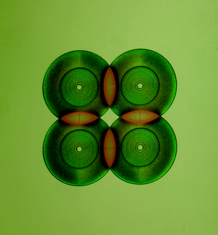
The image on this issue's cover comes from a competition held at the 12th Annual International Conference on Miniaturized Systems for Chemistry and Life Sciences (µTAS 2008) held in San Diego, California, USA, October 12–16, 2008. This competition, called ‘Under the Looking Glass: Art from the World of Small Science’, was sponsored by Lab on a Chip and the US National Institute of Standards and Technology (NIST). Yu-Wen Huang, of Texas A&M University created this micrograph, entitled ‘City Lights.’ A panel of five µTAS scientists, including Harp Minhas (Royal Society of Chemistry, UK), Andreas Manz (UK), Michael Gaitan (NIST, USA), Tae Song Kim (Korea Institute of Science & Technology, Korea) and Julio M. Ottino (Northwestern University, USA) selected City Lights (Fig. 1) from amongst 70 images that were submitted to this competition. The image is a micrograph of a solution of unlabelled double-stranded DNA, electrophoretically concentrated near a 50 µm-wide electrode. The high DNA concentration results in an increased intensity of white light reflected off the microelectrode. Additionally, a stable layer of electrolytically produced microbubbles, produce a colorful speckled effect near the tip of the electrode. The combination of these optical effects produces an image reminiscent of a city skyscraper on a foggy night. This image, which was of a technically functioning device, was evocative of mystic images of urban landscapes at the early dawn, when fog still fills the air and the first rays of sunlight pierce through the darkness, illuminating the beginning of a new day. | ||
| Fig. 1 City Lights, image reproduced by permission of Yu-Wen Huang and Victor M. Ugaz. | ||
This page also features a few of the other images submitted for this award, exemplifying the high quality of images submitted. (Fig. 2) Polymer diffractive lens array—optical elements for the focusing and collection of light inside microfluidic channels. The colour in the figure is not false, but from thin film effects in the SU8 polymer; (Fig. 3) Branched 3-D microchannel network—a branched 3-D microchannel network is formed inside an acrylic rod by harnessing electrostatic discharge phenomena that arise when a dielectric medium is energized by irradiation from an electron beam. Sudden release of the accumulated charge produces a discharge of energy powerful enough to locally vaporize and fracture the substrate, leaving behind a network of branched channels arranged in a tree-like fractal arrangement that bears remarkable similarity to naturally occurring branched architectures. The 1.5 in diameter by 4.5 in long acrylic rod shown here was irradiated using a 5 MeV electron beam, then discharged by contact with the sharp tip of a grounded electrode; (Fig. 4) Electro-active hydrodynamic single-particle traps—scanning electron microscopy (SEM) image of an array of electro-active hydrodynamic traps. Each trap is electrically addressable enabling the loading of arbitrary patterns of bioparticles for the study of fundamental processes at the level of single particles and the effects of patterns of particles on ensemble behaviour. These single-particle traps were realized by insights from multiphysics finite element modeling and numerical methods to the incorporation of new materials in bioMEMS fabrication. These traps present an exciting new avenue for the design of experiments to probe fundamental biological processes at the single-cell level; and (Fig. 5) Art-on-a-Chip—to celebrate the artist inside of all engineers, we have created a ‘painting’ using microfluidic technology, called Art-on-a-Chip. Combining standard microfluidic fabrication techniques with a few novel tricks, we have developed a method to create permanent micro-scale paintings. An intricate network of microfluidic channels in the shape of the Golden Gate Bridge, some smaller than 10 microns, are filled with dyed epoxy using novel techniques to create a tiny piece of art. These techniques have applications ranging from creating complex tissue engineering scaffolds to preserving devices for display. We believe that engineers and non-engineers alike will appreciate this work.
 | ||
| Fig. 2 Polymer diffractive lens array, image reproduced by permission of Ethan Schonbrun. | ||
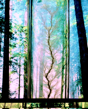 | ||
| Fig. 3 Branched 3-D microchannel network, image reproduced by permission of Jen-Huang Huang and Victor M. Ugaz. | ||
 | ||
| Fig. 4 Electro-active hydrodynamic single-particle traps, image reproduced by permission of Salil Desai. | ||
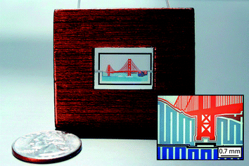 | ||
| Fig. 5 Art-on-a-Chip, image reproduced by permission of Joshua Tanner Nevill and Albert Mach. | ||
The presentation of this award at the conference, along with 2500 cash (USD), was preceded by a plenary lecture from Prof. Julio M. Ottino that cited many examples for the creative process in the visual arts and drew parallels between the creative processes in art and in science. Hollywood has often portrayed the artist or the musician, sitting with his muse, and in a spurt of creative fervor, produces his masterpiece in a short period of time. The scientific process is often portrayed in a similar way; a scientist, in a flash of inspiration, uncovers the missing piece to a problem, he then runs into the laboratory and in a brief period conquers the scientific conundrum before him. As scientists, we realize that the latter portrayal is far from reality and of the hours of sweat and perseverance necessary to realize scientific success. The same expenditure of time and effort is true of creation in the arts. As scientists, we start an experiment or model, encounter unforeseen challenges, and incrementally overcome them; a similar process occurs in the arts—the artist starts with a familiar technique of style and slowly adjusts and perfects it until it is a new creation. It was in this spirit that this award was created, to draw attention to the non-technical aspects of doing beautiful science. In other words it was intended to spotlight ideas, inventions and theories that have both a practical utility and applicability and convey an element of aesthetic appeal—a chrysalis from which the aesthetics of scientific appeal can be discussed. Our planet's most renowned scientists often assert that the aesthetic appeal of a particular experiment or theory was a major guiding force in its development. Examples include: Richard Feynman, in commenting about adhering to one of his theories in the face of apparently contradicting experimental evidence ‘It [the theory] had elegance and beauty. The goddamn thing was gleaming.’ Einstein's concluding remark about his initial announcement of the equations of general relativity ‘Scarcely anyone who fully understands this theory can escape from its magic.’
We hope that the concept and principles of this award will be appreciated by the wider scientific and non-scientific community as the intention is to reach out to all disciplines to demonstrate that scientific research is so much more than that portrayed in the general media and that science too can be an art form.
The µTAS conference hosted over 1000 attendees from Europe, the Americas, Asia and Oceania. This year, a number of awards, including several new awards, were presented at the conference to recognize important contributors to the field of microfluidics. In addition to ‘Under the Looking Glass: Art from the World of Small Science’, other awards included ‘Pioneers in Miniaturization’ sponsored by Corning Inc. and Lab on a Chip, ‘Young Innovator Award’ sponsored by Analytical Chemistry, ‘Best Poster Awards’ (one for each day of the poster exhibition) sponsored by The Society for Chemistry and Micro-Nano Systems (CHEMINAS) and the ‘Widmer Poster Award’ sponsored by Lab on a Chip.
The 13th Annual International Conference on Miniaturized Systems for Chemistry and Life Sciences (µTAS 2009) will be held in Jeju, South Korea from 1 to 5 November 2009. Details can be found at: http://www.microtas2009.org/.
Wyatt Vreeland, Ph. D.
Biochemical Science Division, NIST, USA
| This journal is © The Royal Society of Chemistry 2009 |