Inorganic nanoparticles for biological sensing, imaging and therapeutics
For the past decade, a variety of inorganic nanoparticles have been newly created or modified to provide superior materials properties with functional versatility. Simultaneously, due to their size features similar to biological species (e.g. genes, proteins, viruses) and potential advantages over existing chemical imaging agents or drugs, these nanoparticles have been examined for their uses as new tools not only for investigation of biological processes but also for sensing and treating diseases. Upon addition of specific biological functionalities such as DNAs and peptides to the nanoparticles, these nanoparticle-conjugates have shown a number of new promises and breakthroughs in the biomedical fields. Accordingly, a new research area called ‘nanomedicine’ is rapidly emerging.In this themed issue, important aspects of recent developments of inorganic nanoparticles, which are mostly based on gold, magnetic materials, quantum dots, and silica, are presented in conjunction with their applications ranging from cellular uptake to imaging of biological targets and highly effective drug delivery.
In the highlight article (DOI: 10.1039/b902462j), Zink and Stoddart show that mesoporous silica becomes one of the most advanced imaging and drug releasing systems when combined with molecular machines whose activity is triggered by external stimuli (Fig. 1). In the reviews, an excellent overview of nanomedicine (by Kim and Dobson (DOI: 10.1039/b902711b)) and a series of current perspectives of T2 type superparamagnetic magnetic resonance imaging (MRI) agents (by Gao (DOI: 10.1039/b902394a) and Zhang (DOI: 10.1039/b902182e)) and T1 type nanoparticles (by Hyeon (DOI: 10.1039/b902685a)) with potential applications in biomedical fields are presented. Also, general aspects of silica nanoparticles as a gene delivery system are discussed by Tamanoi (DOI: 10.1039/b904197d).
 | ||
| Fig. 1 A molecular machine controlled drug release system tethered to mesoporous silica (J. Zink, DOI: 10.1039/b902462j). | ||
Metallic gold nanoparticles are important in imaging, as drug carriers, and also for thermotherapy of biological targets. Morphological (rods) and structural (hollow cages) motif variations as well as surface modifications of Au nanoparticles for tuning their SPR imaging properties and their surface interactions for cells and collagens are discussed by Wei (DOI: 10.1039/b823389f), Murphy (DOI: 10.1039/b902760b), and Xia (DOI: 10.1039/b901817d) (Fig. 2), respectively.
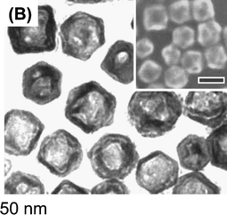 | ||
| Fig. 2 Microscope images of hollow Au-Ag nanocages (Y. Xia, DOI: 10.1039/b901817d). | ||
Quantum dots (QDs) have been especially useful for multiplexed and long term optical imaging of biological processes. While surface ligand driven photocatalytic enzyme activities are reported by Niemeyer (DOI: 10.1039/b902187f), QDs with robust polymeric surface coating technology for in vivo studies (by Taton (DOI: 10.1039/b902275a)) (Fig. 3) and new quantification technology of QDs using phage display processes (by Chan (DOI: 10.1039/b906466d)) are demonstrated.
 | ||
| Fig. 3 Epifluorescence image of zebrafish embryo with QDs (A. Taton, DOI: 10.1039/b902275a). | ||
Magnetic nanoparticles are particularly important for magnetic resonance imaging (MRI) and drug delivery. The atomic pair distribution function study of bare iron oxide nanoparticles (by Sadun (DOI: 10.1039/b911062c)), the stability, toxicity, and cellular uptake phenomena of protein coated iron oxide (by Rotello (DOI: 10.1039/b901616c)), and monocyte and macrophage uptake of anionic nanoparticles (by Wilhelm (DOI: 10.1039/b903306h)) are presented. MR imaging relaxation time measurements in conjunction with new magnetic materials properties of FePt systems are given by Parak (DOI: 10.1039/b906455a) and new screening protocols of polymer coated magnetic nanoparticles for enhanced contrast effects, colloidal stability and biocompatibility are reported by Josephson (DOI: 10.1039/b902170a). T1 based positive MRI contrast agents of gadolinium phosphates are reported to be superior to conventional ones (by Hifumi (DOI: 10.1039/b902134e). Application studies include antibiofouling polymer coated magnetic nanoparticles for cancer imaging (by Jon (DOI: 10.1039/b902445j)) and αvβ6 targeted lung cancer molecular imaging (by Gao (DOI: 10.1039/b902358e)), while imaging and cell killing effects of anticancer drug methotrexate containing iron oxide nanoparticles are presented by Sun (DOI: 10.1039/b902373a) (Fig. 4). Dual functional nanocomposites of magnetic materials and QDs are shown to be excellent in the separating and labelling of cancer cells (by Matsunaga (DOI: 10.1039/b900693a)).
 | ||
| Fig. 4 Microscope image of iron oxide nanoparticles (S. Sun, DOI: 10.1039/b902373a). | ||
Silica nanoparticles also have multi-modal features as smart materials for imaging and therapeutics. Organic dye-doped silica nanoparticles are proven to be excellent near IR probes for imaging applications (by Wiesner (DOI: 10.1039/b902286d)) and also lanthanide doped silica nanoparticles are successfully utilized for ultra-sensitive spore detection (by Lin (DOI: 10.1039/b900866g)).
I am truly grateful to all the authors of this themed issue who have contributed their state-of-the-art high quality articles. Many more breakthroughs are expected in this exciting research field at a relatively fast pace, with the anticipation of bigger impacts of materials chemistry on the biomedical sciences.

Jinwoo Cheon, Horace G. Underwood Professor of Chemistry, Yonsei University, Seoul, Korea
| This journal is © The Royal Society of Chemistry 2009 |