Multifunctional nanostructured materials for multimodal imaging, and simultaneous imaging and therapy
Jaeyun Kim, Yuanzhe Piao and Taeghwan Hyeon*
National Creative Research Initiative Center for Oxide Nanocrystalline Materials and School of Chemical and Biological Engineering, Seoul National University, Seoul 151-744, South Korea. E-mail: thyeon@snu.ac.kr
First published on 1st December 2008
Abstract
Nanotechnology offers tremendous potential for future biomedical technology. Due to their unique characteristics including superparamagnetic or fluorescent properties, and small size comparable to biomolecules, nanostructured materials have emerged as novel bioimaging, diagnostic, and therapeutic agents for the future medical field. Especially, the combinations of various nanostructured materials with different properties can offer synergetic multifunctional nanomedical platforms, which make it possible to accomplish multimodal imaging, and simultaneous diagnosis and therapy. Moreover, the conjugation of targeting moieties on the surface of these multifunctional nanomaterials gives them specific targeted imaging and therapeutic properties. In this tutorial review, we will summarize the recent reports on the fabrication strategies of multifunctional nanoplatforms and their applications to targeted multimodal imaging and simultaneous diagnosis and therapy.
 Jaeyun Kim | Jaeyun Kim received his BS (2001), MS (2003) and PhD (2007) (supervisor: Prof. Taeghwan Hyeon) in the School of Chemical and Biological Engineering (CBE) of Seoul National University (SNU). Currently, he is working as a postdoctoral researcher at the Center for Oxide Nanocrystalline Materials (CONMR) of SNU. His research area is multifunctional nanomaterials for biomedical applications. He received an MRS Graduate Student Award in 2007. |
 Yuanzhe Piao | Yuanzhe Piao received his BS (1993) and MS (1996) in chemistry from Yanbian University, China, and his PhD from SNU (2004) under the supervision of Prof. Hasuck Kim in the area of electrochemistry. Since 2004, he has been a postdoctoral researcher at the CONMR of SNU working on the synthesis of nanomaterials for catalytic and biomedical applications. |
 Taeghwan Hyeon | Taeghwan Hyeon received his BS (1987) and MS (1989) from SNU, and his PhD in chemistry from the University of Illinois (1996). Since he joined the CBE of SNU in 1997, he has been working on uniform nanocrystals and nanoporous materials. He is currently director of the CONMR, and serves on the editorial boards of Advanced Materials, Chemical Communications, and Small. |
1. Introduction
Nanotechnology, one of the critical technologies of the 21st century, is multidisciplinary and interdisciplinary and covers diverse fields including chemistry, physics, materials science, engineering, biology, and even medicine. Nanotechnology makes use of the unique chemical and physical properties of nanoscale (1–100 nm) materials that cannot be achieved in their bulk counterparts. For example, CdSe nanoparticles, known as quantum dots (QDs), exhibit photoluminescence with a controllable wavelength ranging from the visible to near infrared according to their size.1 Colloidal gold nanoparticles exhibit unique surface plasmon resonance (SPR) properties derived from the interaction of electromagnetic waves with the electrons in the conduction band.2 Magnetite (Fe3O4) nanoparticles are superparamagnetic and exhibit high magnetization but no residual magnetization in the absence of an external applied magnetic field.3 Diverse nanostructured materials with these interesting properties have found various biomedical applications including imaging, separation, and drug delivery.The major applications of nanomaterials in the biomedical field can be mainly divided into imaging and therapy. Most of the clinically used imaging and therapeutic modalities are small molecules, such as the gadolinium complexes used as T1 magnetic resonance imaging (MRI) contrast agents and anticancer chemical drugs. The limitations of these small molecules are their very short blood circulation time and non-specific biodistribution, which causes many unwanted side effects. Nanostructured materials can be employed to overcome these limitations.4 For example, the blood circulation times can be increased significantly by the size control and surface modification of nanomaterials and by conjugating targeting molecules, such as antibodies and peptides, on their surface. Various kinds of nanomaterials have been developed for imaging and therapeutic applications and Table 1 summarizes the diverse imaging and therapeutic modalities based on nanomaterials.
| Modality | Component | |
|---|---|---|
| a MRI: magnetic resonance imaging; OCT: optical coherence tomography; PET: positron emission tomography; SPECT: single photon emission computed tomography; CT: computed tomography. | ||
| Imaging | Optical imaging | Organic dye |
| Dye-doped silica | ||
| Quantum dots | ||
| Lanthanide atom | ||
| Carbon nanotube | ||
| MRIa | Paramagnetic ion (Gd3+, Mn2+) | |
| Nanoparticles of paramagnetic ion (Gd2O3, MnO) | ||
| Superparamagnetic nanoparticle (Fe3O4, MnFe2O4, FeCo) | ||
| OCTa | Gold nanostructure (nanoparticle, nanorod, nanoshell, nanocage) | |
| PETa, SPECTa | Radioisotope (18F, 124I, 64Cu, 99mTc, 11In) | |
| CTa | Iodine | |
| Gadolinium | ||
| Gold nanoparticle | ||
| Bismuth sulfide nanoplate | ||
| Ultrasound | Microbubble | |
| Perfluorocarbon nanoparticle | ||
| Therapy | Chemotherapy | Anticancer drug (doxorubicin, camptothecin, paclitaxel, etc.) |
| Photodynamic therapy | Photosensitizer | |
| Quantum dots | ||
| Photothermal therapy | Gold nanostructure (nanoparticle, nanorod, nanoshell, nanocage) | |
| Carbon nanotube | ||
| Graphite carbon shell | ||
| Neutron-capture therapy | Boron | |
| Gadolinium |
The use of combinations of different nanostructured materials will allow the development of novel multifunctional nanomedical platforms for multimodal imaging, and simultaneous diagnosis and therapy. For example, the combination of MRI contrast agent and fluorescent organic dye can allow the detection of cancer through non-invasive MRI and the optical guide of surgery. The encapsulation of MRI contrast agent and anticancer drug in a nanostructured matrix has the potential to allow for simultaneous diagnosis and targeted chemotherapy. There are many possible combinations of the various imaging and therapeutic modalities. In this tutorial review, we will summarize the recent papers on multifunctional nanoplatforms for biomedical applications. In the following two sections, (1) multimodal imaging probes and (2) simultaneous diagnostic and therapeutic agents will be discussed.
2. Multimodal imaging probes
Most popular nanostructured multimodal imaging probes are combinations of MRI and optical imaging modalities. MR imaging can offer high spatial resolution and the capacity to simultaneously obtain physiological and anatomical information, whereas optical imaging allows for rapid screening. In dual imaging modalities, by combining MR and optical imaging probes, it is possible to obtain multiple imaging data using the advantages of both methods. Trimodal imaging probes have also been designed by adding another imaging modality to the dual MRI and optical imaging probes. In particular, positron emission tomography (PET) isotopes such as 111In or 64Cu, which emit gamma rays from their decay, have been most frequently used as the third probes.MRI is one of the most powerful and non-invasive diagnostic techniques for living organisms based on the interaction of protons with the surrounding molecules of tissues. The current MRI contrast agents are in the form of either paramagnetic complexes5 or superparamagnetic nanoparticles.6 Paramagnetic complexes, which are usually gadolinium (Gd3+) or manganese (Mn2+) chelates, accelerate the longitudinal (T1) relaxation of water protons and therefore exhibit bright contrast where they localize. Gadolinium diethylenetriaminepentaacetate (Gd-DTPA) has been the most widely used T1 contrast agent. On the other hand, superparamagnetic iron oxide nanoparticles accelerate the transverse (T2) relaxation of water protons and exhibit dark contrast. Commercially used T2 contrast agents of iron oxide nanoparticles are synthesized in the aqueous phase via a co-precipitation method and stabilized with hydrophilic polymers, such as dextran.
Optical imaging probes include various dye molecules, dye-doped silica materials, quantum dots, lanthanide compounds with up- or down-conversion properties, and near infrared (NIR) emitting nanostructures. All optical imaging modalities can be coupled with MRI contrast agents. The methods of combining these two modalities include the synthesis of multimodal probes with inherently combined properties, for example, heterostructured nanoparticles of Fe3O4–CdSe and Fe3O4–Au, and the assembly of different components in a single nanomatrix, for example, quantum dots stabilized with gadolinium-chelated phospholipids.
Based on the component of the MRI contrast agent, we will categorize the multimodal imaging probes into (1) superparamagnetic iron oxide nanoparticle (SPIO) based and (2) paramagnetic gadolinium (Gd) complex based multifunctional nanoplatforms.
2.1 SPIO based multimodal imaging probes
As described above, the representative T2 MRI contrast agent is superparamagnetic iron oxide nanoparticles (SPIO). There are several ways to chemically synthesize iron oxide nanoparticles, for example, co-precipitation, microemulsion, thermal decomposition, and hydrothermal synthesis.7 Commercially available T2 contrast agents, such as Ferridex and Combidex, have been synthesized using the co-precipitation method, which involves the simultaneous precipitation of Fe2+ and Fe3+ species in aqueous media, and is known to be simple and environmentally friendly. The synthesized nanoparticles are generally coated with hydrophilic polymers, making them highly stable under physiological conditions. However, iron oxide nanoparticles synthesized by the co-precipitation method are generally polydisperse and poorly crystalline. Recently, hydrophobic iron oxide nanoparticles synthesized by the thermal decomposition method have been extensively studied as MRI contrast agents, because they are uniform and highly crystalline.8 These high quality hydrophobic iron oxide nanoparticles have been transferred to the aqueous phase using amphiphilic polymers, phospholipids, and silica shells.9Table 2 summarizes the dual imaging probes based on iron oxide nanoparticles. Iron oxide nanoparticles synthesized using the precipitation method are more frequently used and, more recently, uniform iron oxide nanoparticles synthesized by the thermal decomposition method have begun to be used as MRI contrast agents for dual MRI-optical probes.| Multimodal nanoparticles based on SPIO | Particle size/nm | λem/nm | Relaxivity per Fe/mM−1 s−1 | B0a/T | Test | Reference | Year | ||
|---|---|---|---|---|---|---|---|---|---|
| TEMa | DLSa | r1a | r2a | ||||||
| a TEM: transmission electron microscopy; DLS: dynamic light scattering; r1: longitudinal relaxivity; r2: transverse relaxivity; B0: magnetic field strength. | |||||||||
| CLIO-SS-R4-Cy5.5 | 68 ± 13 | 695 | 27.8 | 91.2 | 9.4 | In vivo | Josephson10 | 2002 | |
| CLIO-Cy5.5-EPPT | 35.8 | 695 | 26.43 | 53.44 | 9.4 | In vivo | Moore12 | 2004 | |
| Fe3O4-SiPEG-Cltx-Cy5.5 | 10.5 ± 1.5 | 695 | 4.7 | In vitro | Veiseh15 | 2005 | |||
| Fe3O4-SiPEG/TCL-Cy5.5 | 32.6 ± 3.7 | 695 | 1.5 | In vivo | Lee16 | 2007 | |||
| CoFe2O4@SiO2(FITC) | 60 | ∼90 | 518 | 4.7 | In vitro | Yoon19 | 2006 | ||
| Fe3O4@SiO2(FITC) | 50 | 128 | 1.5 | In vitro | Lu20 | 2007 | |||
| DySiO2-(Fe3O4)n | 46 | 580 | 397 | 9.4 | In vitro | Lee21 | 2006 | ||
| Fe3O4@mesoporous silica(FITC) | length ∼154 | 518 | 153 | 4.7 | In vitro | Lin22 | 2006 | ||
| Fe2O3/CdSe@silica | 11∼14 | 12∼14 | 600 | In vitro | Selvan24 | 2007 | |||
| Au–Fe3O4 | 8(Au)–20(Fe3O4) | ∼60 | 530 Abs | 3.56 | 105 | 3 | In vitro | Xu25 | 2007 |
| FePt-Au | 6(FePt)–10(Au) | 530 Abs | 58.7 | 9.4 | In vitro | Choi26 | 2006 | ||
| Fe2O3-SWNT | >1000 | NIR Abs | 14.1 | In vitro | Choi27 | 2007 | |||
The representative multimodal imaging probe is superparamagnetic iron oxide nanoparticles coupled with fluorescent organic dyes. The crosslinked iron oxide nanoparticles (CLIO) are chemically crosslinked dextran coated iron oxide nanoparticles and have been widely used for in vivo as well as in vitro MR imaging, because of their biocompatibility, small hydrodynamic size, and stable hydrophilic polymer coating. Josephson et al. developed multimodal imaging probes composed of CLIO conjugated with NIR fluorescent dyes.10 Optical imaging in the NIR region between 700 and 900 nm has low absorption by intrinsic photoactive biomolecules, allows light to penetrate several centimetres into the tissue, and has minimal tissue autofluorescence. They conjugated arginyl peptides to CLIO, followed by the attachment of the indocyanine dye, Cy5.5. Following the subcutaneous injection of this probe into nude mice, the lymph nodes were darkened on the MR images and simultaneously detectable by NIR fluorescent imaging. Kircher et al. of the same research group demonstrated the potential of this multimodal MRI and optical probe using an in vivo brain tumor model.11 They conjugated Cy5.5 directly with aminated CLIO to prepare the multimodal probe and injected CLIO-Cy5.5 via the tail vein into rats in which a brain tumor was developed by the xenograft of 9L gliosarcoma cells with GFP expression. In the T2-weighted MRI images, the tumor region exhibited a reduction of the signal intensity, indicating that the brain tumor could be diagnosed, and the tumor site was specified by preoperative MRI. The delineation of the brain tumor in intraoperative fluorescent imaging was examined after craniotomy. The brain tumor was clearly visualized by Cy5.5 NIR fluorescence and the imaged region was correlated with the actual tumor extent that was determined by GFP fluorescence. These multimodal MRI and NIR fluorescent probes permitted the preoperative visualization of brain tumors by serving as T2 MRI contrast agents and allowed for the intraoperative discrimination of tumors from brain tissue using their NIR fluorescence. Using CLIO-Cy5.5 as a multimodal imaging probe, active cancer targeting has been demonstrated by the conjugation of targeting molecules including peptides and small molecules. Moore et al.12 synthesized cancer targeted multimodal imaging probes consisting of CLIO-Cy5.5 attached to synthetic EPPT peptides on a dextran coating to target the underglycosylated mucin-1 (uMUC-1) antigen of various cancer cells (Fig. 1a). In vivo MR imaging was performed on animals bearing bilateral uMUC-1-positive and uMUC-1-negative tumors in their legs before and 24 h after the intravenous injection of the probe. After administration of the probe, a significant signal reduction in T2 MRI and bright NIR emission was observed in the uMUC-1-positive tumors compared to those in the uMUC-1-negative tumors (Fig. 1b–d). Instead of peptides, Weissleder and co-workers investigated whether the multivalent attachment of small molecules can increase the specific binding affinity of the nanoprobes.13 First, they conjugated 146 different small molecules (MW < 500 Da) with functional groups consisting of primary amines, alcohols, carboxylic acids, sulfhydryls, and anhydrides on CLIO. The library of CLIO attached to small molecules was rapidly screened against different cell lines and it was discovered that the specificity of the nanoparticles for the cells depended on the attached small molecules. Based on the specificity of the small molecules for different cells, they prepared CLIO-Cy5.5 conjugated with pancreatic cancer specific small molecules and confirmed the targeting of nanoparticles in the tumor site in vivo.
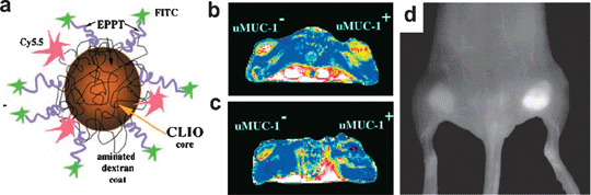 | ||
| Fig. 1 (a) The scheme of the probe. Representative T2 maps of the animals bearing underglycosylated mucin-1 antigen (uMUC-1)-negative (U87) and uMUC-1-positive (LS174T) tumors on their legs (b) before and (c) 24 h after the intravenous injection of the probe. (d) Near-infrared fluorescence (NIRF) image of mouse bearing bilateral underglycosylated mucin-1 antigen (uMUC-1)-negative (U87) and uMUC-1-positive (LS174T) tumors. Reproduced with permission from ref. 12. | ||
Trimodality imaging probes having MRI, NIR fluorescent imaging, and PET functionalities were designed using CLIO as a platform. The Weissleder group decorated NIR fluorescent dyes on an aminated dextran coating of CLIO and conjugated DTPA to chelate PET tracer 64Cu, resulting in a PET, MRI, and optically detectable imaging agent.14 The advantage of the PET imaging capability is that it can potentially provide detection sensitivities an order of magnitude higher than that of MRI. This trimodality nanoprobe showed the in vivo synergetic detection of macrophages in atherosclerotic plaques. Because the macrophages in atherosclerosis have an affinity for polysaccharide-containing supramolecular structures, the dextran-coated nanoparticles could target the macrophages.
Instead of a dextran coating, poly(ethylene glycol) (PEG) is an alternative and representative biocompatible coating material for iron oxide nanoparticles used in biomedical applications. As the iron oxide nanoparticles synthesized in the aqueous phase via the co-precipitation process have many hydroxyl groups on their surface, trialkoxysilane (–Si(OCH3)3 or –Si(OCH2CH3)3)-terminated with PEG was used for coating the nanoparticles via the hydrolysis of the trialkoxysilane groups to –Si(OH)3 and subsequent condensation with the surface hydroxyl groups of the nanoparticles. Zhang and co-workers modified the surface of iron oxide nanoparticles with trifluoroethylester-terminal-PEG-silane, which was then converted to an amine-terminated PEG.15 The terminal amine groups were used for the conjugation of Cy5.5 and chlorotoxin, a targeting peptide for glioma tumors, which are currently known as the most common and lethal type of primary brain tumor. In vitro MRI and confocal fluorescence microscopy showed the strong preferential uptake of the multimodal probes by glioma cells compared to the control nanoparticles. A significantly higher degree of internalization of the probes by glioma cells compared to the control non-cancerous cells was observed, indicating the cancer-targeting abilities of the probes for gliomas (Fig. 2).
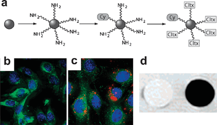 | ||
| Fig. 2 (a) Schematic diagram of the surface modification and preparation of SPIO-chlorotoxin-Cy5.5 conjugates. Confocal fluorescent images of 9L cells cultured with (b) control SPIO-Cy5.5 and (c) SPIO-chlorotoxin-Cy5.5. (d) MR phantom image of 9L cells cultured with control SPIO-Cy5.5 (left) and SPIO-chlorotoxin-Cy5.5 (right) and embedded in agarose (4.7 T, spin-echo pulse sequence, TR 3000 ms, TE 15 ms). Reproduced with permission from ref. 15. | ||
Recently, Lee et al. reported multimodal probes consisting of iron oxide nanoparticles with thermally crosslinked polymer shells.16 They synthesized triblock polymers having –Si(OCH3)3 groups for the purpose of bonding to iron oxide and the crosslinking of the polymer shells, PEG for biocompatibility, and N-hydrosuccinimide-activated carboxylic acid for the further conjugation of fluorescent dyes. The silane groups were attached to the surface of iron oxide nanoparticles synthesized by co-precipitation and the residual silane groups were thermally crosslinked via heating at 80 °C. The in vivo T2 MR and NIR fluorescent imaging of tumor-bearing mice after the intravenous injection of the Cy5.5 conjugated multimodal probes revealed that the probes were accumulated at the tumor site via the enhanced permeation and retention effect, which made it possible to detect tumors by MRI and optical imaging. The ex vivo NIR fluorescent images of several organs and tumors indicated that the highest fluorescence intensity was observed in the tumor. Compared to the Cy5.5 conjugated multimodal probes, when the free Cy5.5 dye was intravenously injected into a tumor-bearing mouse as a control experiment, only a faint fluorescence signal was observed in the tumor.
The Hyeon group reported a simple synthesis of PEG-derivatized phosphine oxide ligands to make water dispersible metal oxide nanoparticles.17 The ligands were synthesized from a simple reaction between POCl3 and PEG. The phosphine group can be exchanged with the stabilizing ligands of various hydrophobic metal oxide nanoparticles of Fe3O4, NiO, MnO, and TiO2. A fluorescent dye, fluorescein isothiocyanate (FITC), was attached to the activated PEG terminal group of Fe3O4 nanoparticles to synthesize fluorescent magnetic nanoparticles. The bifunctional nanoparticles were shown to act as simultaneous T2 MRI and optical cell labeling agents.
In general, fluorescent dyes suffer from a low fluorescence quantum yield and severe photobleaching. To overcome these problems, the dye molecules were incorporated in a protective silica matrix.18 The silica nanomatrix embedded with dye molecules exhibited more intense fluorescence and better photostability than conventional molecular dyes. Because a large number of dye molecules were confined in the relatively small volume of a single silica nanoparticle, a stronger fluorescence signal was observed than that obtained with a single dye molecule under the same excitation conditions. The silica nanomatrix also serves as a shield to protect the dyes from quenching. In addition, silica is known to be biocompatible and the silica surface can be easily modified with various organic functional groups, thereby allowing for the conjugation of targeting molecules. Based on these advantages, iron oxide nanoparticles have been assembled with fluorescent silica to make multimodal imaging probes. Various silica nanostructures including nanoshells, mesoporous silicas, and silica nanoparticles have been employed for multimodal probes using dye-doped silica.
Lee and co-workers reported magnetic fluorescent core–shell nanoparticles.19 Magnetic cobalt ferrite (CoFe2O4) nanoparticles were synthesized by co-precipitation and they were coated with a dye-doped silica shell via the modified Stöber method. The surface of the silica shells was further functionalized with PEG and conjugated with an antibody to target cancer cells. The core–shell nanoparticles in aqueous solution exhibited T2 contrast enhancing properties in the phantom test and the confocal images of the cancer cells treated with the nanoparticles showed strong fluorescence, indicating that the probes have specificity to cancer cells. Huang and co-workers also synthesized core–shell nanoparticles composed of iron oxide nanoparticles and FITC-doped silica shells.20 In this case, the iron oxide nanoparticles were synthesized by the thermal decomposition method and stabilized with oleic acid and oleyl amine. The hydrophobic iron oxide nanoparticles were coated with dye-doped silica shells via a reverse microemulsion system. The surface of the core–shell nanoparticles was not further functionalized and, therefore, the functional groups on the surface might be the hydroxyl group along with the amine group. The use of core–shell nanoparticles was demonstrated for human stem cell tracking. The probes were easily internalized into human mesenchymal stem cells (hMSCs). These labeled stem cells were injected subcutaneously into the dorsal flank of a mouse and could be visualized in a clinical 1.5 T MRI imager.
Heterostructured nanoparticles composed of iron oxide nanoparticles and dye-doped silica particles can also render multimodality. The Cheon and Shin groups fabricated core–satellite structured hybrid nanoparticles composed of dye-doped silica as the core and multiple iron oxide nanoparticles as the satellite (Fig. 3).21 The stabilizing agent of the hydrophobic nanoparticles, oleic acid, was exchanged with 2,3-dimercaptosuccinic acid to prepare water-dispersible nanoparticles. Thiolated iron oxide nanoparticles were assembled on the aminated rhodamine B isothiocyanate (RITC)-doped silica particles using sulfosuccinimidyl 4-(N-maleimidomethyl)cyclohexane-1-carboxylate (Sulfo-SMCC) as a crosslinker. Interestingly, compared to free water-soluble iron oxide nanoparticles with the same size, this probe exhibited superior T2 MRI capabilities due to the synergistic magnetism of the multiple Fe3O4 satellites surrounding the core silica nanoparticles (Fig. 3c). The in vitro MR and optical imaging of neuroblastoma cells using the antibody-conjugated core–satellite nanoparticles revealed that this probe could be used as a targeted dual modal imaging probe. Heterostructured nanoparticles comprised of iron oxide nanoparticles and dye-doped mesoporous silica was reported by the Mou group.22 The hydrophobic iron oxide nanoparticles were coated with silica using a reverse microemulsion system. Using these core–shell nanoparticles as the seed, FITC-doped mesoporous silica was synthesized in the presence of the mesostructure-directing agent, cetyltrimethylammonium bromide (CTAB) to produce the heterostructure of silica coated Fe3O4/dye-doped mesoporous silica.
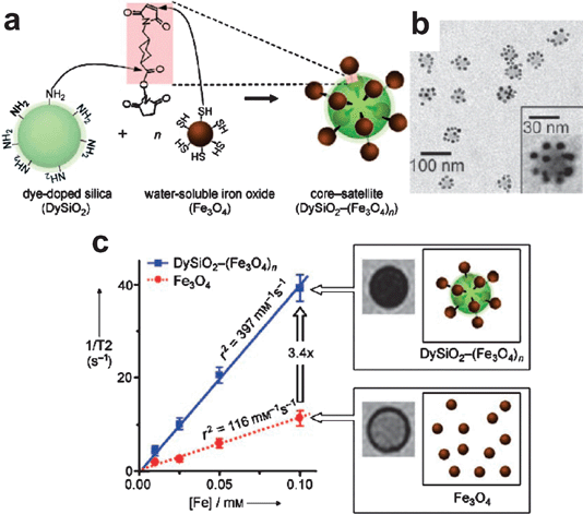 | ||
| Fig. 3 (a) Schematic diagram of the synthesis of core–satellite DySiO2–(Fe3O4)n nanoparticles. (b) Core–satellite DySiO2–(Fe3O4)n nanoparticles. (c) Synergistic MR enhancement effect of DySiO2–(Fe3O4)n. Reproduced with permission from ref. 21. | ||
Instead of an organic dye species, inorganic quantum dots could be used as the optical modality in multifunctional nanoparticles. Hyeon and co-workers reported magnetic fluorescent mesoporous silica nanoparticles composed of mesoporous silica nanoparticles embedded with iron oxide nanoparticles and quantum dots.23 Hydrophobic magnetic and fluorescent nanoparticles were transferred into the aqueous phase using CTAB as a secondary surfactant. Because CTAB also acted as a structure directing agent of the mesostructures, the subsequent silica sol-gel reaction resulted in the formation of mesoporous silica spheres embedded with magnetic and fluorescent nanoparticles. The nanoparticles exhibited superparamagnetic and fluorescent properties. These particles could be used as drug delivery vehicles due to the high surface area and large pore volumes of the mesoporous silica and drug loading and controlled release was demonstrated using them.
Heterodimers of inorganic nanoparticles can impart multimodal imaging capability. For example, Fe3O4–CdSe and Fe3O4–Au nanoparticles not only have magnetic properties, but also optical properties which can be used for cell imaging. The Ying group synthesized Fe3O4–CdSe heterodimer nanoparticles by growing CdSe on the surface of the as-synthesized Fe3O4 nanoparticles.24 The emission of the QD part could be controlled by adjusting the reaction time in the QD growing step. The resulting Fe3O4–CdSe nanoparticles were coated with silica shells via the silica sol-gel reaction in a reverse microemulsion system. The silica surface was functionalized with the oleyl-PEG group and the optical imaging of live cells was demonstrated using these probes. Although no MRI capability was demonstrated in their paper, it might be possible to use this probe as an MRI contrast agent.
Heterodimers of magnetic nanoparticles and gold nanoparticles can also serve as MRI contrast agents and optical probes. The Sun group demonstrated multimodal imaging using dumbbell shaped Fe3O4–Au nanoparticles.25 Fe3O4 nanoparticles were grown on the surface of the as-prepared Au nanoparticles in the presence of oleic acid and oleyl amine. PEG conjugated with dopamine and thiol groups was attached to the surface of Fe3O4 and Au moieties via the surfactant-exchange reaction, respectively, and an antibody was conjugated at the terminal group of PEG on Fe3O4. Cancer targeted MRI images were obtained using these antibody conjugated Fe3O4–Au nanoparticles. In addition, the cancer cells could be visualized with a scanning confocal microscope using the surface plasmon resonance of the Au nanostructure. FePt–Au heterostructured nanoparticles were also used as dual modal detection probes.26 Antibody conjugated FePt–Au nanoparticles were used as both a T2 MRI contrast agent and biosensor. Neutravidin conjugated nanoparticles were employed as biotin detecting probes in a patterned biochip via subsequent silver-staining.
Single walled carbon nanotubes (SWCNTs) exhibit NIR fluorescence derived from their intrinsic graphene structure. Using this characteristic of SWCNTs, Strano and co-workers demonstrated the multifunctionality of Fe3O4–SWCNT heteronanostructures as dual magnetic and fluorescent imaging agents.27 The SWCNTs were synthesized by the chemical vapor deposition of CO in the presence of Fe(CO)5 as a catalyst. During their synthesis, Fe3O4 nanoparticles were generated on the end of the SWCNTs. DNA was wrapped on the heteronanostructures, making them water-dispersible. The in vitro T2 MRI of murine macrophage cells cultured with the Fe3O4–SWCNT heteronanostructures showed a signal decrease compared to the control sample. Furthermore, the NIR mapping of the cells clearly revealed the cell boundaries where the bright red emission of the SWCNTs was observed, while a dark blue area was observed outside the cells.
2.2 Paramagnetic gadolinium ion (Gd3+) based multimodal imaging probes
Due to its seven unpaired electrons, large magnetic moment, and suitably long electronic relaxation time, gadolinium(III) complexes have been most popularly used as MRI contrast agents. Because of the toxicity of the Gd3+ ion, chelate ligands are coordinated to the ions to form metal complexes. The resulting complexes are thermodynamically and kinetically stable and much less toxic. Among them, gadolinium diethylenetriaminepentaacetate (Gd-DTPA) is the most widely used T1 contrast agent. To develop MR contrast agents carrying high amounts of magnetic centers, Gd species are incorporated into various nanostructured materials including quantum dots, dendrimers, silica nanoparticles and mesoporous silica materials, generating new multimodal probes. Using these Gd3+ incorporated nanostructured materials, several multimodal T1 MR and optical imaging probes have been developed. Instead of metal complexes, Gd2O3 nanoparticles have also been used as nanoplatforms. Table 3 shows a comparison between the various multimodal imaging probes based on Gd3+ ions.| Multimodal nanoparticles based on Gd3+ ions | Particle size/nm | λem/nm | Relaxivity per Gd/mM−1 s−1 | Number of Gd per particle | Relaxivity per particle/mM−1 s−1 | B0/T | Test | Ref. | Year | |||
|---|---|---|---|---|---|---|---|---|---|---|---|---|
| TEM | DLS | r1 | r2 | r1 | r2 | |||||||
| Gd-DTPA-phospholipid stabilized QD | 5 | 560 | 12.0 | 170 | 2000 | In vitro | Mulder28 | 2006 | ||||
| Gd-TSPETE-QD@silica | 10∼20 | 590 | 20.5 | 151 | 107 | 2190 | 16![[thin space (1/6-em)]](https://www.rsc.org/images/entities/char_2009.gif) 157 157 | 4.7 | Phantom | Yang29 | 2006 | |
| cf. Gadoteridol | 5.8 | 7.4 | 1 | 5.8 | 7.4 | |||||||
| Gd-TSPETE-silica nanoparticle-[Ru(bpy)3]2+ | 100 | 600 | 9.0 | 116 | 16000 | 140000 | 1800000 | 4.7 | Phantom | Santra30 | 2005 | |
| Gd-DTPA-PAMAM G6 dendrimer-Cy5.5 | 5∼6 | 699 | 13.9 | 36.5 | 145 | 2020 | 5290 | 3 | In vivo | Talanov31 | 2006 | |
| Gd-Si-DTTA-silica nanoparticle-[Ru(bpy)3]2+ | 40 | 615 | 19.7 | 60.0 | 10200 | 201000 | 612000 | In vitro | Rieter32 | 2007 | ||
| cf. Gd-Si-DTTA | 6.8 | 7.0 | 1 | 6.8 | 7.0 | |||||||
| Gd-LbL(3 layers)-silica nanoparticle-[Ru(bpy)3]2+ | 40∼50 | 615 | 19.0 | 55.0 | 17070 | 324000 | 939000 | In vitro | Kim33 | 2007 | ||
| Gd-DTTA-mesoporous silica nanoparticle-RITC | 60∼120 | 575 | 28.8 | 65.5 | 24000 | 700000 | 1600000 | 3 | In vivo | Taylor34 | 2008 | |
| Gd-DTTA-mesoporous silica nanorod-FITC | Length 320∼540 | 520 | 22.0 | 41.0 | 26637 | 586000 | 1090000 | 0.47 | In vitro | Tsai35 | 2008 | |
| Gd2O3@Cy5-doped silica | 3.3 | 690 | 8.8 | 11.4 | 420 | 3700 | 4800 | 7 | In vivo | Bridot36 | 2007 | |
| cf. Gd-DTTA | 4.1 | 4.9 | 1 | 4.1 | 4.9 | |||||||
Mulder et al. fabricated quantum dots coated with paramagnetic Gd3+ ions and pegylated lipids (Fig. 4).28 First, CdSe–ZnS core–shell quantum dots were synthesized in the presence of organic surfactants. Then, pegylated, maleimide-functionalized, and gadolinium-chelated phospholipids were immobilized onto the hydrophobic QDs, rendering them water-soluble, cancer-targetable, and paramagnetic. The hydrophobic chains of the phospholipids and the organic surfactants were interdigitated via their hydrophobic interaction when they were sonicated in an aqueous phase. After separating the empty micelles from the micelles containing QDs by ultracentrifugation, cyclic arginine-glycine-aspartic acid (RGD) peptide sequences were conjugated with the maleimide groups on the surface of the resulting QDs. RGD peptides are known to act as targeting molecules for αvß3-integrins overexpressed on the surface of angiogenic endothelial cells and tumor cells. The in vitro targeted dual MR and fluorescent imaging capability of paramagnetic QDs (pQDs) was demonstrated for angiogenic human umbilical vein endothelial cells (HUVECs).
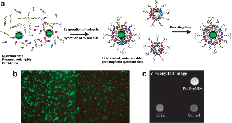 | ||
| Fig. 4 (a) Schematic representation of the preparation of QDs with a paramagnetic micellular coating. (b) Fluorescence microscopy of HUVEC incubated with RGD-pQDs and bare pQDs. (c) T1-weighted image of cells that were incubated with RGD-pQDs, pQDs, or without contrast agent. Reproduced with permission from ref. 28. | ||
Gd3+-functionalized QDs embedded in a silica nanomatrix were investigated as dual modal imaging probes by Yang, Santra and their co-workers.29 They synthesized water-dispersible silica-coated ZnS-passivated CdS:Mn QDs functionalized with Gd3+ ions. After applying a silica coating to the QDs via the reverse microemulsion method, the surface of the silica shells was functionalized with n-(trimethoxysilylpropyl)ethyldiamine triacetic acid trisodium salt (TSPETE) and gadolinium(III) acetate. TSPETE, a metal chelating carboxylate silane coupling agent, is known to have five reactive coordination sites for the capture of Gd3+ ions. Santra et al. also fabricated dye-doped silica nanoparticles via the reverse microemulsion method, and functionalized the silica surface with TSPETE and chelated Gd3+ ions.30
Due to their well-defined structures, distinct sizes, and large number of accessible reactive amine functional groups on the periphery, dendrimers have been investigated as potential imaging probes. The representative water-soluble and biocompatible dendrimers, polyamidoamine (PAMAM) dendrimers, were used as nanoplatforms for multimodal imaging probes. Talanov et al. used generation 6 (G6) PAMAM dendrimers as nanoplatforms on which 256 surface amino groups were conjugated with Gd3+-chelating ligands and NIR fluorescent Cy5.5 (Fig. 5a).31 They used this nanoprobe for in vivo lymph node imaging. The nanoprobe was injected into the mammary fat pad of a normal mouse. Within approximately 30 min after the injection, the agent was taken up by the draining axillary mammary lymph node, which was verified simultaneously by T1 MRI and NIR fluorescent imaging. Visualization of the sentinel lymph node in both the magnetic resonance and fluorescence images was clearly achieved (Fig. 5b). Due to the much better spatial resolution in MRI, both the lymph node and lymphatic channel were visualized. As described in relation to the SPIO-Cy5.5 nanoprobe, the combination of MRI and NIR fluorescent imaging could potentially allow lymph nodes to be mapped with MRI prior to surgical resection and subsequent fluorescent imaging can guide the surgeon during the surgery.
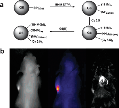 | ||
| Fig. 5 (a) Scheme for the preparation of the dual modality dendrimer-based nanoprobe. (b) Sentinel lymph node images of a normal mouse. Reproduced with permission from ref. 31. | ||
Although the nanoplatforms introduced above have a sufficiently high surface area to load a large amount of paramagnetic ions, several strategies have been pursued to further increase the payloads of Gd3+ ions in order to develop high relaxivity MRI contrast agents. The accessibility of the Gd centers to water molecules is the key to design highly efficient T1 MR contrast agents in nanoparticulate form. Lin and co-workers used dye-doped silica nanoparticles as fluorescent nanoplatforms to load Gd3+ ions.32 They synthesized silica nanoparticles containing Ru(bpy)3via the reverse microemulsion method and functionalized the surface of the silica with Gd3+-chelating ligands (Fig. 6a). They synthesized silane agents containing Gd3+-chelating ligands, for example, silated-Gd-DTTA (Gd-Si-DTTA, DTTA = diethylenetriaminetetraacetate). The nanoparticles modified with Gd-Si-DTTA have higher r1 and r2 relaxivity values compared to that of the Gd-Si-DTTA complex itself. Moreover, the Gd-Si-DTTA-modified silica nanoparticles showed much higher r1 and r2 relaxivities per particle compared to the other types of nanoparticles described above. The same group increased the relaxivities of Gd3+-chelated dye-doped silica nanoparticles using the layer-by-layer (LbL) method.33 They deposited the Gd-DOTA (DOTA = 1,4,7,10-tetraazacyclododecane-1,4,7,10-tetraacetic acid) complex on the above Gd-Si-DTTA-modified silica nanoparticles via the electrostatic interactions between the positively charged complex and the negatively charged polystyrenesulfonate (PSS) group (Fig. 6b). As the number of layers was increased, the r1 and r2 values per particle were increased proportionally. This indicates that water molecules could readily access the Gd centers, thus allowing for efficient water proton relaxivity, because of the hydrophilic nature of the layer composed of Gd-DOTA and PSS. The in vitro targeted dual imaging of T1 MRI and fluorescent imaging was also demonstrated by the noncovalent functionalization of RGD peptides via electrostatic interaction.
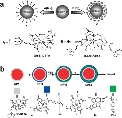 | ||
| Fig. 6 (a) Synthesis of hybrid silica nanoparticles. Reproduced with permission from ref. 32. (b) LbL self-assembly strategy for magnetic fluorescent nanoparticles. Reproduced with permission from ref. 33. | ||
Mesoporous silica can be another strategy to increase the Gd3+ loading in nanoplatforms owing to its high surface area and highly accessible mesopores. Recently, two research groups reported dual imaging agents using mesoporous silica.34,35 The Lin group synthesized dye-doped mesoporous silica nanoparticles with an average size of 75 nm and modified their surface via post-functionalization with Gd-Si-DTTA.33 The N2 adsorption/desorption measurements indicated that the surfactant-extracted mesoporous silica nanoparticles are highly porous with a surface area of 1633 m2 g−1 and a pore diameter of 2.4 nm, whereas the mesoporous silica post-functionalized with Gd-Si-DTTA has a reduced surface area of 1470 m2 g−1 and a pore diameter of 0.9–1.0 nm. The relaxivities per particle of the mesoporous silica nanoparticles loaded with Gd3+ were even higher than those of non-porous silica nanoparticles loaded with Gd3+via the LbL method.33 The enhanced MR relaxivity was attributed to the ready access of water molecules through the mesopores of the Gd3+-loaded mesoporous silica particles. Using the Gd3+-loaded mesoporous silica nanoparticles, in vivo MR contrast enhancing efficiency was demonstrated upon the tail vein injection of 2.1 μmol kg−1 of body weight of the nanoprobes. A significant T1-weighted enhancement was clearly visible in the aorta of the mouse 15 min post injection. Moreover, after the tail injection of 31 μmol kg−1, a significant loss of the MR signal was observed in the liver of the mouse 1 h post injection. This indicated the ability of the Gd3+-loaded mesoporous silica nanoparticles to enhance the T1-weighted images through the phagocytosis of the Gd3+-loaded mesoporous silica nanoparticles by the liver macrophage cells. On the other hand, the Mou group synthesized Gd3+-loaded and fluorescent mesoporous silica nanorods with a diameter of 100 nm and length of 400 nm.35 In a similar manner to the above mesoporous silica nanoparticles, the mesoporous silica nanorods also showed high relaxivities per particle and provided T1 and T2 contrast enhancing capabilities along with optical imaging in vitro. Although mesoporous silica nanorods can be used for in vitro dual imaging, mesoporous silica nanoparticles would be better for in vivo applications because of their small particle size.
The paramagnetic ions on the surface of the Gd2O3 nanoparticles could be responsible for the shortening of the T1 relaxation times. Bridot et al. reported that gadolinium oxide nanoparticles themselves, without chelating ligands, can act as a T1 contrast agent and that the dye-doped silica shells can serve as a fluorescent imaging probe.36 They synthesized three types of Gd2O3 nanoparticles with diameters of 2.2, 3.8, and 4.6 nm, coated with 2 nm silica shells, and evaluated the effect of the size of the Gd2O3 core on the T1 relaxivity. Interestingly, the r1 relaxivities of the nanoparticles with different Gd2O3 sizes were similar, for example, the r1 relaxivities of the core–shell nanoparticles with 2.2, 3.8, and 4.6 nm Gd2O3 cores were 8.8, 8.8, and 4.4 mM−1 s−1, respectively, which were higher than the value of 4.1 mM−1 s−1 for Gd-DOTA. This indicated that the silica shells might not prevent the Gd3+ ions inside the cores from exerting their influence on the water protons. To explain this phenomenon, the authors proposed that the silica shell was porous enough to allow the circulation of water molecules between the outside environment and the surface of the Gd2O3 nanoparticles and that the Gd3+ ions inside the cores as well as those at the surface could affect the r1 relaxivity. After the pegylation of the 2.2 nm Gd2O3/Cy5-doped silica shell nanoparticles whose hydrodynamic diameter was 3.3 nm, in vivo fluorescent imaging and T1 MRI were conducted via the intravenous injection of the nanoprobe. The fluorescent imaging and T1 MRI after the injection revealed that the pegylated particles accumulated in the kidney and in the bladder and no uptake in the lung or liver was detected, whereas the non-pegylated nanoparticles were almost accumulated in the liver and lung. This behavior of the nanoparticles in terms of their biodistribution is attributable not only to the small hydrodynamic size of the nanoparticles, but also to their pegylation.
3. Simultaneous imaging and therapeutic agents
In recent years, there has been a tremendous amount of research into the use of multifunctional nanostructured materials for simultaneous imaging and therapy. The representative therapeutic modalities are various chemical drugs, such as anticancer agents and photosensitizers. Because, in general, they are small molecules and have no targeting properties, they have low therapeutic efficacies for cancers and many side effects. For the same reason as that mentioned in the discussion of multimodal imaging probes, the nanoparticulated forms of therapeutic modalities can afford many advantages, such as a long circulation time and targeting capability. The nanomaterials investigated in cancer therapy can be categorized into two classes according to their role. The first category is the carriers of therapeutic molecules including anticancer drugs, photosensitizers, and therapeutic oligonucleotides. The loaded molecules have the capability to destroy the cancer cells, while the nanomaterials are used as vehicles to carry the therapeutic molecules into them. The second category is therapeutic nanomaterials that have intrinsic properties related to therapy, for example, iron oxide or gold nanoparticles that have the ability to induce hyperthermia. The multifunctional nanomaterials used for simultaneous imaging and therapy can be produced by assembling the imaging modalities with the above therapeutic agents. In this section, we will present the recent ideas and applications of these multifunctional nanomaterials to combined imaging and therapy.3.1 Simultaneous imaging and chemotherapy
The use of polymer nanoparticles with therapeutic modalities can afford many advantages, such as a long circulation time and targeting capability, as described above. Moreover, the uptake of polymeric nanoparticles containing drugs within tumor cells is believed to circumvent the multidrug resistance (MDR) effect. MDR means that when the cancer cells are treated with the free drug, the P-glycoprotein pump in the cell membrane is expressed and hampers the drug action by pumping out the drug molecules from the cytosol to the extracellular area. In this context, polymeric nanoparticles have been studied in an attempt to synthesize drug delivery vehicles. To develop multifunctionality, iron oxide nanoparticles or fluorescent probes were combined with drug-loaded polymeric particles.Gao and co-workers employed amphiphilic block copolymers of methoxy- and maleimide-terminated poly-(ethylene glycol)-block-poly(D,L-lactide) (PEG-PLA) to fabricate polymer micelles.37 During the evaporation of the organic solvents, hydrophobic iron oxide nanoparticles and hydrophobic doxorubicin (DOX) were spontaneously incorporated into the hydrophobic PLA core part and hydrophilic PEG was exposed to the outer environment. RGD peptides were conjugated at the terminal maleimide group to target the αvβ3 integrins of SLK cancer cells. Several iron oxide nanoparticles were clustered in the hydrophobic core part of 50 nm polymer micelles. The amounts of magnetic nanoparticles and DOX in the polymer micelles were 6.7 wt% and 2.7 wt%, respectively. Cancer cells incubated with RGD-functionalized polymer micelles were more detectable in T2 MRI than the non-targeted ones. In addition, the cancer-targeted polymer micelles exhibited higher toxicity in cancer cells via the release of loaded doxorubicin.
Yang et al. also synthesized multifunctional polymer micelles using a similar method and further demonstrated their in vivo use for simultaneous diagnosis and therapy.38 Superparamagnetic MnFe2O4 nanoparticles and DOX were embedded in carboxyl-terminated poly(ethylene glycol)-block-poly(D,L-lactic-co-glycolic acid) (PEG-PLGA) micelles and the HER2 antibody was conjugated at the terminal carboxyl groups. The size of the polymer micelles was around 70 nm, as determined by DLS, and the amounts of magnetic nanoparticles and DOX in the polymer micelles were 41.7 wt% and 3.3 wt%, respectively. Nude mice with the tumor model of the NIH3T6.7 cell lines were injected intravenously with the HER2-conjugated multifunctional polymer micelles to confirm the efficacy of T2 MRI and drug delivery. After the injection of the polymer micelles, a signal decrease in T2 MRI was observed at the tumor site, whereas there was an insignificant change when the control IgG antibody conjugated polymer micelles were administrated. The therapeutic efficacy was also increased remarkably, owing to the targeted release of DOX at the tumor site. The HER2-conjugated multifunctional polymer micelles were target-specifically delivered to the overexpressed HER2/neu receptors of the NIH3T6.7 cells in the mouse model and were taken up by a receptor-mediated endocytosis process. Instead of magnetic nanoparticles, organic dyes can be employed to prepare optical imaging and therapeutic particles.
Hsiue and co-workers designed polymer micelles functionalized with the organic dye, FITC, using a grafted polymer and diblock copolymer.39 They synthesized FITC-conjugated PEG-PLA copolymers and synthesized DOX-loaded polymer micelles. The HepG2 cells incubated with the FITC-conjugated polymer micelles could not only be used as optical imaging probes, but also exhibited cytotoxicity toward the cells. As the authors mentioned, multifunctional micelles combined with an NIR dye in place of FITC can be extended to animal models in order to evaluate the distribution in the body and for cancer therapy.
Hyeon, Park and their co-workers developed multifunctional polymer nanoparticles consisting of 200 nm PLGA nanoparticles, inorganic nanoparticles, and DOX (Fig. 7).40 The biodegradable PLGA nanoparticles, synthesized by the oil-in-water microemulsion method using F127 as a surfactant, were used as a matrix for loading the inorganic nanoparticles and DOX. Two kinds of inorganic nanocrystals were incorporated into the PLGA matrix. Superparamagnetic magnetite nanocrystals were employed as both magnetically guided delivery and T2 MRI contrast agents and semiconductor nanoparticles (quantum dots) were incorporated into the polymer particles for optical imaging. The density of the iron oxide nanoparticles embedded in the polymer nanoparticles could be easily controlled. Cancer-targeting folate was conjugated onto the PLGA nanoparticles by means of the PEG groups to target KB cancer cells that have over-expressed folate receptors on their cell surface. Interestingly, the synergetic cancer-targeting of multifunctional polymer nanoparticles was demonstrated by using an external magnetic field and folate targeting. The high loading of the iron oxide nanoparticles per polymer nanoparticle made it possible to concentrate the diagnostic and therapeutic vehicles near the cancer cells by means of an external magnetic field. The use of both magnetic targeting and folate targeting enhanced the MRI signal and cytotoxicity toward the cancer cells in vitro compared to the use of only folate targeting. On the other hand, PLGA particles embedded with quantum dots along with DOX could be used as simultaneous optical imaging probes and drug delivery vehicles.
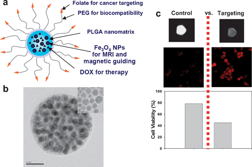 | ||
| Fig. 7 (a) Multifunctional nanomedical platforms. (b) TEM images of PLGA(SPIO/DOX) nanoparticles embedded with Fe3O4 nanocrystals. (c) Control vs. targeting. Reproduced with permission from ref. 40. | ||
Quantum dots themselves can be used as carriers of drugs and optical imaging agents. Bagalkot et al. synthesized quantum dot-aptamer (Apt)-doxorubicin conjugates (QD-Apt-DOX) as a targeted cancer imaging, therapy, and sensing system.41 The CdSe–ZnS core–shell QDs are surface functionalized with the A10 PSMA aptamer to target PSMA cancer cells. The intercalation of DOX within the A10 PSMA aptamer on the surface of the QDs results in the formation of the QD-Apt-DOX conjugates and quenching of both the QD and DOX fluorescence through a fluorescence resonance energy transfer (FRET) mechanism (Fig. 8a). This is the “OFF” state, in which the fluorescence of the QDs is quenched by DOX while, simultaneously, the fluorescence of DOX is quenched by intercalation within the A10 PSMA aptamer. When the conjugates were internalized into the cancer cells through PSMA mediate endocytosis, the release of DOX from the conjugates induced the recovery of the fluorescence from both QD and DOX, resulting in them entering the “ON” state (Fig. 8b). Using this smart conjugate system, the cancer targeted intracellular delivery of DOX and synchronous fluorescent localization in the cancer cells was achieved.
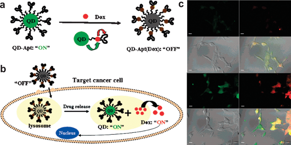 | ||
| Fig. 8 (a) Schematic illustration of QD-Apt-Dox FRET system. (b) Schematic illustration of specific uptake of QD-Apt-Dox conjugates into a target cancer cell through PSMA mediated endocytosis. (c) Confocal laser scanning microscopy images of PSMA expressing LNCaP cells after incubation with 100 nM QD-Apt-Dox conjugates for 0.5 h at 37 °C, washed two times with PBS buffer, and further incubated at 37 °C. Dox and QD are shown in red and green, respectively, and the lower right images of each panel represents the overlay of Dox and QD fluorescence. The scale bar is 20 μm. Reproduced with permission from ref. 41. | ||
Due to their high porosity mesoporous silica materials constitute an attractive drug delivery system. As organic dye molecules can be incorporated in the silica walls, the resultant fluorescent mesoporous silica materials have the potential to be used simultaneously for optical imaging and drug delivery. Zink and co-workers used FITC-doped mesoporous silica spheres to deliver the anticancer drug, camptothecin, into different types of human cancer cells to induce cell death.42 Because camptothecin is insoluble in water, which is a major obstacle to chemotherapy, the mesoporous silica spheres acted as good carriers of the drug, allowing it to enter into the cells without losing its therapeutic efficiency. The hydrophobic drugs were adsorbed into the mesopores and released within the cancer cells after internalization. The intracellular localization of the mesoporous silica spheres in the cytoplasm was confirmed by fluorescent microscopy.
Recently, the Hyeon group reported the synthesis of hollow iron oxide nanocapsules and demonstrated their in vitro T2 MRI and drug delivery capabilities to cancer cells (Fig. 9).43 Hollow iron oxide nanocapsules were fabricated from akagenite (β-FeOOH) nanorods via the wrap–bake–peel process, which involves silica coating, heat treatment and finally the removal of the silica layer. During heat treatment at 500 °C, first in air and then in 10% hydrogen atmospheres, the β-FeOOH nanorods were transformed into magnetite (Fe3O4) capsules. Removing the silica shells resulted in the formation of water-dispersible hollow Fe3O4 nanocapsules. The silica shells protected the β-FeOOH nanorods from aggregation and hollow cavities were developed in the nanorods. To increase their colloidal stability and biocompatibility, the surface of the hollow iron oxide nanocapsules was pegylated. In vitro MRI after incubating the nanocapsules with SKBR3 cells for 24 h indicated that the signal intensity of the MRI signal decreases as the concentration of the magnetite nanocapsules increases, demonstrating that the magnetite nanocapsules can be used as a T2 MRI contrast agent. The pores could be used to load chemical drugs and release them within the cancer cells. DOX-loaded hollow iron oxide nanocapsules were incubated with SKBR3 breast cancer cells. The growth inhibition of the cancer cells was observed when the cells were treated with either free DOX or DOX-loaded nanocapsules, demonstrating that the magnetite nanocapsules can be used as a drug delivery vehicle.
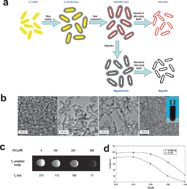 | ||
| Fig. 9 (a) Schematic illustration of the procedure for the synthesis of uniform and water-dispersible iron oxide nanocapsules. (b) TEM images of each product. (c) T2-weighted magnetic resonance images of the magnetite nanocapsules and in vitro cytotoxicity of free DOX and DOX-loaded magnetite capsules against SKBR3 cells. Reproduced with permission from ref. 43. | ||
3.2 Simultaneous imaging and photodynamic therapy
Photodynamic therapy (PDT) is a form of light activated chemotherapy using light-sensitive drugs, that is, photosensitizers (PSs). The commonly used photosensitizers are usually of porphyrinic origin and, once excited by light of the appropriate wavelength, are capable of producing cytotoxic reactive oxygen species (ROS), such as single oxygen (1O2), free radicals or peroxides, through interactions between the excited porphyrinic molecule and molecular oxygen which are present under these circumstances. This leads to the irreversible destruction of diseased cells and tissues. One of the main advantages of PDT is that treatment takes place only under the irradiation of light, which can be localized. Recently, multifunctional imaging and therapeutic agents were developed by combining PDT and imaging agents to achieve the diagnosis and treatment of tumor cells simultaneously.In a similar manner to the chemotherapeutic drugs, in order to overcome the various disadvantages of small molecules, PSs were incorporated into polymeric nanoparticles. Kopelman and co-workers developed 30–60 nm polyacrylamide (PAA) nanoparticles encapsulating iron oxide nanoparticles and Photofrin, a kind of commercial PS (Fig. 10a).44,45 The multifunctional polymer nanoparticles were synthesized via a microemulsion system. Acrylamide was polymerized in the presence of iron oxide and Photofrin using Brij 30 and sodium bis(2-ethylhexyl) sulfosuccinate (AOT) as surfactants. Using the surface amine group, the resulting polymer nanoparticles were pegylated and conjugated with the targeting peptides, F3. The exposure of the nanoparticles to 630 nm laser light produced significant amounts of singlet oxygen species in a concentration-dependent manner. In vitro studies revealed that the F3-targeted nanoparticles were specifically internalized and concentrated within the tumor cell nuclei and that the photoactivation of the nanoparticles resulted in higher cytotoxicity in the cancer cells compared to the non-targeted nanoparticles. After the intravenous injection of nanoparticles into mice with 9L glioma brain tumors, T2 MRI revealed that the tumors could be clearly imaged when the F3-targeted nanoparticles were used. The administration of laser light through a fiber optic applicator into the brain tumor site resulted in significant therapeutic benefit, with the F3-targeted nanoparticles providing a significantly increased survival time compared to that of the non-targeted Photofrin encapsulated nanoparticles or Photofrin alone (Fig. 10b).
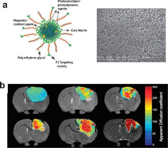 | ||
| Fig. 10 (a) Schematic representation and characterization of a multifunctional nanoparticle. (b) T2-weighted magnetic resonance images. Reproduced with permission from ref. 45. | ||
As most PSs absorb in the visible range below 700 nm, the penetration depth of light into tissue is limited. To circumvent this limitation, the two photon absorption (TPA) induced excitation of PSs is a promising approach to increase the penetration depth. The Prasad group designed a fluorescence resonance energy transfer (FRET) system composed of silica nanoparticles possessing TPA dyes and PSs.46 The organically modified silica (ORMOSIL) nanoparticles were synthesized via the co-precipitation of dyes and polymeric organically modified silica sol using AOT as a surfactant. The silica nanoparticles served as biocompatible nanoplatforms to overcome the poor water solubility and aggregation under physiological conditions of the PSs. A TPA dye, 9,10-bis[4′-(4″-aminostyryl)styryl]anthracene (BDSA), acted as an energy donor and 2-devinyl-2-(1-hexyloxyethyl)pyropheophorbide (HPPH) served as a PS and energy receptor. The HPPH absorption in the ORMOSIL nanoparticles showed significant spectral overlap with the fluorescence of BDSA, resulting in an energy transfer between them. The in vitro fluorescent imaging of Hela cells incubated with the nanoparticles under two photon excitation at 850 nm revealed the fluorescence from the cytoplasm exhibited the acceptor HPPH fluorescence. Two photon irradiation of the Hela cells incubated with the nanoparticles showed drastic changes in the morphology of the cells, while the reactive oxygen species were generated by HPPH, which is excited as a result of the two-photon excitation of BDSA and subsequent energy transfer to HPPH.
Iron oxide nanoparticles with the crosslinked dextran coating, CLIO, were used as nanoplatforms for simultaneous imaging and PDT. The McCarthy and Weissleder group conjugated a potent PS, 5-(4-carboxyphenyl)-10,15,20-triphenyl-2,3-dihydroxychlorin (TPC), and a NIR fluorescent dye, Alexa Fluor 750, onto the surface amine groups of the dextran coating.47 The energy transfer was minimized by the 100 nm difference between the longest wavelength absorption for TPC (650 nm) and that for Alexa Fluor 750 (750 nm), thus minimizing the risk of killing the cells while performing fluorescent imaging. The uptake of the nanoparticles within the macrophage cells was observed by fluorescent microscopy. When the cells incubated with the TPC-loaded nanoparticles were irradiated with light at 650 nm, a dose-dependent phototoxicity was observed. These magnetofluorescent nanoparticles conjugated with PSs have the potential to be used for simultaneous MRI, NIR fluorescent imaging, and PDT.
Usually, when magnetic nanoparticles are conjugated with dye molecules, the close contact between these species generally results in significant fluorescent quenching. In the case of PSs, the production of cytotoxic singlet oxygen might be decreased by quenching. To address this issue, Lai et al. fabricated silica-coated iron oxide nanoparticles coupled with a phosphorescent iridium complex instead of organic PS.48 The hydrophobic iron oxide nanoparticles synthesized by the thermal decomposition method were coated with silica shells in the presence of iridium-coupled silane agents via a reverse-microemulsion system. Using two-photon laser scanning fluorescence microscopy, they demonstrated that the co-encapsulated nanoparticles are actively taken up by tumor cells in vitro, and that the excitation efficiency of HPPH through energy transfer from the two-photon excited BDSA is preserved in the intracellular environment. The cellular uptake and imaging ability were further confirmed by MRI. A decrease in the T2 MRI signal was observed in a dose-dependent manner, indicating that the cellular uptake could be imaged by MRI. The PDT of Hela cells showed the formation of bubbles and fragmented cytoplasm, indicating that the singlet oxygen induced the deformation of the cellular membrane and loss of organelles.
3.3 Simultaneous imaging and gene delivery
Nanoparticles have the potential to deliver DNA or siRNA molecules into cells for therapeutic purposes. In general, polymer and lipid systems have been used as nonviral gene delivery vehicles. Recently, several strategies have been reported to track the gene delivery using nanoparticles of imaging modality such as of iron oxide nanoparticles combined with therapeutic genes. Gene silencing using small interfering RNA (siRNA) has emerged as an attractive approach for therapy. Using the well-known CLIO system as a platform, simultaneous imaging and gene delivery agents were developed. Moore and co-workers reported the development of a dual purpose nanoplatform for simultaneous noninvasive imaging and siRNA delivery to tumors (Fig. 11).49 They synthesized dual purpose nanoplatforms using CLIO (iron oxide nanoparticles with crosslinked dextran coatings) as the core, with NIR fluorescent Cy5.5 dyes and siRNA molecules decorated on the surface of the dextran coating. Additionally, these nanoparticles were modified with myristoylated polyarginine peptide (MPAP), a membrane translocation peptide, for intracellular delivery. Upon the intravenous injection of the nanoparticles into nude mice bearing subcutaneous human colorectal carcinoma tumors, the tumor was able to be imaged via T2 MR and NIR fluorescent imaging (Fig. 11a,b). The nanoparticles might have been accumulated at the tumor site via passive targeting through the permeable tumor vasculature and the nanoparticles were also observed in other organs such as the liver and spleen. Gene silencing was observed only when the multimodal nanoparticles having siRNA were used (Fig. 11c,d,e). The combination of the favorable biodistribution of these nanoparticles to tumors and their imaging properties represents an exciting possibility for the simultaneous delivery and detection of siRNA-based therapeutic agents in vivo.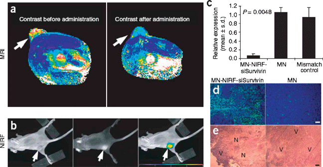 | ||
| Fig. 11 Application of MN-NIRF-siSurvivin in a therapeutic tumor model. (a) In vivo MRI of mice bearing subcutaneous LS174T human colorectal adenocarcinoma (arrows). (b) A high-intensity NIRF signal on in vivo optical images associated with the tumor following injection of MN-NIRF-siSurvivin confirmed the delivery of the probe to this tissue (left, white light; middle, NIRF; right, color-coded overlay). (c) Quantitative RT-PCR analysis of survivin expression in LS174T tumors after injection with either MN-NIRF-siSurvivin, a mismatch control or the parental magnetic nanoparticle (MN). (d) Note distinct areas with a high density of apoptotic nuclei (green) in tumors treated with MN-NIRF-siSurvivin (left). (e) H&E staining of frozen tumor sections revealed considerable eosinophilic areas of tumor necrosis (N) in tumors treated with MN-NIRF-siSurvivin (left). Reproduced with permission from ref. 49. | ||
Bartlett et al. used PET to quantify the in vivo biodistribution and siRNA delivery of multifunctional nanoparticles formed with cyclodextrin-containing polycations and siRNA.50 They conjugated DOTA to the 5′ end of the siRNA molecules. DOTA allowed the labeling of the nanoparticles with 64Cu for PET imaging. The multifunctional nanoparticles used for PET and siRNA delivery were synthesized through the electrostatic interaction between cyclodextrin-containing polycations and 64Cu-DOTA-siRNA conjugates. The in vivo PET imaging of mice bearing luciferase-expressing Neuro2A subcutaneous tumors indicated that both the non-targeted and transferrin-targeted siRNA nanoparticles exhibit a similar biodistribution and tumor localization via the enhanced permeability and retention (EPR) effect. However, the transferrin-targeted siRNA nanoparticles reduced the tumor luciferase activity by ≈50% relative to the non-targeted siRNA nanoparticles 1 d after injection, indicating that the transferrin-targeted nanoparticles delivered siRNA into the tumor cells owing to their enhanced internalization.
Viruses have been used as gene delivery vehicles owing to their facile cellular transfection and gene expression efficacies. Huh et al. developed target-specific viral gene delivery vehicles combined with magnetic nanoparticles to achieve simultaneous MRI and gene delivery.51 The adenovirus used possessed not only the enhanced green fluorescent protein (eGFP) gene inside the capsid, but also specificity to the Coxsackievirus B adenovirus receptor (CAR) of U251N cells. About five MnFe2O4 nanoparticles were covalently linked with the surface of the adenovirus between the lysine groups on the surface of the adenovirus and thiol groups of the nanoparticles using a chemical linker. The resulting multifunctional virus showed higher eGFP expression and a T2 MR signal decrease in the targeted cells compared to the control cells.
Chen et al. utilized fluorescent silica nanotubes for gene delivery.52 The hollow structure of nanotubes allows biomolecules to be loaded and protected. The hollow silica nanotubes were synthesized by the silica sol-gel reaction using anodic aluminium oxide as a template. After modification of the inner surface of the silica nanotubes with amine groups, CdSe–ZnS nanoparticles stabilized with mercaptoacetic acid were coated on the inner surface of the silica nanotubes through the electrostatic interaction between the positively charged amine groups and negatively charged QDs. The subsequent silica coating and amine-modification of the inner surface of the silica nanotubes made it possible to incorporate negatively-charged DNA molecules inside the fluorescent silica nanotubes. The transfection of nanotubes having plasmid DNA encoding GFP into the cells led to successful GFP expression in the cells and the existence of the nanotubes in cytoplasm was confirmed by their observation in the confocal laser scanning microscopy (CLSM) images.
3.4 Simultaneous imaging and photothermal therapy
Recently, a great deal of attention has been paid to noninvasive photothermal therapy for the selective treatment of tumor cells. This type of therapy utilizes the large absorption cross section of nanomaterials in the NIR region. Owing to its weak absorption by tissues, NIR radiation is able to penetrate the skin without causing much damage to normal tissues and, thus, can be used to treat specific cells targeted by the nanomaterials. Several nanomaterials that strongly absorb NIR radiation, including gold nanoshells, gold nanorods, and single-walled carbon nanotubes, were recently demonstrated to have potential therapeutic applications. The radiation that is absorbed by these nanomaterials is converted efficiently into heat, causing cell destruction. Much of the promise for nanotechnology in medicine has focused on the development of multifunctional agents for integrated imaging and photothermal therapy.West and co-workers successfully demonstrated the use of gold nanoshells in vivo to increase the optical contrast in tumors for optical coherence tomography (OCT) imaging and to treat the tumors using the nanoshell absorption of NIR light for photothermal ablation.53 Gold nanoshells consist of a dielectric silica core surrounded by a thin gold nanoshell with tunable plasmon resonances according to the ratio of the core and the shell. Gold nanoshells exhibit both absorption and scattering in the NIR range. In their study, gold nanoshells were used as contrast agents for enhanced OCT imaging, based on their backscattering properties, as well as cancer therapeutics, due to their absorption properties. The pegylated gold nanoshells were injected intravenously into mice bearing tumors. The skin and underlying muscle at the tumor site were visualized by OCT upon injection of the gold nanoshells, while there was no contrast enhancement in the controls. The gold nanoshells might be accumulated in the tumors, due to the leakiness of the tumor vasculature via the EPR effect. Nanoshells also allow the photothermal therapy of tumors with increased survival. The in vivo irradiation of NIR light onto the tumor resulted in a decrease of the tumor size and the enhanced survival of the mice 21 days after irradiation.
Gold nanorods can be used for simultaneous optical imaging and photothermal therapy. Gold nanorods absorb NIR light owing to their longitudinal resonance ascribed to their aspect ratio. The El-Sayed group reported in vitro cancer cell imaging and photothermal therapy using gold nanorods.54 The gold nanorods were synthesized via seed-mediated growth and conjugated with anti-epidermal growth factor receptor (anti-EGFR) monoclonal antibodies. The light scattering images of the anti-EGFR antibody-conjugated gold nanorods taken after their binding to the malignant cells showed that the nanorods strongly scatter orange to red light, due to their strong longitudinal surface plasmon oscillation with a frequency in the NIR region, while yellow scattering is observed in the visible range when using gold nanoparticles. After their exposure to a continuous NIR laser at 800 nm, the malignant cells incubated with the anti-EGFR antibody-conjugated gold nanorods required about half the laser power for them to be photothermally destroyed compared to the nonmalignant cells. Using single gold nanorods, both the optical imaging and selective photothermal therapy of cancer cells were demonstrated at the same time.
Gold nanocages also scatter and absorb NIR light due to their hollow and porous structure. The Xia group reported OCT imaging and photothermal therapy using gold nanocages as multifunctional probes.55 The Au nanocages were synthesized via the galvanic replacement reaction using Ag nanocubes as the starting materials. The surface plasmon resonance peak of the Au nanocages can easily be tuned by adjusting the extent of replacement of Ag with Au atoms. Similar to gold nanoshells and gold nanorods, Au nanocages showed good OCT contrast and photothermal therapeutic properties at the same time.
To address the problem of the limited penetration depth of optical imaging using gold nanoshells, Hyeon and co-workers proposed using a combination of magnetic nanoparticles and gold nanoshells to develop a novel nanomedical platform for diagnostic MRI and simultaneous photothermal therapy (Fig. 12).56 They fabricated magnetic gold nanoshells (Mag-GNS) consisting of gold nanoshells embedded with magnetic Fe3O4 nanoparticles (Fig. 12a). Monodisperse 7-nm Fe3O4 nanoparticles stabilized with 2-bromo-2-methylpropionic acid (BMPA) were covalently attached to amino-modified silica spheres through a direct nucleophilic substitution reaction between the bromo groups and the amino groups. Gold seed nanoparticles were attached to the residual amino groups of the silica spheres. Finally, complete gold nanoshells with embedded Fe3O4 nanoparticles were formed around the silica spheres, resulting in the formation of the Mag-GNS. Anti-HER2/neu was linked on the surface of the Mag-GNS for the targeted MRI and NIR photothermal therapy of cancer cells. SKBR3 breast cancer cells targeted by these Mag-GNS were able to be detected by a clinical MRI system and selectively killed by NIR radiation (Fig. 12b,c). Especially, the targeted cancer cells were rapidly destroyed upon their short exposure to femtosecond laser pulses with an NIR wavelength and a low power compared to the use of a continuous laser. In a similar approach, the Huh and Haam group fabricated a multifunctional nanocomposite of MnFe2O4 nanoparticles and gold nanoshells for simultaneous MRI and photothermal therapy.57 The gold nanoshells were formed on silica particles possessing several MnFe2O4 nanoparticles inside them. The antibody-conjugated nanocomposites showed synchronous therapeutic efficacy stemming from the therapeutic antibody and NIR laser-induced hyperthermia, as well as T2 MRI contrast enhancing properties.
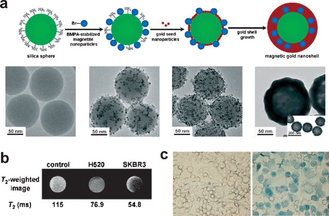 | ||
| Fig. 12 (a) Synthesis of the magnetic gold nanoshells (Mag-GNS). (b) T2-weighted MR images of control SKBR3 cells, HER2/neu-negative H520 cells incubated with Mag-GNS-AbHER2/neu, and HER2/neu-positive SKBR3 cells incubated with Mag-GNS-AbHER2/neu; the corresponding T2 relaxation times are indicated. (c) Optical microscope images of control SKBR3 cells and SKBR3 cells incubated with Mag-GNS-AbHER2/neu after irradiation at a power of 80 mW and subsequent staining. Reproduced with permission from ref. 56. | ||
The photothermal properties of gold nanoshells were used for the controlled release of chemical drugs. Yoo and co-workers synthesized a nanocomposite consisting of a rhodamine-encapsulated PLGA nanoparticle core on which Au and Mn were physically deposited in sequence.58 The nanocomposites exhibited photothermal properties and T2 decreases due to the Au and Mn half shells, respectively. Furthermore, the burst release of rhodamine, a model drug, was induced upon NIR irradiation. The release rate of rhodamine from the PLGA nanoparticles was about twice as high as that observed without NIR irradiation.
Graphitic carbon species absorb light in the range from the visible to NIR. Previously, Dai and co-workers reported that single wall carbon nanotubes could be used as photothermal therapeutic agents.59 Selective cancer cell destruction can be achieved by the functionalization of SWNTs with a folate moiety, the selective internalization of the SWNTs inside cells labeled with folate receptor tumor markers, and NIR-triggered cell death, without harming the receptor-free normal cells. Thus, the combination of the transporting capabilities of carbon nanotubes combined with suitable functionalization chemistry and their intrinsic optical properties can lead to new classes of novel nanomaterials for drug delivery and cancer therapy. Recently, the same group developed FeCo/graphitic shell nanoparticles for use as advanced MRI and NIR agents.60 The nanoparticles were fabricated by methane chemical vapor deposition on the FeCo nanoparticles formed on silica powder. After removing the silica supporter using HF, the resulting FeCo/graphitic shells were functionalized with phospholipid-PEG to obtain the water-dispersible nanoparticles (Fig. 13a). Interestingly, the core FeCo nanoparticles were highly stable against HF etching. The nanoparticles exhibited ultrahigh saturation magnetization and high r1 and r2 relaxivities. Upon the intravenous injection of the nanoparticles into rabbits, the long-lasting positive-contrast intravascular MRI of the blood pool was observed in the rabbits (Fig. 13b). The nanoparticles also exhibited photothermal conversion properties, indicating their potential to be used for integrated diagnosis and therapeutic applications (Fig. 13c).
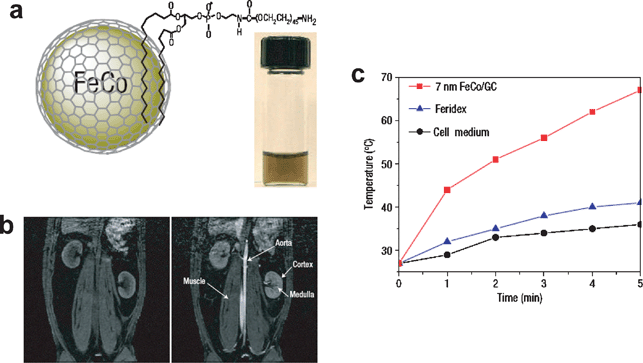 | ||
| Fig. 13 (a) Schematic diagram of a FeCo/GC nanocrystal, structure of the phospholipid molecule used for functionalization and a photograph of a PBS suspension of functionalized FeCo nanocrystals taken after heating to 80 °C for 1 h. (b) T1-weighted MR images of a rabbit before (left) and 30 min after (right) initial injection of a solution of ∼4 nm FeCo/GC nanocrystals (metal dose ∼9.6 μmol kg−1 for the ∼5 kg rabbit). (c) Temperature-evolution curves for solutions of FeCo/GC nanocrystals and Ferridex (all in Dulbecco's modified eagle's medium (DMEM) cell medium) and cell medium alone (as a control) under continuous radiation of an 808 nm NIR laser at 3.5 W cm−2. Reproduced with permission from ref. 60. | ||
4. Conclusions and outlook
Multifunctional nanomaterials have been synthesized by combining different types of nanomaterials. There has been a considerable amount of research on the development of multifunctional nanoplatforms for either multimodal imaging or simultaneous imaging and therapy. The ultimate goal in creating these multifunctional nanomaterials is the efficient and specific treatment as well as the diagnosis of diseases. In order for them to be used in clinics in the future, many issues including the biocompatibility, toxicity, in vivo targeting efficacy, and long term stability of the multifunctional nanoparticles need to be addressed. Especially, toxicological studies of the nanoparticles are necessary before they can be used in humans.61 The undesirable effects of the nanoparticles should be removed prior to their use. The development of novel multifunctional nanomaterials and the exact investigation of their characteristics in a living organism will open the way to the diagnosis and therapy of diseases in the future.Acknowledgements
We would like to acknowledge financial support from the Korean Ministry of Education, Science and Technology through the National Creative Research Initiative Program of the Korea Science and Engineering Foundation (KOSEF).References
- I. L. Medintz, H. T. Uyeda, E. R. Goldman and H. Mattoussi, Nat. Mater., 2005, 4, 435–446 CrossRef CAS.
- M. C. Daniel and D. Astruc, Chem. Rev., 2004, 104, 293–346 CrossRef CAS.
- Y.-W. Jun, J.-W. Seo and J. Cheon, Acc. Chem. Res., 2008, 41, 179–189 CrossRef CAS.
- D. Peer, J. M. Karp, S. Hong, O. C. Farokhzad, R. Margalit and R. Langer, Nat. Nanotechnol., 2007, 2, 751–760 Search PubMed.
- P. Caravan, Chem. Soc. Rev., 2006, 35, 512–523 RSC.
- J. W. M. Bulte and D. L. Kraitchman, NMR Biomed., 2004, 17, 484–499 CrossRef CAS.
- A.-H. Lu, E. L. Salabas and F. Schüth, Angew. Chem., Int. Ed., 2007, 46, 1222–1244 CrossRef CAS.
- J. Park, J. Joo, S. G. Kwon, Y. Jang and T. Hyeon, Angew. Chem., Int. Ed., 2007, 46, 4630–4660 CrossRef CAS.
- J. M. Klostranec and W. C. W. Chan, Adv. Mater., 2006, 18, 1953–1964 CrossRef CAS.
- L. Josephson, M. F. Kircher, U. Mahmood, Y. Tang and R. Weissleder, Bioconjugate Chem., 2002, 13, 554–560 CrossRef CAS.
- M. F. Kircher, U. Mahmood, R. S. King, R. Weissleder and L. Josephson, Cancer Res., 2003, 63, 8122–8125 CAS.
- A. Moore, Z. Medarova, A. Potthast and G. Dai, Cancer Res., 2004, 64, 1821–1827 CrossRef CAS.
- R. Weissleder, K. Kelly, E. Y. Sun, T. Shtatland and L. Josephson, Nat. Biotechnol., 2005, 23, 1418–1423 CrossRef CAS.
- M. Nahrendorf, H. Zhang, S. Hembrador, P. Panizzi, D. E. Sosnovik, E. Aikawa, P. Libby, F. K. Swirski and R. Weissleder, Circulation, 2008, 117, 379–387 CrossRef CAS.
- O. Veiseh, C. Sun, J. Gunn, N. Kohler, P. Gabikian, D. Lee, N. Bhattarai, R. Ellenbogen, R. Sze, A. Hallahan, J. Olson and M. Zhang, Nano Lett., 2005, 5, 1003–1008 CrossRef CAS.
- H. Lee, M. K. Yu, S. Park, S. Moon, J. J. Min, Y. Y. Jeong, H.-W. Kang and S. Jon, J. Am. Chem. Soc., 2007, 129, 12739–12745 CrossRef CAS.
- H. B. Na, I. S. Lee, H. Seo, Y. I. Park, J. H. Lee, S.-W. Kim and T. Hyeon, Chem. Commun., 2007, 5167–5169 RSC.
- A. Burns, H. Ow and U. Wiesner, Chem. Soc. Rev., 2006, 35, 1028–1042 RSC.
- T.-J. Yoon, K. N. Yu, E. Kim, J. S. Kim, B. G. Kim, S.-H. Yun, B.-H. Sohn, M.-H. Cho, J.-K. Lee and S. B. Park, Small, 2006, 2, 209–215 CrossRef CAS.
- C.-W. Lu, Y. Hung, J.-K. Hsiao, M. Yao, T.-H. Chung, Y.-S. Lin, S.-H. Wu, S.-C. Hsu, H.-M. Liu, C.-Y. Mou, C.-S. Yang, D.-M. Huang and Y.-C. Chen, Nano Lett., 2007, 7, 149–154 CrossRef CAS.
- J.-H. Lee, Y.-w. Jun, S.-I. Yeon, J.-S. Shin and J. Cheon, Angew. Chem., Int. Ed., 2006, 45, 8160–8162 CrossRef.
- Y.-S. Lin, S.-H. Wu, Y. Hung, Y.-H. Chou, C. Chang, M.-L. Lin, C.-P. Tsai and C.-Y. Mou, Chem. Mater., 2006, 18, 5170–5172 CrossRef CAS.
- J. Kim, J. E. Lee, J. Lee, J. H. Yu, B. C. Kim, K. An, Y. Hwang, C.-H. o. Shin, J.-G. Park, J. Kim and T. Hyeon, J. Am. Chem. Soc., 2006, 128, 688–689 CrossRef CAS.
- S. T. Selvan, P. K. Patra, C. Y. Ang and J. Y. Ying, Angew. Chem., Int. Ed., 2007, 46, 2448–2452 CrossRef CAS.
- C. Xu, J. Xie, D. Ho, C. Wang, N. Kohler, E. G. Walsh, J. R. Morgan, Y. Eugene Chin and S. Sun, Angew. Chem., Int. Ed., 2008, 47, 173–176 CrossRef CAS.
- J.-s. Choi, Y.-w. Jun, S.-I. Yeon, H. C. Kim, J.-S. Shin and J. Cheon, J. Am. Chem. Soc., 2006, 128, 15982–15983 CrossRef CAS.
- J. H. Choi, F. T. Nguyen, P. W. Barone, D. A. Heller, A. E. Moll, D. Patel, S. A. Boppart and M. S. Strano, Nano Lett., 2007, 7, 861–867 CrossRef.
- W. J. M. Mulder, R. Koole, R. J. Brandwijk, G. Storm, P. T. K. Chin, G. J. Strijkers, C. de M. Donega, K. Nicolay and A. W. Griffioen, Nano Lett., 2006, 6, 1–6 CrossRef CAS.
- H. Yang, S. Santra, G. A. Walter and P. H. Holloway, Adv. Mater., 2006, 18, 2890–2894 CrossRef CAS.
- S. Santra, R. P. Bagwe, D. Dutta, J. T. Stanley, G. A. Walter, W. Tan, B. M. Moudgil and R. A. Mericle, Adv. Mater., 2005, 17, 2165–2169 CrossRef CAS.
- V. S. Talanov, C. A. S. Regino, H. Kobayashi, M. Bernardo, P. L. Choyke and M. W. Brechbiel, Nano Lett., 2006, 6, 1459–1463 CrossRef CAS.
- W. J. Rieter, J. S. Kim, K. M. L. Taylor, H. An, W. Lin, T. Tarrant and W. Lin, Angew. Chem., Int. Ed., 2007, 46, 3680–3682 CrossRef CAS.
- J. S. Kim, W. J. Rieter, K. M. L. Taylor, H. An, W. Lin and W. Lin, J. Am. Chem. Soc., 2007, 129, 8962–8963 CrossRef CAS.
- K. M. L. Taylor, J. S. Kim, W. J. Rieter, H. An, W. Lin and W. Lin, J. Am. Chem. Soc., 2008, 130, 2154–2155 CrossRef CAS.
- C.-P. Tsai, Y. Hung, Y.-H. Chou, D.-M. Huang, J.-K. Hsiao, C. Chang, Y.-C. Chen and C.-Y. Mou, Small, 2008, 4, 186–191 CrossRef CAS.
- J.-L. Bridot, A.-C. Faure, S. Laurent, C. Rivie`re, C. Billotey, B. Hiba, M. Janier, V. Josserand, J.-L. Coll, L. V. Elst, R. Muller, S. Roux, P. Perriat and O. Tillement, J. Am. Chem. Soc., 2007, 129, 5076–5084 CrossRef CAS.
- N. Nasongkla, E. Bey, J. Ren, H. Ai, C. Khemtong, J. S. Guthi, S.-F. Chin, A. D. Sherry, D. A. Boothman and J. Gao, Nano Lett., 2006, 6, 2427–2430 CrossRef CAS.
- J. Yang, C.-H. Lee, H.-J. Ko, J.-S. Suh, H.-G. Yoon, K. Lee, Y.-M. Huh and S. Haam, Angew. Chem., Int. Ed., 2007, 46, 8836–8839 CrossRef CAS.
- C.-K. Huang, C.-L. Lo, H.-H. Chen and G.-H. Hsiue, Adv. Funct. Mater., 2007, 17, 2291–2297 CrossRef CAS.
- J. Kim, J. E. Lee, S. H. Lee, J. H. Yu, J. H. Lee, T. G. Park and T. Hyeon, Adv. Mater., 2008, 20, 478–483 CrossRef CAS.
- V. Bagalkot, L. Zhang, E. Levy-Nissenbaum, S. Jon, P. W. Kantoff, R. Langer and O. C. Farokhzad, Nano Lett., 2007, 7, 3065–3070 CrossRef CAS.
- J. Lu, M. Liong, J. I. Zink and F. Tamanoi, Small, 2007, 3, 1341–1346 CrossRef CAS.
- Y. Piao, J. Kim, H. B. Na, D. Kim, J. S. Baek, M. K. Ko, J. H. Lee, M. Shokouhimehr and T. Hyeon, Nat. Mater., 2008, 7, 242–247 CrossRef CAS.
- R. Kopelman, Y.-E. L. Koo, M. Philbert, B. A. Moffat, G. R. Reddy, P. McConville, D. E. Hall, T. L. Chenevert, M. S. Bhojani, S. M. Buck, A. Rehemtulla and B. D. Ross, J. Magn. Magn. Mater., 2005, 293, 404–410 CrossRef CAS.
- G. R. Reddy, M. S. Bhojani, P. McConville, J. Moody, B. A. Moffat, D. E. Hall, G. Kim, Y.-E. L. Koo, M. J. Woolloscroft, J. V. Sugai, T. D. Johnson, M. A. Philbert, R. Kopelman, A. Rehemtulla and B. D. Ross, Clin. Cancer Res., 2006, 12, 6677–6686 CrossRef CAS.
- S. Kim, T. Y. Ohulchanskyy, H. E. Pudavar, R. K. Pandey and P. N. Prasad, J. Am. Chem. Soc., 2007, 129, 2669–2675 CrossRef CAS.
- J. R. McCarthy, F. A. Jaffer and R. Weissleder, Small, 2006, 2, 983–987 CrossRef CAS.
- C.-W. Lai, Y.-H. Wang, C.-H. Lai, M.-J. Yang, C.-Y. Chen, P.-T. Chou, C.-S. Chan, Y. Chi, Y.-C. Chen and J.-K. Hsiao, Small, 2008, 4, 218–224 CrossRef CAS.
- Z. Medarova, W. Pham, C. Farrar, V. Petkova and A. Moore, Nat. Med. (N. Y.), 2007, 13, 372–377 Search PubMed.
- D. W. Bartlett, H. Su, I. J. Hildebrandt, W. A. Weber and M. E. Davis, Proc. Natl. Acad. Sci. U. S. A., 2007, 104, 15549–15554 CrossRef CAS.
- Y.-M. Huh, E.-S. Lee, J.-H. Lee, Y.-w. Jun, P.-H. Kim, C.-O. Yun, J.-H. Kim, J.-S. Suh and J. Cheon, Adv. Mater., 2007, 19, 3109–3112 CrossRef CAS.
- C.-C. Chen, Y.-C. Liu, C.-H. Wu, C.-C. Yeh, M.-T. Su and Y.-C. Wu, Adv. Mater., 2005, 17, 404–407 CrossRef CAS.
- A. M. Gobin, M. H. Lee, N. J. Halas, W. D James, R. A. Drezek and J. L. West, Nano Lett., 2007, 7, 1929–1934 CrossRef CAS.
- X. Huang, I. H. El-Sayed, W. Qian and M. A. El-Sayed, J. Am. Chem. Soc., 2006, 128, 2115–2120 CrossRef CAS.
- S. E. Skrabalak, J. Chen, L. Au, X. Lu, X. Li and Y. Xia, Adv. Mater., 2007, 19, 3177–3184 CrossRef CAS.
- J. Kim, S. Park, J. E. Lee, S. M. Jin, J. H. Lee, I. S. Lee, I. Yang, J.-S. Kim, S. K. Kim, M.-H. Cho and T. Hyeon, Angew. Chem., Int. Ed., 2006, 45, 7754–7758 CrossRef CAS.
- J. Lee, J. Yang, H. Ko, S. J. Oh, J. Kang, J.-H. Son, K. Lee, S.-W. Lee, H.-G. Yoon, J.-S. Suh, Y.-M. Huh and S. Haam, Adv. Funct. Mater., 2008, 18, 258–264 CrossRef CAS.
- H. Park, J. Yang, S. Seo, K. Kim, J. Suh, D. Kim, S. Haam and K.-H. Yoo, Small, 2008, 4, 192–196 CrossRef CAS.
- N. W. S. Kam, M. O’Connell, J. A. Wisdom and H. Dai, Proc. Natl. Acad. Sci. U. S. A., 2005, 102, 11600–11605 CrossRef CAS.
- W. S. Seo, J. H. Lee, X. Sun, Y. Suzuki, D. Mann, Z. Liu, M. Terashima, P. C. Yang, M. V. McConnell, D. G. Nishimura and H. Dai, Nat. Mater., 2006, 5, 971–976 CrossRef CAS.
- N. Lewinski, V. Colvin and R. Drezek, Small, 2008, 4, 26–49 CrossRef CAS.
| This journal is © The Royal Society of Chemistry 2009 |