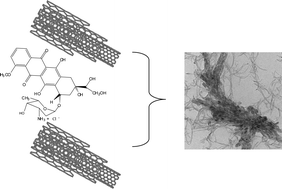
Multiwalled carbon nanotubedoxorubicin supramolecular complexes for cancer therapeutics

Hanene Ali-Boucetta a, Khuloud T. Al-Jamal a, David McCarthy a, Maurizio Prato b, Alberto Bianco c and Kostas Kostarelos *a
aNanomedicine Laboratory, Centre for Drug Delivery, The School of Pharmacy, University of London, 29-39 Brunswick Square, London, UK WC1N 1AX. E-mail: kostas.kostarelos@pharmacy.ac.uk; Fax: +44 20 7757 5942; Tel: +44 20 77535861
bDipartimento di Scienze Farmaceutiche, Università di Trieste, 34127, Trieste, Italy
cInstitut de Biology Molèculaire et Cellulaire, UP R9021 CNRS, Immunologie et Chimie Thèrapeutiques, 67084, Strasbourg, France

Received (in Cambridge, UK) 10th August 2007 , Accepted 5th November 2007

First published on 14th November 2007


Abstract

Multiwalled carbon nanotube aqueous dispersions using block copolymers are able to form supramolecular complexes with the aromatic chromophore and anticancer agent doxorubicinvia π–π stacking and enhance its cytotoxic activity.


The discovery of novel nanomaterials such as carbon nanotubes (CNTs) capable of traversing the plasma membrane1 and promoting the cellular uptake of small molecules and macromolecules (e.g.nucleic acids and peptides2,3) has offered new opportunities for various biological applications.4 Although most of the existing anticancer drugs are very potent small molecules, their efficacy is constrained not only by their systemic toxicity and narrow therapeutic window but also as a result of drug resistance and limited cellular entry. For this reason the development of efficient delivery systems with the ability to enhance cellular uptake of existing potent drugs is needed. Functionalised CNTs have shown great promise as novel delivery systems especially based on their ability to cross biological barriers independently of the cell type they interact with and the functional group at their surface.1 In addition, the high aspect ratio of CNTs offers great advantages over existing delivery vectors, as the high surface area provides multiple attachment sites for drug targeting. Pastorin et al. have developed CNT–methotrexate conjugates via covalent linkage for the use of CNTs as multimodal drug delivery systems.5 Though such conjugates seem promising and are actively explored in our laboratories, the efficiency of drug activity is dependent on the nature of the covalent bond between the CNT and the small molecule. As an alternative strategy, we wanted to explore the hypothesis of any prevalent non-covalent interactions between anticancer drug molecules and CNTs.

In this communication, we describe a previously unreported non-covalent multiwalled nanotube (MWNT)–doxorubicin supramolecular complex that can be developed for cancer therapy. We have investigated the ability of doxorubicin to interact non-covalently with very thin, pristine MWNT 1 at various mass ratios and evaluated their capability to kill human breast cancer cells. Doxorubicin belongs to a clinically-used family of anthracyclines, therefore constitutes one of the best candidates to test non-covalent complexation with MWNTs. Moreover, doxorubicin 3 is a fluorescent molecule with a chromophore composed of three planar and aromatic hydroxyanthraquinonic rings that are used to monitor its supramolecular interaction with MWNTs. MWNTs were dispersed in water using the tri-block copolymer (Pluronic F127) 2.

Pristine MWNTs 1 (Nanocyl® 3150) were dispersed using a 1% Pluronic F127 2 (Sigma Ltd) solution to a final MWNT concentration of 1 mg ml–1 by bath sonication for 30 min as previously described by others.6Doxorubicin 3 and pluronic–MWNT were allowed to interact by mixing equal volumes of doxorubicin hydrochloride (20 µg ml–1) with increasing MWNT aqueous dispersion concentrations (10, 20 and 40 µg ml–1). The complexes formed contained from 0.5 × 10¹⁸ to 2 × 10¹⁸ molecules of doxorubicin per mg of MWNT. The interaction between doxorubicin and MWNT was studied by monitoring the emission spectrum of doxorubicin by fluorescence spectrophotometry (Perkin Elmer Luminescence Spectrometer LS 50B).

ugraphic, filename = b712350g-u1.gif

As can be seen from Fig. 1, the fluorescence intensity of doxorubicin dramatically decreased as the final concentration of MWNT was increased from 5 to 20 µg ml–1. Maximum quenching was occurring at 0.5 × 10¹⁸doxorubicin molecules per mg MWNT indicating that optimum interaction between the drug and the MWNT occurs at this mass ratio. In a control experiment, when the same number of doxorubicin molecules were mixed with the equivalent Pluronic F127 concentration in the absence of MWNTs, no decrease in fluorescence intensity compared to that of doxorubicin in water (Fig. 1) was observed, suggesting that no interaction was taking place between the drug and the copolymer molecules.


Normalised fluorescence intensity of MWNT–doxorubicin complexes. Final concentration of doxorubicin was fixed to 10 µg ml–1 while MWNT final concentration was increased (5, 10 and 20 µg ml–1) which is equivalent to doxorubicin : MWNT mass ratios of 1 : 0.5, 1 : 1 and 1 : 2.
Fig. 1 Normalised fluorescence intensity of MWNT–doxorubicin complexes. Final concentration of doxorubicin was fixed to 10 µg ml–1 while MWNT final concentration was increased (5, 10 and 20 µg ml–1) which is equivalent to doxorubicin : MWNT mass ratios of 1 : 0.5, 1 : 1 and 1 : 2.

The dramatic decrease in fluorescence intensity seen when doxorubicin interacted with the MWNT can be explained by the static quenching of doxorubicin molecules due to the π–π stacking of its aromatic chromophore groups and the carbon nanotube backbone. It is well known that doxorubicin exhibits a unique fluorescence emission spectrum that is susceptible to changes in its microenvironment.7 Interaction with MWNTs resulted in changes in its molecular conformation leading to loss in fluorescence signal, similar to that described when binding to human α-1 glycoprotein.7 The affinity of the doxorubicin chromophore for self- and hetero-association with various compounds with planar aromatic ring systems has also been shown before.8,9 Moreover, the binding and intercalation of doxorubicin between the aromatic bases of nucleic acids is well described, and is also shown to cause considerable reduction in doxorubicin fluorescence intensity.7 The possibility of electrostatic or hydrogen bond formation between the doxorubicin molecule and the non-ionic block copolymer may also be enhancing the interaction between doxorubicin and MWNTs, however it is not dominant as shown in Fig. 1 when doxorubicin interacted with block copolymer alone. This indicated that the structure of the CNT backbone can act as a platform for the formation of supramolecular complexes with small drug molecules, similar to what has been described before with the aromatic bases of nucleic acids.10

In order to study the structural characteristics of the doxorubicin–MWNT complexes, transmission electron microscopy (TEM) was used. Complexes were prepared by simple mixing as described above, by keeping the final concentration of MWNT constant at 0.5 mg ml–1, in order to be able to image the structure of the complexes, while the same number of doxorubicin molecules per mg MWNT as in the fluorescence analysis were added. Fig. 2A shows copolymer-wrapped MWNTs as well-individualised and dispersed nanotubes, clean from any impurities, and confirming the ability of the polymer molecules to disperse the CNTs effectively. Doxorubicin alone showed crystal-like structures at low magnification (Fig. 2B). Overall, the TEM images revealed that MWNTs were strongly interacting with doxorubicinvia formation of supramolecular clusters. These MWNT–doxorubicin clusters were very well visualised as the number of doxorubicin molecules was decreased from 2 × 10¹⁸ to 0.5 × 10¹⁸ molecules per mg MWNT (Fig. 2C, D, E, and F). The observed clustering between the MWNTs and doxorubicin also correlated well with the maximum fluorescence quenching of doxorubicin (Fig. 1).


          TEM images of doxorubicin–MWNT complexes. (A) MWNTs alone (scale bar 100 nm), (B) doxorubicin alone (scale bar 2 µm), (C) 2 × 10¹⁸, (D) 1 × 10¹⁸, (E) 0.5 × 10¹⁸doxorubicin molecules per mg MWNT with scale bar of 500 nm and (F) 0.5 × 10¹⁸doxorubicin molecules per mg MWNT at higher magnification (scale bar corresponds to 100 nm). The final MWNT concentration was kept constant at 0.5 mg ml–1.
Fig. 2 TEM images of doxorubicin–MWNT complexes. (A) MWNTs alone (scale bar 100 nm), (B) doxorubicin alone (scale bar 2 µm), (C) 2 × 10¹⁸, (D) 1 × 10¹⁸, (E) 0.5 × 10¹⁸doxorubicin molecules per mg MWNT with scale bar of 500 nm and (F) 0.5 × 10¹⁸doxorubicin molecules per mg MWNT at higher magnification (scale bar corresponds to 100 nm). The final MWNT concentration was kept constant at 0.5 mg ml–1.

Next we studied the cytotoxic capability of these MWNT–doxorubicin supramolecular assemblies in in vitro cytotoxicity studies using the MCF7 human breast cancer cells. The MTT assay used is a well-established toxicological assay to assess cell viability based on the activity of mitochondrial enzymes.11–14 Moreover, this assay has been used by several groups to assess the cytotoxic response of cell cultures to carbon nanotubes.15–18 In this assay the conversion and cleavage of the tetrazolium salt MTT (3-(4,5-dimethylthiazol-2-yl)-2,5-diphenyl tetrazolium bromide) into a purple formazan product is monitored at 570 nm. This conversion occurs by mitochondrial reductase only present in living and metabolically active cells. The epithelial breast cancer derived MCF-7 cells were seeded and the cell viability following interaction for 24 h with the doxorubicin–MWNT complex (mass ratio 1 : 2) is shown in Fig. 3. There was a statistically significant enhancement in the cytotoxic capability of the doxorubicin–MWNT complex compared to that of the doxorubicin alone (p = 0.016724) and the equivalent pluronicdoxorubicin (p = 0.000173) after 24 h. Interestingly, cells treated with the equivalent pluronic-coated MWNT and pluronic alone demonstrated no difference compared to untreated cells, exhibiting 100% cell viability. The enhanced cytotoxicity obtained with the doxorubicin–MWNT complex suggests that MWNTs can mediate the delivery of doxorubicin and hence improve the cellular uptake of the drug. Importantly, the copolymer does not seem to be contributing to this improved cytotoxic capacity, since mixing doxorubicin with pluronic showed no significant difference as compared to doxorubicin alone (p = 0.493148). This correlated well with the fluorescence spectroscopy data that indicated no complex formation between doxorubicin and the block copolymer molecules (Fig. 1). Recently published data1 indicated that CNT may possess an inherent capacity to interact and translocate within living cells, however the exact contributing mechanisms are not yet fully elucidated. In light of this, the mechanism by which the presently reported complexes between MWNT and doxorubicin are delivered into the breast cancer cells needs to be further investigated.


The percentage cell viability of MCF-7 cells after 24 h incubation at a final concentration of doxorubicin (Dox) 600 nM); CNT (651.5 ng ml–1); Pluronic (6.5 ng ml–1) and Pluronic–Dox and CNT–Dox at CNT : doxorubicin mass ratio of 2 : 1. Untreated cells were used as control. Statistical significance was observed between the CNT–Dox and the † doxorubicin (p < 0.05) and the *** Pluronic–Dox groups (p < 0.005) respectively.
Fig. 3 The percentage cell viability of MCF-7 cells after 24 h incubation at a final concentration of doxorubicin (Dox) 600 nM); CNT (651.5 ng ml–1); Pluronic (6.5 ng ml–1) and Pluronic–Dox and CNT–Dox at CNT : doxorubicin mass ratio of 2 : 1. Untreated cells were used as control. Statistical significance was observed between the CNT–Dox and the † doxorubicin (p < 0.05) and the *** Pluronic–Dox groups (p < 0.005) respectively.

We have demonstrated that copolymer-coated MWNT can form non-covalent supramolecular complexes with doxorubicin. The formation of such complexes is evidenced by a sharp decrease in the intensity of the doxorubicin fluorescence spectrum and takes place presumably via π–π interactions with the MWNT backbone. We have also shown that the doxorubicin–MWNT complex exhibits enhanced cytotoxic activity compared to both doxorubicin alone and doxorubicinpluronic complexes. Following submission of this communication, another group published studies they ran independently and simultaneously with ours also reporting enhanced cell kill efficacy from non-covalent complexation between lipid-coated single-walled nanotubes (SWNTs) and doxorubicin.19 The two studies clearly corroborate each other, indicating that both SWNTs and MWNTs seem to offer available surface area for π–π interactions with the doxorubicin aromatic rings, leading to enhanced cell kill efficacy. The use of carbon nanotubes to facilitate anticancer drug delivery and improve drug activity seems promising given the ability of CNT to cross biological barriers.1 These observations may be a result of this reported CNT behaviour, however more mechanistic work needs to be carried out in order to investigate if the capabilities of CNT to translocate into cells is also the case for the drug–MWNT complex. Moreover, factors such as the timely and effective intracellular release of the drug molecule from the CNT complex that will determine the efficacy of drug action need to be studied. These results are promising and warrant further investigation to reveal the in vitro and in vivo cytotoxic capacity and assess the potential of the CNT–drug complexes in cancer therapy.

This work has been supported by The School of Pharmacy, University of London, CNRS and the Agence Nationale de la Recherche (grant ANR-05-JCJC-0031-01), the University of Trieste, MUR (cofin Prot. 2006035330) and Regione Friuli Venezia-Giulia. H. A-B wishes to acknowledge the Ministére de l'Enseignement Supèrieur et de la Recherche Scientifique (Algeria) for a full PhD scholarship.

Notes and references

  1. K. Kostarelos, L. Lacerda, G. Pastorin, W. Wu, S. Wieckwski, J. Luangsivilay, S. Godefroy, D. Pantarotto, J. P. Briand, S. Muller, M. Prato and A. Bianco, Nat. Nanotechnol., 2007, 2, 108 Search PubMed.
  2. D. Pantarotto, R. Singh, D. McCarthy, M. Erhardt, J. P. Briand, M. Prato, K. Kostarelos and A. Bianco, Angew. Chem., Int. Ed., 2004, 43, 5242 CrossRef CAS.
  3. D. Pantarotto, J. P. Briand, M. Prato and A. Bianco, Chem. Commun., 2004,(1), 16 RSC.
  4. A. Bianco, K. Kostarelos, C. D. Partidos and M. Prato, Chem. Commun., 2005, 571 RSC.
  5. G. Pastorin, W. Wu, S. Wieckwski, J. P. Briand, K. Kostarelos, M. Prato and A. Bianco, Chem. Commun., 2006, 1182 RSC.
  6. P. Wick, P. Manser, L. K. Limbach, U. Dettlaff-Weglikowska, F. Krumeich, S. Roth, W. J. Stark and A. Bruinink, Toxicol. Lett., 2007, 168(2), 121 CrossRef CAS.
  7. N. Husain, R. A. Agbaria and I. M. Warner, J. Phys. Chem., 1993, 97, 10857 CrossRef CAS.
  8. M. Menozzi, L. Valentini, E. Vannini and F. Arcamone, J. Pharm. Sci., 1984, 73(6), 766 CAS.
  9. M. Dalmark and P. Johansen, Mol. Pharmacol., 1982, 22, 158 CAS.
  10. M. Zheng, A. Jagota, E. D. Semke, B. A. Diner, R. S. Mclean, S. R. Lustig, R. E. Richardson and N. G. Tassi, Nat. Mater., 2003, 2, 338 CrossRef CAS.
  11. T. Mosmann, J. Immunol. Methods, 1983, 65, 55 CrossRef CAS.
  12. F. Denizot and R. Lang, J. Immunol. Methods, 2007, 89, 271.
  13. D. T. Vistica, P. Skehan, D. Scudiero, A. Monks, A. Pittman and M. R. Boyds, Cancer Res., 1991, 51, 2515 CAS.
  14. M. B. Hansen, S. E. Nielsen and K. Berg, J. Immunol. Methods, 1989, 119, 203 CrossRef CAS.
  15. M. Davoren, E. Herzog, A. Casey, B. Cottineau, G. Chambers, H. J. Byrne and F. M. Lyng, Toxicol. in Vitro, 2007, 21, 438 CrossRef CAS.
  16. C. M. Sayes, F. Liang, J. L. Husdson, J. Mendez, W. Guo, J. M. Beach, V. C. Moore, C. D. Doyle, J. L. West, W. E. Billups, K. D. Ausman and V. L. Colvins, Toxicol. Lett., 2006, 161, 135 CrossRef CAS.
  17. D. Cui, F. Tian, C. O. Ozkan, M. Wang and H. Gao, Toxicol. Lett., 2005, 155, 73 CrossRef CAS.
  18. A. Magrez, S. Kasas, V. Salicio, N. Pasquier, J. W. Seo, M. Celio, S. Catsicas, B. Schwaller and L. Forro, Nano Lett., 2006, 6, 1121 CrossRef CAS.
  19. Z. Liu, X. Sun, N. Nakayama-Ratchford and H. Dai, ACS Nano, 2007, 1, 50 Search PubMed.

This journal is © The Royal Society of Chemistry 2008