Hot off the Press
Hot off the Press highlights recently published work for the benefit of our readers. Our contributors this month have focused on TRP ion channels and a new method for DNA sequencing. New contributors are always welcome. If you are interested please contact molbiosyst@rsc.org for more information, we’d like to hear from you.
Cysteine knot peptides from spider venom activate rather than inhibit the TRPV1 channel
Studies on the interactions of natural products and peptides with ion channels have advanced neurophysiology and promoted drug discovery for diseases of the nervous system. Transient receptor potential (TRP) channels are generally calcium-permeable and exhibit polymodal activation, such as through temperature change and the binding of certain natural products from plants. For example, the TRPV1 channel is the receptor for vanilloids such as capsaicin, a pungent component of hot chilli pepper; it is also heat-sensitive. Naturally occurring inhibitor cysteine knot (ICK) peptides are often found to be antagonists for ion channels. To identify new activators of sensory neurons, David Julius and colleagues screened 22 different spider venoms by calcium imaging of TRP ion channels expressing cells. They found that the venom of Psalmopoeus cambridgei, a tarantula native to the West Indies gave a positive readout. Three previously unknown ICK peptides named vanillotoxins were extracted from the venom, isolated by HPLC and sequenced by Edman degradation. To confirm that these peptides account for the TRPV1 channel activation, one of the purified toxins was chemically synthesized and the synthetic peptide indeed had similar activity to the native material. The interactions of these peptides with ion channels were investigated in more detail both at the cellular and intact animal levels. Whole-cell patch clamp analysis of TRPV1-transfected cells showed that one of the toxins had a very similar effect on the current–voltage relation as capsaicin. However, the toxin and capsaicin interact with distinct regions of the channel as shown by using inside-out and outside-out patch clamp recordings. All three peptides showed specific actions towards TRPV1 channels, as opposed to other TRP family members. One of vanillotoxins also inhibited the voltage-gated potassium channel Kv2.1, suggesting a similarity between the TRP and voltage-gated channel structures in their overall transmembrane topology and tetrameric subunit organization. Moreover, studies of animal behaviour demonstrated that the vanillotoxins are agonists for the endogenous TRPV1 channels responsible for the elicitation of pain and inflammation. Therefore, the new vanillotoxins, as opposed to most known ICK peptides, activate rather than inhibit TRP ion channels. The findings provide new understanding of the molecular mechanism by which pain and inflammation are produced by bites and stings from venomous creatures. Future three dimensional structure determinations of these new peptides and structure-functional studies will help disclose the detailed mechanisms of vanillotoxin-channel interactions, and perhaps lead to new therapies for pain relief.
Siemens J, Zhou S, Piskorowski R, Nikai T, Lumpkin EA, Basbaum AI, King D, Julius D., Nature, 2006, 444, 208-212
Reviewed by: Wen-Wu Li, University of Oxford, UKAn innovative solution for non-Sanger DNA sequencing
The means to rapidly and inexpensively sequence DNA would bring about major advances in science, medicine and the pharmaceutical industry. In particular, the ability to sequence the entire human genome at an affordable price would allow healthcare to be tailored to the specific needs of an individual, trigging a revolution in personalised medicine (the $1000 human genome project).J. Ju and co-workers present an interesting improvement in DNA sequencing based on the Sequencing by Synthesis (SBS) methodology. Their approach is based on a new class of cleavable fluorescent nucleotides that are selectively incorporated in a growing self-primed DNA strand by a mutated DNA polymerase (9oN DNA polymerase (exo-) A485L/Y409V mutant). The 3′ ends of the nucleotides are protected by an allyl moiety so that only one nucleotide per cycle is incorporated. This nucleotide is then recognised by an inexpensive four-colour fluorescence scanner. After detection, the fluorescent tag of the nucleotide and the 3′-O-allyl protecting group are cleaved simultaneously by using a Pd-catalysed deallylation reaction, which prepares the DNA template for a new elongation cycle. In order to keep the sequencing in phase, a synchronisation step is added where 3′-O-allyl dNTPs (without the fluorescent tag) are incorporated between each reading cycles.
Any new DNA sequencing technique must show potential for automation. To demonstrate this, the authors immobilise an alkyne-labelled DNA template on an azide functionalised chip. To avoid non-specific adsorption of unincorporated fluorescent nucleotides on the chip surface, a PEG linker is introduced between the DNA and the surface. In this way, the authors are able to determine 20 consecutive bases in the immobilised DNA template by self-primed SBS. A major advantage of using the allyl protected-fluorescent nucleotides is the ability to detect several bases in a homopolymeric sequence, a known weak point of several other SBS techniques that are under development.
Undoubtedly, the ability to sequence the human genome at an affordable price would bring about a revolution in science and the healthcare industry. Ju and co-workers demonstrated an interesting approach, which overcomes a technical problem in SBS. However, the methodology must be much improved to be practicable. The authors must reduce the time between cycles (they quote 10 minutes for each deallylation cycle) and they need to immobilise and image millions of DNAs at once. Finally and crucially, the authors must be able to sequence more than 20 DNA bases in one run, i.e. hundreds of bases, if they want to match current technology or eventually meet the target of 0.000003 cent per base required for the $1000 Human Genome Project (5× coverage).
Ju J, Kim DH, Bi L, Meng Q, Bai X, Li Z, Li X, Marma MS, Shi S, Wu J, Edwards JR, Romu A, Turro NJ. Proc. Nat. Acad. Sci., U. S. A., 2006, 103(52), 19635–40
Reviewed by: Giovanni Maglia, Oxford University, UKHot off the RSC Press
Gut feeling for antibody detection
A protein-coated electrode offers a sensitive test for people with gluten intolerance.Thomas Balkenhohl and Fred Lisdat at the University of Applied Sciences Wildau in Germany have invented a sensor that detects antibodies involved in coeliac disease. Coeliac disease is an autoimmune reaction to gluten – found in wheat, rye and barley – that prevents the absorption of essential nutrients in the gut.
The method works by immobilising gliadins, proteins found in gluten, on the surface of gold electrodes. People with coeliac disease produce antigliadin antibodies in reaction to gluten. When the electrodes are immersed in blood serum samples from coeliac sufferers, these antibodies bind to the gliadins and the electrodes’ electrical properties change in proportion to the antibody concentration. The method is even sensitive enough to detect antigliadin antibodies in samples taken from people who do not suffer from coeliac disease. Balkenhohl and Lisdat have transferred their system to screen-printed electrodes, which will allow the sensors to be mass produced.
But antigliadin antibodies are not the whole story. Anti-tissue transglutaminase antibodies, which are currently not detected by the system, are also implicated in coeliac disease. Being able to detect both antibodies will guarantee improved sensitivity and specificity of the test, said Lisdat. And more work needs to be done on the electrical measurements, which are ‘still limited to an advanced laboratory,’ he warned. The duo hopes that more practical methods will be developed as the technique is used for different kinds of biochemical detection.
 | ||
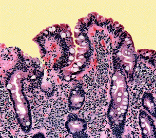
| Fig. 1 Coeliac disease prevents essential nutrients being absorbed in the gut. | ||
T Balkenhohl and F Lisdat, Analyst, 2007, DOI: 10.1039/b609832k.
Reviewed by: Colin Batchelor, Royal Society of Chemistry, Cambridge, UK.Manipulating microcoils
A prototype chip can be used to make cells hop along a magnetic field.Robert Westervelt and his colleagues at Harvard University, US, have combined microelectronics and microfluidic technologies to develop a hybrid chip that can manipulate individual biological cells.
In Westervelt’s chip, cells are contained within microfluidic channels. The microfluidic system is built on top of a custom-designed integrated circuit (IC) that controls microcoils on the chip’s surface. By tagging cells with peptide-coated magnetic beads, their motion can be controlled using local magnetic fields generated in the microcoils.
‘The microcoils are matched in size to an individual cell which makes it possible for a microcoil to trap a single cell,’ explained Westervelt. Since a single microcoil or a number of microcoils can be activated simultaneously, either single or multiple cells can be held.
In the single cell case, because only one microcoil is magnetically active at any given moment, applying a current pulse to the hybrid chip forces the cell to hop from one microcoil to another. Time-sharing the current source generates magnetic fields in more than one microcoil, allowing multiple cells to be trapped and moved independently of each other. ‘It is this capability to individually control many cells in parallel that allows experiments to be conducted with single-cell level precision,’ said Westervelt.
According to Westervelt, these chips offer a powerful tool for biotechnology because they use standard IC technology and can be produced cheaply. They will allow tests, assays and diagnoses to be performed reliably on a scale and at speeds not previously possible, he said.
‘Preventing damage to the integrated circuits by the biological fluids is a challenge,’ said Westervelt. ‘In particular, we need to find coatings that will protect them from salts and organic compounds in biological samples.’
 | ||
| Fig. 2 A single cell moves across a chip (bottom) as the surrounding magnetic field changes (top). | ||
H Lee et al, Lab Chip, 2007, 7, 331.
Reviewed by: Janet Crombie, Royal Society of Chemistry, UK.| This journal is © The Royal Society of Chemistry 2007 |
