Detection of airborne Legionella while showering using liquid impingement and fluorescent in situ hybridization (FISH)
Magali
Deloge-Abarkan
a,
Thi-Lan
Ha
b,
Enric
Robine
b,
Denis
Zmirou-Navier
a and
Laurence
Mathieu
*c
aDépartement Environnement et Santé Publique, INSERM ERI no 11, Faculté de Médecine, 9 avenue de la Forêt de Haye, BP 184, F-54 505, Vandoeuvre-lès-Nancy, France
bCentre Scientifique et Technique du Bâtiment (C.S.T.B.), Laboratoire de Microbiologie des Environnements Intérieurs, 84 avenue Jean Jaurès, F-77 447, Marne la Vallée cedex 2, France
cLaboratoire d’Hydroclimatologie Santé Environnement, Ecole Pratique des Hautes Etudes, INSERM ERI no 11, Faculté de Médecine, 9 avenue de la Forêt de Haye, BP 184, F-54 505, Vandoeuvre-lès-Nancy, France. E-mail: laurence.mathieu@medecine.uhp-nancy.fr; Fax: 33 383 683 489; Tel: 33 383 683 486
First published on 10th November 2006
Abstract
Aerosols of water contaminated with Legionella bacteria constitute the only mode of exposure for humans. However, the prevention strategy against this pathogenic bacteria risk is managed through the survey of water contamination. No relationship linked the Legionella bacteria water concentration and their airborne abundance. Therefore, new approaches in the field of the metrological aspects of Legionella bioaerosols are required. This study was aimed at testing the main principles for bioaerosol collection (solid impaction, liquid impingement and filtration) and the in situ hybridization (FISH) method, both in laboratory and field assays, with the intention of applying such methodologies for airborne Legionella bacteria detection while showering. An aerosolization chamber was developed to generate controlled and reproducible L. pneumophila aerosols. This tool allowed the identification of the liquid impingement method as the most appropriate one for collecting airborne Legionella bacteria. The culturable fraction of airborne L. pneumophila recovered with the liquid impingement principle was 4 and 700 times higher compared to the impaction and filtration techniques, respectively. Moreover, the concentrations of airborne L. pneumophila in the impinger fluid were on average 7.0 × 10⁵ FISH-cells m−3 air with the fluorescent in situ hybridization (FISH) method versus 9.0 × 10⁴ CFU m−3 air with the culture method. These results, recorded under well-controlled conditions, were confirmed during the field experiments performed on aerosols generated by hot water showers in health institutions. This new approach may provide a more accurate characterization of aerobiocontamination by Legionella bacteria.
Introduction
First described in 1977,1Legionella bacteria are still of topical interest, and constitute a significant problem of public health. The main mode of transmission of Legionella bacteria is inhalation of a contaminated aerosol produced by colonized hydrous sources. Exposure to Legionella sp., bacteria naturally found in hydrous and humid environments, is increasing because of the number of man-made habitats (air conditioning systems, cooling towers, whirlpools, spas, fountains, dental devices, shower heads), all potentially producing aerosols.2–7 Such multiplication of artificial hydrous environments can involve epidemics of legionellosis.8–10Thus, in France, the management of the risk associated with Legionella bacteria consists of preventing microbial proliferation, and keeping concentrations below the lawful threshold, fixed at 103 CFU l−1 for hot water networks.11 This approach is unreliable due to the underestimation of the culture-based method because of the presence of viable/active but non-culturable bacteria, and Legionella bacteria within biofilms or intra-amoebas that are not accessible with the standardized cultural technique. Moreover, culture analysis is time-consuming and could be affected by stresses arising during aerosolization of microorganisms which could lead to a loss of their culturability.3,12 Even if some old works have focused on detecting airborne Legionella,4,6,13–17 any relationship between the Legionella concentration present in water and bacteria dispersed into aerosols has not yet been established. It is therefore difficult to assess the Legionella bacteria health risk based only on water measurements. To have a more representative exposure assessment, it would be helpful to determine airborne Legionella bacteria generated from suspicious aquatic environments.
Our objective was to perform an environmental study on the feasibility of the detection of airborne Legionella bacteria while showering in two health care centres in northern France. This monitoring needed efficient sampling and adequate methods for analysis. So, a first step, performed in the laboratory with controlled Legionella bacteria aerosols, aimed to identify the methodologies to be used, by testing: (i) three principles of bioaerosol collection (solid impaction, liquid impingement and filtration) to evaluate their influence on the culturability of aerosolized Legionella, and (ii) the in situ hybridization (FISH) method to detect airborne Legionella bacteria.
In the second part of the study, field trials were performed in two health institutions chosen because of their recurrent problems with water Legionella spp. contamination. These field trials have enabled us to appreciate: (i) the abundance of airborne Legionella bacteria generated by hot-water showers, and (ii) the relevance of the aerosol’s collection principles and the analytical methods for airborne Legionella spp. detection.
Material and methods
Laboratory trials
To perform the aerosolization procedure, L. pneumophila colonies were harvested and grown in a specific nutrient broth (ACES buffer 6.0 g l−1, α-ketoglutarate 1.0 g l−1, brain heart 11.5 g l−1, L-cysteine 0.4 g l−1, ferric pyrophosphate 0.25 g l−1, deionized water, pH = 6.8–7.0) at 37 °C (±1 °C).
After 2 days’ incubation, bacterial cells were further harvested and were washed three times by centrifugation at 8 × 10²g for 20 min (model EBA 8S, Hettich) in sterile deionized water, also used as diluent for the aerolization suspension. Deionized water was preferred to a buffer solution because salts were expected to affect the microbial viability and the results obtained with the optical particle counter. Bacterial density was determined by turbidity using a spectrometer (model HI 93703, HANNA Instruments) calibrated with a standard of Formazine Turbidity Unit tubes.
 | ||
| Fig. 1 Schematic representation of the laboratory experimental setup (F: flowmeter; Qbubbl: bubbling airflow; Qdil: dilution airflow; Qsampl: sampling airflow). | ||
In order to assess the methodologies to be used, the bioaerosol generated during the laboratory trials was sampled using three collection principles: (i) impaction onto agar (Andersen sampler; cut-off diameter: 0.65 μm; flow rate: 28.3 l min−1; sampling volume: 8.5 l of air; collection medium: BCYEα); (ii) impingement into liquid (SKC Biosampler (Arelco); cut-off diameter: 0.3 μm; flow rate: 12.5 l min−1; sampling volume: 125 l of air; collection medium: sterile distilled water); and (iii) filtration (collectron MD8 (Sartorius), cut-off diameter: >0.5 μm; flow rate: 10 l min−1; sampling volume: 10 l of air; collection medium: 0.8 μm pore-size nitrocellulose membrane). The impaction and liquid impingement methods remove particles from the airstream by utilizing their inertia to depose them onto an agar medium or into a liquid surface.19 Filtration collects particles by inertial forces, interception and diffusion.19
In previous studies performed in this aerosolization chamber in controlled conditions (data not shown), the aerodynamic diameter of airborne L. pneumophila was evaluated at 0.9 μm, suggesting that all aerosolized cells will be sampled by the three methods used.
Three independent assays were conducted in the laboratory, for each sampling technique. The samples collected on BCYEα medium were kept at 37 °C (±1 °C). For the impinger fluid, aliquots (0.1 ml) of an appropriate ten-fold dilution were spread on BCYEα agar before incubation. After filtration sampling, the membrane was transferred aseptically to the Petri dish containing the BCYEα agar.
The bacterial recovery rate (R) was measured as the concentration of L. pneumophila in aerosols (Cpart)AIR (measured using the optical partile counter), relative to its culturable fraction (CCFU)AIR, as following:
 | (1) |
Field investigation procedures
The environmental study, about the feasibility of airborne Legionella bacteria detection while showering, was performed in two health care centres in northern France, chosen because of their recurrent problems with water Legionella spp. contamination. Both hot water and bioaerosols were sampled as described in Fig. 2. | ||
| Fig. 2 Field sampling protocol. | ||
Eighteen showers were sampled, with the hot water faucet opened to its maximum flow; the bathrooms’ ventilation was systematically shut down and the doors and windows closed during the investigations.20
One litre of hot water, collected in sterile bottles, was systematically taken after about 7 min of water flushing (average duration of a shower21). The bioaerosol sampling began only after the faucet of hot water was closed, in order to prevent the sampling of water droplets. The bioaerosols were sampled using the same collection principles as in the laboratory experiments, except the air volume sampled which were higher than those used in laboratory assays with a pure strain of L. pneumophila at high concentration (∼106 CFU l−1). Field sampling characteristics are summarized in Table 1. During the sampling, the water and air temperatures and relative humidity (RH) (Testo 452, Testo) were monitored.
| Collection principles | Samplers and collection media | Flow rate/l min−1 | Sampled air volume/l |
|---|---|---|---|
| Impaction onto agar | MAS 100 (Merck); BCYEα agar | 100 | 500 |
| Liquid impingement | SKC Biosampler (Arelco); sterile distilled water | 12.5 | 195 |
| Filtration | Millipore cassette; 0.2 μm pore size polycarbonate membrane | 10 | 600 |
The three principles of aerosol sampling were used simultaneously at a fixed point (oriented towards the shower at 80 cm height and at one metre in front of it), and started at the same time. Using solid impaction, the samples collected on the BCYEα agar medium were kept at 37 °C (±1 °C) ( they permit solely the detection of the culturable Legionella spp). For the impinger and filtration methods, the samples were kept for in situ hybridization.
Determination of the culturable Legionella fraction
The detection of the culturable Legionella bacteria was adapted from the French standard method AFNOR,22 using the selective nutrient agar BCYE (Buffer Charcoal Yeast Extract) (Oxoïd, UK). For laboratory assays, L. pneumophila colonies were enumerated after 7 days’ incubation at 37 + 1 °C. For field assays, characteristic Legionella spp. colonies were counted and tested for their cysteine dependence (CD) by inoculation onto BCYE agar with cysteine (0.4 g l−1) and without cysteine, and incubated for 48 h at 37 °C ± 1 °C. Colonies that grew on both media were considered CD negative and were reported as non-Legionella bacteria. Colonies that grew only on BCYE with cysteine (CD positive colonies) were identified as Legionella spp. The results were expressed in CFU l−1 of water or in CFU m−3 of air. The detection limit was 1.0 × 10² CFU l−1 water and 1.2 × 10⁴ CFU m−3 air.Application of the FISH technique for detection of airborne Legionella bacteria
The fluorescent in situ hybridization (FISH) protocol was performed as suggested by Manz et al.23 The oligonucleotide probe (Molecular Probes) LEG705 (5′CTGGTGTTCCTTCCGATC-3′) specific for Legionellaceae 16S RNA and labeled with carbocyanine at the 5′ end, was used to detect Legionella spp.23,24 Each sample was filtered through 0.2 μm pore size white polycarbonate membrane (Millipore) and was fixed with 3.7% formaldehyde (vol/vol) for 30 min. The membrane was washed twice with phosphate-buffered saline (pH 7.4), air-dried and dehydrated with 2 ml of increasing concentrations of ethanol (50, 80, and 95%, 3 min each). Then 50 μl aliquot of hybridization solution (20% formamide, 900 mM NaCl, 0.1% sodium dodecyl sulfate, 20 mM Tris-HCl [pH 7.2]), containing 50 ng of the labeled probe, was applied to the membrane. Hybridization was performed for 2 h at 46 ± 1 °C in a moisture chamber. The filter was then washed twice with 46 °C preheated wash solution for 30 min (20 mM Tris-HCl, 215 mM NaCl, 0.1% sodium dodecyl sulfate, [pH 7.2]), air dried and mounted on a slide with AF87 antifading reagent (Citifluor, Biovalley). Hybridized bacterial cells were visualized by epifluorescence microscopy with an X100 immersion objective lens and a 510 to 550 nm excitation filter and a 590 nm barrier filter. Thirty microscopic fields were counted, and the results were expressed in Legionella-cells m−3 of air or Legionella-cells l−1 of water. The hybridization procedure quality was checked systematically by using species known to hybridize (or not) with the LEG705 probe.Determination of the total cell counts in hot water samples
The total number of bacteria in water samples was determined with the DAPI (4′,6-diamidino-2-phenylindole) staining, using the protocol described by Saby et al.25 The water samples were filtered through 0.2 μm pore size black polycarbonate membranes (Millipore) and were overlaid with 3 ml of a 0.5 μg ml−1 DAPI solution for 15 min. After washing with sterile distilled water, the membranes were air dried, then mounted on a slide with AF87 antifading reagent (Citifluor, Biovalley). The fluorescence microscopy conditions were the same as described for the FISH method, except that the filter sets were appropriate for DAPI staining detection. Thirty randomly chosen microscopic fields were counted for each water sample. The results were expressed in bacterial cells l−1 of water. The detection limit was 6.7 × 10⁴ bacterial cells l−1 for 20 ml of water analyzed.Statistical analysis
All data are analyzed with the program Stat View 5.0 (SAS Institute Inc., Cary, NC). The Mann–Whitney rank sum test was used to assess the association between Legionella bacteria detection by the culture method and by the FISH method.Results
Laboratory experiments
 | ||
| Fig. 3 Bacterial recovery rates between average L. pneumophila concentrations in the aerosol and their culturable fraction, according to the three collection methods used in the laboratory trials—the bars correspond to the minimal and maximal values (n = 3). | ||
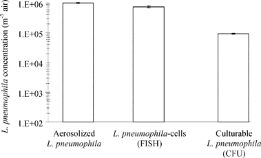 | ||
| Fig. 4 Concentrations of aerosolized L. pneumophila in the aerosol chamber (expressed as particles m−3) and after collection using impingement and detection by FISH and culture methods during the laboratory trials—the bars correspond to the standard deviation (n = 3). | ||
Detection of Legionella in field samples
| Hot water shower temperature/°Ca | Culturable Legionella spp./CFU l−1 | Hybridized Legionella spp./cells l−1 | Total bacteria/cells l−1 | Relative humidity (%) | Air temperature/°C | |
|---|---|---|---|---|---|---|
| a Monitored during shower (from 2.5 to 7 min). b Arithmetic mean. c Geometric mean. | ||||||
| Positive samples (%) | — | 18/18 (100) | 18/18 (100) | 18/18 (100) | — | — |
| Average value | 37.4b | 4.03 × 10⁴c | 4.65 × 10⁵c | 9.69 × 10⁷c | 89.3b | 25.0b |
| Standard deviation | 3.7 | 1.92 × 10⁵ | 1.11 × 10⁶ | 4.10 × 10⁷ | 12.3 | 1.9 |
| Minimum | 27.0 | 6.70 × 10³ | 3.60 × 10⁴ | 4.33 × 10⁷ | 64.2 | 21.8 |
| Maximum | 41.0 | 5.00 × 10⁵ | 3.55 × 10⁶ | 1.80 × 10⁸ | 99.9 | 28.2 |
To perform airborne Legionella bacteria detection, impinger fluids and the membranes used for filtration were analyzed with the FISH method (Fig. 5). Hybridized Legionella spp. were detected in 50% and in 72.2% of the aerosol samples with filtration and impingement, respectively. The liquid impingement method allowed a significantly higher detection of airborne Legionella bacteria (on average 2.71 × 10⁶Legionella-cells m−3 air) than filtration (on average 1.4 × 101Legionella-cells m−3 air). Filtration appeared to be harmful to airborne Legionella bacteria, unlike impingement.
 | ||
| Fig. 5 Hybridized Legionella spp. concentrations (FISH method) in hot water shower aerosols according to the collection method—the bars correspond to the standard deviation (n = 18). | ||
Discussion
In this study, we attempted to characterize the airborne Legionella contamination while showering by testing (i) the three main principles adapted for bioaerosol sampling (impaction on solid medium, liquid impingement and filtration) and (ii) the FISH method for Legionella spp. detection in aerosols. Two sets of experiments were performed: laboratory assays in controlled conditions using an aerosolization chamber, and in field conditions with contaminated showers in two health institutions.Performance of sampling methods for airborne Legionella detection
A number of studies with contradictory findings have been published about the samplers’ performance, both under laboratory and field settings26–28 for detection of fungi, or total or culturable heterotrophic airborne bacteria. The performance of these biocollection methods has previously been reported in few studies but using the culture method for bacterial detection. They showed that impingement can detect more bacteria than agar impaction6,15,29 or than filtration.30Using controlled, well-characterized and reproducible L. pneumophila aerosols generated in an aerosolization chamber,31 our results, specifically dedicated to airborne Legionella collection, showed that the impingement technique improved the aerosolized Legionella bacteria detection, measured through their culturability, by 4 to 700 times compared to impaction and filtration, respectively.
This is in agreement with the study of Ishimatsu et al.,6 where impingement technique also improved the culturability of aerosolized Legionella pneumophila detection, by 2 to 20 times compared to impaction.
This difference of samplers’ performance could be explained by their design and physical performances. It is generally assumed that all microorganisms above the particle cut-size are collected and an impinger might be more efficient in collecting small bioaerosol particles compared to impaction and filtration.28,32 It should be noted that the evaluation of bioaerosol sampling performance includes inevitably the combined effects of three factors: aerosolization, collection and environment. Wang et al.33 estimated that the aerosolization and collection by filtration accounted for more than 40% of the culturability loss of bacterial cells: the process of aerosolization, in addition to killing a large number of vegetative cells, causes damage to the cell envelope.34 For Hambleton et al.,16 this evaluation may be affected by the composition of the spray and collecting fluids: an aerosolized L. pneumophila strain was tolerant of distilled water and Page’s saline. The effects of spray fluid additives and of collecting fluids were not investigate here, but we have studied the survival in the best conditions, which achieved 80% of aerosolized Legionella pneumophila with an intact membrane (Baclight kit, data not shown).
Another factor could also explain the differences between the airbone Legionella bacteria recoveries: the medium onto which bioaerosol particles were collected (filter, agar or liquid).
During bioaerosol collection, microbial stress is influenced by the medium used for their recovery.35–37 For collecting fluids, Ishimatsu et al.6 reported, for instance, that a BYE broth allowed better trapping and survival rates of Legionella pneumophila compared to Page’s saline and 2% yeast extract solution. For Stewart et al.,37 microbial injuries occurred during impaction on solid media, thus preventing the culturability of airborne bacteria. For aerosol collection onto filter, Li and Lin38 showed that the relative survival of E. coli collected by filtration was lower than that observed when collected by impingement, presumably because of the dryness of the agar plate or filter during the sampling time.39 Indeed, desiccation stress is of great importance when collecting bioaerosols3,12 and microorganisms do fail to grow when the collection support dries out.40,41 In this study, the liquid impingement method is a relevant tool for Legionella bacteria detection, by culture and FISH methods.
Application of the FISH method for airborne Legionella detection
Culture-based approaches are commonly used to detect and characterize biological particles of aerosols. Even if culture is the standard conventionnal method for environmental samples, numbers of studies have proved that it underestimates the real number of viable microorganisms in environments,24,42–45 including aerosols.46–47 Several authors reported detection of non-culturable environmental bacteria by non-conventional techniques.44,48–49 Nyström50 reported the unculturability of Legionella bacteria from water samples but also confirmed the presence of viable L. pneumophila after guinea pig inoculation.These low culturability of environmental bacteria, and especially of airborne microorganisms, require usage of alternative methods such as molecular techniques, in combination with culture-based techniques.51 Thus, studies using PCR-based methods are widespread for aerosolized Legionella detection6,13,52,53 and to our knowledge, only one study was performed using in situ hybridization (FISH) to detect airborne bacteria,54 and none for aerosolized Legionella bacteria detection.
In the laboratory, as in the field assays, our results showed that airborne Legionella spp. could be detected with FISH whereas a low fraction or none was culturable, which is in agreement with the recent work done during the Pas-de-Calais Legionnaires’ Disease outbreak.55 Thereby a fraction of Legionella bacteria has already lost its culturability while its remains physiologically active. This suggests the existence of bacteria which have enough intact rRNA to be detected by FISH, but which are not able to replicate on agar medium within 10 days. This lead to the concept of active non-culturable bacteria (ANC) developed by Lebaron et al.49 for the hydrous environment. It could be considered that bacterial detection with oligonucleotide probes may be related to their physiological states, which is dependent on their growth rate,56,57 and to their rRNA content,24,49,58,59 as already shown with environmental samples.60,61 Therefore FISH might yield useful information on the bacterial physiology even if it is not considered as a viability marker.62,63
In our experimental conditions, the FISH method is suitable for aerosolized Legionella spp. detection without any cultivation64 or selective purification or amplification steps.45 The analysis can be performed within 12 h, as against 7 to 10 days for the culture method. Because of the utilization of a nucleic probe targetted at Legionella spp., FISH is a specific method which allows the identification of nonculturable Legionella bacteria among the environment’s bacterial populations. The FISH method could be therefore considered as a relevant tool for auto-controls and rapid monitoring of installations at risk (cooling tower, hot water supply network...), as well as the PCR, with therefore the advantage of results expressed in numbers of Legionella bacterial cells. This point is particularly important with respect to risk management, which can then be done almost in real time and not a posteriori, as it is currently the case.
Some improvement could be realized, in particular combining the FISH method with a viability measurement, which is not used today, neither for airborne bacteria in general, nor for Legionella bacteria. In the future, the application of image analysis could enhance the hybridized Legionella bacteria detection, making it less fastidious.
Conclusions
Our objective was to perform an environmental study on the feasibility of the Legionella bacteria detection while showering, using the three main principles of bioaerosol collection. The liquid impingement technique has been identified as the most appropriate method for collecting airborne Legionella in well-controlled conditions (aerosolization chamber) and during the field experiments (hot water shower aerosols). Moreover, this study showed an improved assessment of airborne Legionella bacteria using the impingement technique combined with the FISH detection method. It may provide a more accurate characterization of aerobiocontamination by this pathogenic bacteria.Acknowledgements
This research was supported by the Agence Française de Sécurité Sanitaire Environnementale (AFSSE), Veolia Environment and the Direction Générale de la Santé (DGS). This work was sponsored in part by the French Agency for Environment and Energy Management (ADEME) and Electricité de France (EDF).References
- J. E. McDade, C. C. Shepard, D. W. Fraser, T. R. Tsai, M. A. Redus and W. R. Dowdle, N. Engl. J. Med., 1977, 297, 1197–1203 CAS.
- R. S. Bhopal, J. Public Health Med., 1991, 13, 281–289 Search PubMed.
- G. E. Bollin, J. F. Plouffe, M. F. Para and B. Hackman, Appl. Environ. Microbiol., 1985, 50, 1128–1131 CAS.
- P. J. L. Dennis, A. E. Wright, D. A. Rutter, J. E. Death and B. P. C. Jones, J. Hyg., 1984, 93, 349–353 Search PubMed.
- M. Hodgson and B. J. Casey, http://www.claytongrp.com/legion_art.html, 1998.
- S. Ishimatsu, H. Miyamoto, H. Hori, I. Tanaka and S. I. Yoshida, Ann. Occup. Hyg., 2001, 45, 421–427 CAS.
- D. W. Keller, R. Hajjeh, A. DeMaria, B. S. Fields, J. M. Pruckler, R. S. Benson, P. E. Kludt, S. M. Lett, L. A. Mermel, C. Giorgio and R. F. Breiman, Clin. Infect. Dis., 1996, 22, 257–261 CAS.
- Centers for Disease Control and Prevention, Morbidity Mortality Wkly. Rep., 2005, 54, 165–168 Search PubMed.
- C. A. Joseph, Epidemiol. Infect., 2004, 132, 417–424 Search PubMed.
- T. M. N. Nguyen, D. Ilef, S. Jarraud, L. Rouil, C. Campese, D. Che, S. Haeghebaert, F. Ganiayre, F. Marcel, J. Etienne and J. C. Desenclos, J. Infect. Dis., 2006, 193, 102–111 CrossRef.
- Le Risque Lié aux Légionelles, Guide d’Investigation et d’Aide à la Gestion, Conseil Supérieur d’Hygiène Publique de France (CSHPF), Paris, 2005 Search PubMed.
- B. Z. Predicala, J. E. Urban, R. G. Maghirang, S. B. Jerez and R. D. Goodband, Curr. Microbiol., 2002, 44, 136–140 CrossRef CAS.
- A. J. Alvarez, M. P. Buttner, G. A. Toranzos, E. A. Dvorsky, A. Toro, T. B. Heikes, L. E. Mertikas-Pifer and L. D. Stetzenbach, Appl. Environ. Microbiol., 1994, 60, 374–376 CAS.
- R. F. Berendt, H. W. Young, R. G. Allen and G. L. Knutsen, J. Infect. Dis., 1980, 141, 186–192 CAS.
- P. J. Dennis and J. V. Lee, J. Appl. Bacteriol., 1988, 65, 135–141 CAS.
- P. Hambleton, M. G. Broster, P. J. Dennis, R. Henstridge, R. Fitzgeorge and J. W. Conlan, J. Hyg., 1983, 90, 451–460 Search PubMed.
- L. Pascual, S. Perez-Luz, A. Amo, C. Moreno, D. Apraiz and V. Catalan, Can. J. Microbiol., 2001, 47, 341–347 CrossRef CAS.
- V. Ulevicius, K. Willeke, S. A. Grinshpun, L. Donnelly, X. Lin and G. Mainelis, Aerosol Sci. Technol., 1997, 26, 175–190 CAS.
- P. A. Jensen, B. Lighthart, A. J. Mohr and B. T. Shaffer, in Atmospheric Microbial Aerosols Theory and Applications, ed. B. Lighthart and A. J. Mohr, Chapman and Hall Inc., New York, 1994, pp. 226–284 Search PubMed.
- B. Marshall, P. Flynn, D. Kamely and S. B. Levy, Appl. Environ. Microbiol., 1988, 54, 1776–1783 CAS.
- United States Environmental Protection Agency (U.S. EPA), Exposure Factors Handbook, U.S. EPA, Washington, DC, 1997 Search PubMed.
- Association Française de Normalisation, Norme AFNOR T90-431, Association Française de Normalisation, Paris, France, 2003 Search PubMed.
- W. Manz, R. Amann, R. Szewzyk, U. Szewzyk, T. A. Strenström, P. Hutzler and K. H. Schleifer, Microbiology, 1995, 141, 29–39 CrossRef CAS.
- R. I. Amann, W. Ludwig and K. H. Schleifer, Microbiol. Rev., 1995, 59, 143–169 CAS.
- S. Saby, I. Sibille, L. Mathieu, J. L. Paquin and J. C. Block, Appl. Environ. Microbiol., 1997, 63, 1564–1569 CAS.
- P. A. Jensen and M. P. Schafer, in NIOSH Manual of Analytical Methods, 1998, ch. J, pp. 82–112 Search PubMed.
- S. K. Mehta, D. M. Robinson, T. O. Groves, L. D. Stetzenbach and D. L. Pierson, AIHA J., 2000, 61, 850–854 Search PubMed.
- S. D. Pillai and S. C. Ricke, Can. J. Microbiol., 2002, 48, 681–696 CrossRef CAS.
- L. L. Lembke, R. N. Kniseley, R. C. Van Nostrand and M. D. Hale, Appl. Environ. Microbiol., 1981, 42, 222–225 CAS.
- C. Duchaine, P. S. Thorne, A. Meriaux, Y. Grimard, P. Whitten and Y. Cormier, Appl. Environ. Microbiol., 2001, 67, 2775–2780 CrossRef CAS.
- T. L. Ha, M. Deloge-Abarkan, L. Mathieu and E. Robine, Metrology of Legionella aerosols: from a laboratory approach to a field evaluation of sampling and detection methods, 6th International Conference on Legionella, Chicago, IL, USA, 16th–20th October 2005, American Society for Microbiology, board 36, p. 87, ISBN 1-555-81-367-4 Search PubMed.
- S. Terzieva, J. Donnelly, V. Ulevicius, S. A. Grinshpun, K. Willeke, G. N. Stelma and K. P. Brenner, Appl. Environ. Microbiol., 1996, 62, 2264–2272 CAS.
- Z. Wang, T. Reponen, S. A. Grinshpun, R. L. Gorny and K. Willeke, J. Aerosol Sci., 2001, 32, 661–674 CrossRef CAS.
- L. A. White, D. J. Hadley, D. E. Davids and R. Naylor, Appl. Microbiol., 1975, 29, 335–339 Search PubMed.
- M. P. Buttner and L. D. Stetzenbach, Appl. Environ. Microbiol., 1991, 57, 1268–1270.
- B. Marthi, V. P. Fieland, M. Walter and R. J. Seidler, Appl. Environ. Microbiol., 1990, 56, 3463–3467 CAS.
- S. L. Stewart, S. A. Grinshpun, K. Willeke, S. Terzieva, V. Ulevicius and J. Donnelly, Appl. Environ. Microbiol., 1995, 61, 1232–1239 CAS.
- C. S. Li and Y. C. Lin, Aerosol Sci. Technol., 1999, 30, 280–287 CAS.
- K. T. Durand, M. L. Muilenberg, H. A. Burge and N. S. Seixas, Ann. Occup. Hyg., 2002, 46, 113–118 CrossRef.
- G. Blomquist, G. Strom and L. H. Stromquist, Scand. J. Work Environ. Health, 1984, 10, 109–113 Search PubMed.
- K. Heldal, A. Skogstad and W. Eduard, Ann. Occup. Hyg., 1996, 40, 437–447 CrossRef CAS.
- J. J. Byrd, H. S. Xu and R. R. Colwell, Appl. Environ. Microbiol., 1991, 57, 875–878 CAS.
- R. R. Colwell, J. Infect. Chemother., 2000, 6, 121–125 CrossRef CAS.
- D. Hussong, R. R. Colwell, M. O’Brien, E. Weiss, A. D. Pearson, R. M. Weiner and W. D. Burge, Bio/Technology, 1987, 5, 947–950 Search PubMed.
- A. Moter and U. B. Göbel, J. Microbiol. Methods, 2000, 41, 85–112 CrossRef CAS.
- W. D. Griffiths, I. W. Stewart, A. R. Reading and S. J. Futter, J. Aerosol Sci., 1996, 27, 803–820 CrossRef CAS.
- J. F. Heidelberg, M. Shahamat, M. Levin, I. Rahman, G. Stelma, C. Grim and R. R. Colwell, Appl. Environ. Microbiol., 1997, 63, 3585–3588 CAS.
- F. Joux and P. Lebaron, Microbes Infect., 2000, 2, 1523–1535 CrossRef CAS.
- P. Lebaron, P. Servais, H. Agogué, C. Courties and F. Joux, Appl. Environ. Microbiol., 2001, 67, 1775–1782 CrossRef CAS.
- T. Nyström, Arch. Microbiol., 2001, 176, 159–164 CrossRef CAS.
- T. Paez-Rubio, E. Viau, S. Romero-Hernandez and J. Peccia, Appl. Environ. Microbiol., 2005, 71, 804–810 CrossRef CAS.
- P. Declerck, L. Verelst, L. Duviver, A. Van Damme and F. Ollevier, Water Sci. Technol., 2003, 47, 143–146 Search PubMed.
- H. Miyamoto, H. Yamamoto, K. Arima, J. Fujii, K. Maruta, K. Izu, T. Shiomori and S. Yoshida, Appl. Environ. Microbiol., 1997, 63, 2489–2494 CAS.
- J. L. Lange, P. S. Thorne and N. Lynch, Appl. Environ. Microbiol., 1997, 63, 1557–1563 CAS.
- L. Mathieu, E. Robine, M. Deloge-Abarkan, S. Ritoux, D. Pauly, P. Hartemann and D. Zmirou-Navier, J. Infect. Dis., 2006, 193, 1333–1335 CrossRef.
- F. Fegatella, J. Lim, S. Kjelleberg and R. Cavicchioli, Appl. Environ. Microbiol., 1998, 64, 4433–4438 CAS.
- L. Kerkhof and P. Kemp, FEMS Microbiol. Ecol., 1999, 30, 253–260 CrossRef CAS.
- E. F. DeLong, G. S. Wickham and N. R. Pace, Science, 1989, 243, 1360–1363 CrossRef CAS.
- P. F. Kemp, S. Lee and J. Laroche, Appl. Environ. Microbiol., 1993, 59, 2594–2601 CAS.
- L. K. Poulsen, G. Ballard and D. A. Stahl, Appl. Environ. Microbiol., 1993, 59, 1354–1360 CAS.
- C. Ramos, L. Molbak and S. Molin, Appl. Environ. Microbiol., 2000, 66, 801–809 CrossRef CAS.
- J. L. McKillip, L. A. Jaykus and M. Drake, Appl. Environ. Microbiol., 1998, 64, 4264–4268 CAS.
- A. Villarino, O. Bouvet, B. Regnault, S. Delautre and P. A. Grimont, Int. J. Food Microbiol., 2000, 55, 245–247 CrossRef CAS.
- B. Wullings, R. Voogt, H. Veenendaal and D. Van der Kooij, in Legionella, ed. R. Marre, Y. Abu Kwaik, C. Bartlett, N. P. Cianciotto, B. S. Fields, M. Frosch, J. Hacker and P. C. Lück, American Society for Microbiology Press, Washington, DC, 2002, pp. 263–266 Search PubMed.
| This journal is © The Royal Society of Chemistry 2007 |