Morphology templating of nanofibrous silica through pH-sensitive gels: “in situ” and “post-diffusion” strategies
Guillaume
Roy
a,
Juan F.
Miravet
a,
Beatriu
Escuder
a,
Clément
Sanchez
b and
Mario
Llusar
*a
aDepartament de Química Inorgànica i Orgànica, Universitat Jaume I, 12071-Castelló, Spain. E-mail: mllusar@qio.uji.es; Fax: +34 964 728214; Tel: +34 964 728244
bLCMC, Université Pierre et Marie Curie, Paris, France. E-mail: clems@ccr.jussieu.fr; Fax: +33 1 44274769; Tel: +33 1 44275534
First published on 24th March 2006
Abstract
A pH-sensitive pyridine-based gelator has been found as a flexible and efficient template for the transcription of nanofibrous silica. The morphology and aggregation degree of silica nanofibers and nanotubes (30–60 nm thick, with 3–10 nm inner channels) could be tuned by simply adjusting the pH conditions through the use of “in situ” formed organogels. Interestingly, silica nanotubes with larger inner mesoporous channels (5–35 nm) were obtained by using a “post-diffusion” route instead of the conventional “one pot” or in situ strategy. Preliminary results about the templating of nanofibrous Fe-based hybrid xerogels are also shown, suggesting that this family of gelators could be used to design nanofibers and nanotubes of more complex inorganic phases.
Introduction
Low molecular-weight organogelators (LMWOGs) are molecules capable of forming thermoreversible physical gels in a wide variety of organic solvents. Upon gelation, the 1-D fiber-like self-assembly of these molecules (through π–π stacking, H-bonding, charge transfer, and other supramolecular interactions) results in the formation of highly anisotropic 3-D fibrous networks.1,2 Recently, different families of organogelators have been smartly used as templates to direct the growth of anisotropic fibrous nanoobjects,3–5 with potential applications in multiple fields.6 Through the use of “one-pot” procedures (the organogel is formed “in situ” in the presence of the inorganic reactants, water and catalyst; see Fig. 1-I), it has been possible for instance to replicate fibrous nanoobjects (hollow fibers, ribbons, tubes, helices, etc.) of silica,3 alumina, titania and other transition metal single oxides (of Ta, V, Zr, Zn and W),7 and even of hybrid organosilicas8 or other non-oxide materials (such as CuS).9 Also recently, Jung et al. have tried to extend the organogelator approach to the synthesis of binary (TiO2/ZrO2) nanotubes, though the finally obtained tubular morphologies should be rather ascribed to the microscale domain (0,4 µm thick tubes).10 In all these cases, however, the success in the transcription process and also the morphology (shape and dimensions) of the replicated fibrous nanoobjects are very sensitive to the chemical nature of the gelator molecules (presence of positive charges and/or H-bonding sites, such as amine moieties, length of the aliphatic spacers, etc.) and also to the reaction conditions (pH, catalyst, gelator concentration, temperature, stirring shear flow, etc.), which must be carefully selected.3–5,11 | ||
| Fig. 1 Two different procedures followed for the preparation of nano-fibrous silicas through the use of supramolecular gel templates: I) “one-pot” or “in situ” route, and II) “post-diffusion” or “two-step” route. | ||
With the purpose to generalise this gelator-templating approach to more complex multi-metallic systems (i.e. for the synthesis of mixed-oxide fibrous nanoobjects) we are currently investigating the use of pH-sensitive amino acid derivatives.12 These “universal” gelators may be easily post-functionalised (becoming “reactive” or functional gels),13 and are ideal candidates since they are capable of forming gels in a wide variety of organic solvents (organogels) and/or even in aqueous media (hydrogels) and, moreover, their self-aggregation may be controlled by tuning the pH conditions (pH-responsive gels).14 In this respect, Hanabusa's group have recently developed some organogelators and hydrogelators based on L-amino acids (such as L-valine, L-isoleucine and L-lysine),5,11,15 and used their fibrous supramolecular gels for the sol–gel transcription of inorganic materials; for instance, using organogels of a L-lysine-based gelator as templates, they have prepared titania nanotubes with different sizes controllable by the organic solvent.5 They have also recently prepared nanofibrous silica with inner helical channels through the use of hydrogels of L-valine as templates.11
Herein we report how the morphology and self-aggregation (or co-alignment) degree of nanofibrous silica can be tuned with a pyridine-based gelator by simply adjusting the pH–catalyst conditions through the use of a conventional “one pot” or in situ templating procedure. Remarkably, we have also employed a more flexible, two-step or “post-diffusion” route (see Fig. 1-II), in which the organogel template is previously formed in the absence of silicate species, and we have observed interesting differences in the morphology and/or dimensions of the resulting silica nanotubes when comparing both “in situ” and “post-diffusion” strategies.
Moreover, preliminary tests have also confirmed the potential interest and flexibility of this approach by preparing nanofibrous Fe-based hybrid xerogels with these gelator templates.
For the experiments, the amino acid-based gelators (see Scheme 1) 1 (C25H34N6O4, N,N′-bis-[N-nicotinoyl-L-valyl]propylenediamine) and 2 (C27H36N4O4, N,N′-bis-[N-benzoyl-L-valyl]propylenediamine) were prepared as reported previously.12 These compounds are soluble in acidic media (ca. pH < 3, in water) and, by increasing the pH, the self-assembly of the gelator molecules through H-bonding becomes more and more dominant (an energy minimised model of a dimer of gelator 1 is presented in the graphical abstract, showing 4 intermolecular H-bonds) forming fibrous organogels or hydrogels. In this respect, previous experiments have shown that gelator 2 exhibits a higher flexibility in organic media, forming organogels in a wide range of both polar and non-polar organic solvents;12a in contrast, the pyridine-based gelator 1 cannot form organogels in conventional polar organic solvents (such as ethanol or 2-propanol),12b though, as an advantage, it can gelate aqueous solutions forming fibrous hydrogels (by simply increasing the pH under exposure to ammonia vapours). As an example, we show in Fig. 2 representative SEM images of a fibrous organogel of 1 in acetonitrile, and also of a hydrogel formed after increasing the pH (with ammonia vapours) of an initially acidic solution of 1 in H2O–HCl.†
 | ||
| Scheme 1 | ||
 | ||
| Fig. 2 Representative SEM images of a fibrous organogel of 1 in acetonitrile (a and b), and of a hydrogel of 1 formed after increasing the pH of an initially acidic solution of 1 in H2O–HCl. | ||
Experimental
Preparation of silica nanofibers through the in situ procedure
In a first experiment, different nanofibrous silicas were prepared through the conventionally used “one-pot” or “in situ” procedure. In a typical preparation, a hot solution (65–70 °C) of the gelator compounds 1 or 2 (2 × 10⁻² M) was prepared in the appropriate solvent (acetonitrile or ethanol), including also the necessary amounts of water and catalyst (the molar ratios are shown in Table 1 for samples prepared with gelator 1). Then, TEOS (Aldrich, 99%) was added to this hot solution under vigorous stirring, and the resulting transparent sol was left homogenising during 5 minutes before proceeding to organogelation by free cooling (closed flask) at room temperature. The formed organogels were then dried at room temperature first in the open air (1–3 days) and then under vacuum (1 day). The resulting dried xerogels were further consolidated by subsequent pre-heating treatments at 40 (3 days) and 90 °C (1–3 days), prior to organogelator removal. The organogelator template was extracted either by calcination (1 °C min−1 heating rate) at 500 °C for 5 h (with 2 h and 4 h soaking times at 150 and 350 °C, respectively) or by Soxhlet washing with acetonitrile (6 h).| Sample | Catalyst | Molar ratios (Si ∶ acetonitrile ∶ catalyst ∶ H2O) | pHa |
|---|---|---|---|
| a Approximate (apparent) pH of the final precursor solution just before organogelation. | |||
| A | CF3CO2H | 1 ∶ 14 ∶ 0.02 ∶ 2 | ca. 3 |
| B | HCl | 1 ∶ 14 ∶ 0.004 ∶ 2 | ca. 4 |
| C | HCl | 1 ∶ 14 ∶ 0.004 ∶ 4 | ca. 5 |
| D | NaOH | 1 ∶ 14 ∶ 0.01 ∶ 2 | ca. 8 |
Preparation through the “post-diffusion” procedure
In this case, the corresponding amount of TEOS (without pre-hydrolysis treatment) was added to a previously formed organogel with composition A (after 2 hours of organogel formation) placed in a conventional chromatography column (see Fig. 1-II). The diffusion process was completed in 6 h approximately. The formed xerogel was then dried and heat-treated similarly to the “in situ” formed samples.Characterization
The morphologies of dried xerogels and of calcined (or washed) nanofibrous materials were examined by scanning electron microscopy (SEM) with a Leo-440i Leyca electron microscope (as grain mounts following conventional preparation and imaging techniques). In Fe-based xerogels, semiquantitative elemental analyses were also obtained with an EDX analyser (Oxford instruments) attached to the microscope. 1H NMR spectra of precursor solutions before organogelation (using CD3CN as solvent) were recorded in a 500 MHz Varian Inova instrument (TMS as internal standard). Further microstructure details of the calcined silica nanotubes were obtained by transmission electron microscopy (TEM) in a JEOL JEM-1010 (100 kV) microscope. For TEM observations, the micronised powders were suspended in EtOH, and deposited after sonication onto conventional holey carbon grids. Moreover, the porosity of representative silica nanotubes (after calcination or Soxhlet washing) was characterised by the conventional nitrogen adsorption–desorption method at 77 K using an ASAP 2010 (Micromeritics Co.) analyzer. The samples were outgassed in vacuo at 150 °C for 12 h, prior to each measurement. Pore size distributions (accumulated volume% with a pore diameter higher than D) were determined by the BJH method (from the desorption branches), and specific surface areas were obtained by using the BET equation between 0.05 and 0.30 relative pressures.Results and discussion
Effect of pH-catalyst conditions on nanofibrous morphologies
Different nanofibrous silicas were prepared through the conventionally used “in situ” procedure, using the pyridine-terminated gelator 1, and acetonitrile as solvent. To gain more understanding of the effect of the apparent pH on the templating process, the pH of the precursor solutions (containing the organogelator, acetonitrile, H2O and the catalyst in the molar ratios shown in Table 1) was fixed at different values (from 3 to 8; samples A to D) using different acid (trifluoroacetic acid or TFA, and HCl) and basic (NaOH) catalysts. In all cases a satisfactory replication of the organogelator fibrils was obtained, and the morphology and self-aggregation (or co-alignment) degree of the resulting nanofibrous silicas (dried xerogels) were found to depend strongly on the pH (see Fig. 3): at more acidic pH values (ca. 3 and 4, samples A and B), and irrespective of the employed acid catalyst (TFA or HCl), a 3-D mesh of single and very thin (20–40 nm) nanofibrils was obtained; thicker flat-like fibers (100–250 nm) and ribbon-like bundles (250 nm–1 µm) made of co-aligned fibrils formed in less acidic conditions (pH ca. 5, sample C), while more entangled and thicker fibrous or ribbon-like bundles (1–2 µm) resulted under more basic conditions (pH ca. 8, sample D). | ||
| Fig. 3 Representative SEM images of the “one-pot”-templated fibrous xerogels B (a, pH ca. 4), C (b and c, pH ca. 5), and D (d, pH ca. 8), before removal of organogelator (compound 1). | ||
However, the transcription process was not so effective when the benzoylated gelator (2) was used under similar acidic conditions, both in ethanol and acetonitrile as solvents (see SEM images in Fig. 4); as may be appreciated, more densely packed or aggregated fibrous bundles were obtained this time, with more abundant non-fibrous regions. Thus, it seems that the presence of the hydrophilic (and pH-sensitive) terminal pyridines in the gelator compound 1 enables a more favourable interaction with the condensing silicate species through electrostatic matching (between protonated pyridines and silicate species present at pH values above 2)3,4,16 and/or through H-bonding interations (R–N: → HO–Si–), which become more dominant the higher the pH.
![SEM images of the SiO2 xerogels obtained using gelator 2 ([2] = 2.10−2 M) as template and HCl as catalyst: a) with acetonitrile as solvent (molar ratios Si ∶ acetonitrile ∶ HCl ∶ H2O = 1 ∶ 13.4 ∶ 0.004 ∶ 4), and b) with ethanol as solvent (molar ratios Si ∶ ethanol ∶ HCl ∶ H2O = 1 ∶ 12.7 ∶ 0.004 ∶ 2).](/image/article/2006/JM/b601561a/b601561a-f4.gif) | ||
| Fig. 4 SEM images of the SiO2 xerogels obtained using gelator 2 ([2] = 2.10−2 M) as template and HCl as catalyst: a) with acetonitrile as solvent (molar ratios Si ∶ acetonitrile ∶ HCl ∶ H2O = 1 ∶ 13.4 ∶ 0.004 ∶ 4), and b) with ethanol as solvent (molar ratios Si ∶ ethanol ∶ HCl ∶ H2O = 1 ∶ 12.7 ∶ 0.004 ∶ 2). | ||
Therefore, the results obtained with gelator 1 suggest that we can control somehow the final morphology by adjusting the pH of the precursor solution before organogelation, and thus the extent (%) of protonation of the terminal pyridines (pKa of gelator 1 = ca. 3.5). The approximate protonation degree (%) was estimated from the 1H NMR spectra of the precursor solution samples (with CD3CN as solvent; see Fig. 5). At the more acidic pH conditions (samples A and B, pH < 4), a considerable amount of the pyridine groups (61 and 22%, respectively) are protonated, and H-bonding interactions are less important. Thus, the strong electrostatic interactions (non-directional) among protonated gelator molecules and silicate species could prevent the self-aggregation of the organogel fibrils and favour an efficient silica coating, being responsible for the formation of round-shaped, very thin, and non-aggregated single fibrils (Fig. 3a). At an intermediate (moderately acidic) pH (ca. 5, sample C), however, the electrostatic component becomes weaker (only 8% of pyridine moieties are protonated), and H-bonding interactions become predominant. These interactions are much more directional and could bring out the self-aggregation and co-alignment of the single fibrils into thicker and straighter flat fibers or ribbon-like fibrous bundles (Fig. 3b and c). This tendency towards self-aggregation would be maximised at neutral or moderately basic conditions (pH ca. 8, sample D, 0% protonation), forming much thicker fibrous bundles (Fig. 3d).
 | ||
| Fig. 5 1H NMR spectra of the precursor solutions (TEOS/CD3CN/catalyst/H2O/gelator 1) of silica xerogels A (pH ca. 3), B (pH ca. 4), C (pH ca. 5) and D (pH ca. 8) before organogelation. The chemical shift of the pyridine Hc protons (see inset) and the estimated % of pyridine protonation is also shown (% values have been obtained from the δ(Hc) position, considering 0% for the basic sample D (δ = 8.21) and 100% for a sample prepared with an excess of trifluoroacetic acid (δ = 8.93)). | ||
Apart from these electrostatic and H-bonding interactions, the kinetics (or rate) of silica polycondensation would also play an important role, in agreement with previous observations.17 Effectively, if the silica condensation takes place very fast (as occurs in sample D due to the more basic pH), most of the silicate species can condense in the spaces left among the organogel fibrils, without being organised around them (owing to the structure-directing effect of the “weaker” forces previously mentioned); this results in a more extended aggregation of fibrils and in the formation of much thicker and mostly entangled fibrous bundles.
Formation of silica nanotubes upon gelator removal
Remarkably, the fibrous morphologies were satisfactorily conserved after removal of the gelator template either by Soxhlet washing (see representative washed samples in Fig. 6a and b) or by calcination; in this last case, the preservation of the fibrous morphologies was even possible after subsequent calcinations at 350, 500, 800 and 1000 °C (Fig. 6c to f). Moreover, a more detailed insight of the calcined materials by means of TEM characterisation confirmed the successful formation of hollow nanofibers or nanotubes after removal of the gelator template. For instance, in the case of 500 °C-calcined A sample (see representative TEM details in Fig. 7), the templated nanotubes may be seen to present average diameters ranging from 25 to 40 nm and inner mesoporous channels around 3–10 nm. This clearly confirms that the original silica nanofibers were template-grown onto the organogel fibrils. Moreover, in this sample the chirality of gelator 1 molecules resulted in the random formation of helical nanofibrils (left- and right-handed nanohelices). This helicity was transcribed in some cases into the silica during the templating process (as an example, some silica nanohelices may be seen in the SEM images of Fig. 6c and e, and some nanotubes conserving the helical inner channels may also be observed in the TEM details of Fig. 7a and b). | ||
| Fig. 6 SEM details of representative Soxhlet-washed fibrous samples A (a) and D (b), and of sample A after subsequent calcination at 350 (c), 500 (d), 800 (e) and 1000 °C (f). | ||
 | ||
| Fig. 7 Representative TEM images of the nanotubes resulting from the calcination at 500 °C of the “in situ”-formed xerogel with composition A. | ||
Silica nanotubes obtained through “post-diffusion” procedure
Interestingly, in a second experiment the dimensions of the replicated silica nanotubes could be also fine-tuned by making the transcription process through a “post-diffusion” (Fig. 1-II) or “two-step” route (diffusing the TEOS through a previously formed organogel with composition A, excluding the TEOS).In comparison to the “in situ” formed nanotubes of sample A (Fig. 7), the nanotubes obtained through the “post-diffusion” procedure (see representative SEM and TEM details in Fig. 8) were considerably thicker (from 40 to 80 nm as average), with also thicker inner mesoporous channels (ca. 5–35 nm against 3–10 nm). Since in this route the organogelation takes place in the absence of silicate species, we consider that in this case the organogelator fibrils can grow or self-aggregate further, allowing the transcription of silica nanotubes with thicker inner diameters. Indeed, and as may be appreciated in the SEM details of the fibrous organogel (Fig. 2a and b) and hydrogel (Fig. 2c) obtained with gelator 1 in the absence of silicate species, the self-stacking of gelator molecules results in very thin fibrils, around 20–40 nm thick, which further aggregate forming highly entangled and thicker fibrous bundles and/or ribbons (from 200 nm to even several microns thick).
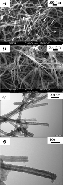 | ||
| Fig. 8 Representative SEM images of the SiO2 nanofibers (a, dried xerogel before calcination) and resulting nanotubes (b, after calcination at 500 °C) obtained by “post-diffusion” of TEOS through a pre-formed organogel with composition A (excluding TEOS); some representative TEM images of the nanotubes are also shown in c and d. | ||
Therefore, these results confirm that the final morphology and dimensions of the silica nanotubes may not only be controlled by adjusting the reaction conditions (i.e. the pH), but are also strongly affected by the presence or absence of condensing silicate species during the self-assembly of gelator molecules. As we have mentioned, the sol–gel condensation of silicate species seems to play a key role inducing an important rearrangement in the aggregation of gelator molecules, in accordance with previous reports.17 Accordingly, when using organogels or hydrogels as templates, the replication of fibrous morphologies into silica through the in situ procedure should be considered as a real co-assembly process (the formed organogel fibrils direct the growth of nanofibrous silica, and vice versa, the condensing silicate species affects the way the gelator molecules and/or the organogel fibrils self-assemble); in contrast, the post-diffusion strategy would rather be a more direct or simple transcription process, the preformed organogel template consisting of more aggregated and thicker fibrous bundles (though some rearrangement of the “flexible” organogel fibrils during the diffusion of siliceous species should not be discarded, since these fibrils are held by relatively weak supramolecular forces).
On the other hand, and complementing the SEM/TEM characterisation, the BET measurements performed with the silica nanotubes (after gelator removal) gave nitrogen adsorption–desorption isotherms representative of mesoporous materials (type IV) in both strategies (see Fig. 9a). The fact that some of the nanotube tips are not perfectly opened or accessible (as may be observed in the TEM images) could explain the rather weak hysteresis loops and the relatively low surface areas obtained by the BET method (149 and 262 m2 g−1 for in-situ and post-diffusion routes, respectively). Also remarkably, in both cases the corresponding accumulated volume (%) vs. pore size curves (Fig. 9b), obtained by the BJH method from the desorption branches, were in good agreement with the SEM/TEM observations previously discussed. Indeed, for the silica nanotubes prepared by the “in situ” procedure, the BJH curve (accumulated volume% with a pore size higher than D) shows a higher mesopore size population below 15 nm (68 against 46%); in contrast, the “post-diffusion” nanotubes present a considerably higher mesopore population with sizes compressed between 15 and 45 nm (31 against 12%), which could be associated with the thicker inner mesoporous channels obtained in this procedure. In both cases, there is also a considerably high population (around 20%) of mesopores with sizes above 45 nm; this should be rather ascribed to secondary (or textural) mesopores which form among the entangled or aggregated nanotubes (as may be appreciated for instance in Fig. 7a and 8c), and is also responsible for the increased nitrogen adsorption observed in the isotherms at relative pressures (P/P0) higher than 0.9 (especially in the post-diffusion sample). In this respect, the higher textural mesoporosity in the post-diffusion nanotubes would be also consistent with the more aggregated nature of the preformed organogel in the absence of silicate species.
 | ||
| Fig. 9 N2 adsorption–desorption BET isotherms (a) of the gelator-extracted SiO2 nanotubes obtained through the “in situ” and “post-diffusion” procedures (composition A), and corresponding accumulated volume% vs. pore size curves (b) obtained by the BJH method. | ||
Preliminary results with fibrous Fe-based xerogels
Finally, the great potential interest of these gelator compounds has been further confirmed in current experiments, that have enabled the successful preparation of fibrous Fe-based hybrid xerogels with gelator 1 as template.‡ The formed xerogel exhibited mostly fibrous morphologies as those presented in the SEM details of Fig. 10a–c. Note that semiquantitative EDAX analyses performed on these fibrous regions (a representative EDAX spectrum is shown in Fig. 10d) gave an average Fe2O3/C weight ratio of ca. 8.7%, which represents approximately 1/3 of the maximum or theoretical quantity (26.6%) considering the amounts of Fe(OEt)3 and gelator 1 in the original formulation (equimolecular quantities). | ||
| Fig. 10 Representative SEM images (a to c) and EDAX spectrum (d) of the Fe-based/gelator hybrid xerogel (40 °C-dried) obtained by the in situ procedure using gelator 1 as template.‡ | ||
Current investigations are being performed to optimize the preparation methodology (by in situ and/or post-diffusion procedures), and also to study the preservation of the fibrous morphologies after template removal and transformation of Fe oxo-hydroxo species into hematite (α-Fe2O3) or magnetite (Fe3O4) oxides. Indeed, hematite-based nanofibers and/or nanotubes have still not been prepared through the use of organogelator templates (α-Fe2O3 nanotubes have been recently prepared, but using porous membranes of anodized alumina as templates),18 and this would be an important step to extend the organogelator approach to the design of more complex (multicationic) oxide-based systems (such as transition metal mixed oxides).
Conclusions
The morphology and aggregation degree of nanofibrous silica have been successfully controlled by simply adjusting the pH conditions through the use as templating agents of “in situ” formed organogels of a pH-sensitive, pyridine-based gelator. Removal of the gelator template allowed the formation of well-formed silica nanotubes (30–60 nm thick, with 3–10 nm inner channels). Thicker silica nanotubes (from 40 to 80 nm as average), exhibiting also thicker inner mesoporous channels (15–35 nm), were prepared using a more flexible two-step or “post-diffusion” strategy.The morphology differences observed when comparing both strategies suggest that in the in situ procedure the templating proceeds via a co-assembly process, with important interactions among the gelator molecules and the condensing silicate species (which are also very dependent on reaction conditions, such as pH and/or catalyst); in contrast, in the “post-diffusion” route the templating would be rather a simple transcription process, the fibrils of the preformed organogel being more aggregated and thicker since they are formed in the absence of silicate species. As far as we know, this is the first report comparing both “in situ” and “post-diffusion” templating strategies to obtain nanofibrous inorganic replicas from organogels. The “post-diffusion” route may be very useful for more complex systems (i.e. multi-metallic) where the use of the “one-pot” procedure is not possible for any reason (insolubility of precursors, inadequate pH or catalyst conditions, disruption of the organogelation capability by metallic species, and so on).
Finally, preliminary tests have also confirmed the potential interest and flexibility of this approach by preparing nanofibrous Fe-based hybrid xerogels with these gelator templates.
Therefore, this work demonstrates the versatility of this family of gelators to pattern nanofibrous silica with different fibrous morphologies. Current investigations are being carried out to optimise the template synthesis of iron oxide-based nanofibers and/or nanotubes with these gelator compounds, thus opening a land of opportunities to design nanofibers and/or nanotubes of any inorganic phase (i.e. transition metal mixed oxides).
Acknowledgements
The authors thank the “Generalitat Valenciana” (GV04B-697 and GV05/089 projects) and “Ministerio de Educación y Ciencia” (MAT2005-00507 project) from Spain, for financial support. The authors also thank Mónica Vicent and Jessica Gilabert (Institute of Ceramic Technology, ITC, Spain) for the BET/BJH measurements.References
- (a) P. Terech and R. G. Weiss, Chem. Rev., 1997, 97, 3133 CrossRef; (b) J. H. van Esch and B. L. Feringa, Angew. Chem., Int. Ed., 2000, 39, 2263 CrossRef; (c) D. Abdallah and R. G. Weiss, Adv. Mater., 2000, 17, 1237 CrossRef CAS; (d) O. Gronwald, E. Snip and S. Shinkai, Curr. Opin. Colloid Interface Sci., 2002, 7, 148 Search PubMed; (e) N. M. Sangeetha and U. Maitra, Chem. Soc. Rev., 2005, 34, 821–836 RSC.
- Molecular Gels: Materials with Self-Assembled Fibrillar Networks, ed. R. G. Weiss and P. Terech, Springer, Berlin, 2006 Search PubMed.
- (a) J. H. Jung, Y. Ono, K. Hanabusa and S. Shinkai, J. Am. Chem. Soc., 2000, 122, 5008 CrossRef CAS; (b) J. H. Jung, Y. Ono and S. Shinkai, Langmuir, 2000, 16, 1643 CrossRef CAS; (c) K. J. C. van Bommel, A. Friggeri and S. Shinkai, Angew. Chem., Int. Ed., 2003, 42, 980 CrossRef.
- J. H. Jung and S. Shinkai, Top. Curr. Chem., 2004, 248, 223–260.
- M. Suzuki, Y. Nakajima, T. Sato, H. Shirai and K. Hanabusa, Chem. Commun., 2006, 377 RSC.
- (a) J. Hu, T. W. Odom and C. M. Lieber, Acc. Chem. Res., 1999, 32, 435 CrossRef CAS; (b) Y. Xia and P. Yang, Adv. Mater., 2003, 15, 351 CrossRef CAS; (c) Y. Xia, P. Yang, Y. Sun, Y. Wu, B. Mayers, B. Gates, Y. Yinn, F. Kim and H. Yan, Adv. Mater., 2003, 15, 353 CrossRef CAS; (d) C. N. R. Rao and M. Nath, Dalton Trans., 2003, 1–24 RSC; C. N. R. Rao, F. L. Deepak, G. Gundiah and A. Govindaraj, Prog. Solid State Chem., 2003, 31, 5 CrossRef CAS; (e) Y. Xiong, B. T. Mayers and Y. Xia, Chem. Commun., 2005, 5013 RSC.
- (a) S. Kobyashi, K. Hanabusa, N. Hamasaki, M. Kimura and H. Shirai, Chem. Mater., 2000, 12, 1523 CrossRef CAS; (b) S. Kobayashi, N. Hamasaki, M. Suzuki, M. Kimura, H. Shirai and K. Hanabusa, J. Am. Chem. Soc., 2002, 124, 6550 CrossRef CAS; (c) M. Llusar, L. Pidol, C. Roux, J. L. Pozzo and C. Sanchez, Chem. Mater., 2002, 14, 5124–5133 CrossRef CAS; (d) G. Gundiah, S. Mukhopadhyay, U. G. Tumkurkar, A. Govindaraj, U. Maitra and C. N. R. Rao, J. Mater. Chem., 2003, 13, 2118 RSC.
- (a) M. Llusar, C. Roux, J. L. Pozzo and C. Sanchez, J. Mater. Chem., 2003, 13, 442 RSC; (b) M. Llusar, G. Monrós, C. Roux, J. L. Pozzo and C. Sanchez, J. Mater. Chem., 2003, 13, 2505 RSC; (c) M. Llusar, G. Monrós, J. L. Pozzo, C. Roux and C. Sanchez, Z. Anorg. Allg. Chem., 2005, 631, 2215–2220 CrossRef CAS.
- P. Xue, R. Lu, D. Li, C. Tan, C. Bao, Z. Wang and Y. Zhao, Langmuir, 2004, 20, 11234 CrossRef CAS.
- J. H. Jung, T. Shimizu and S. Shinkai, J. Mater. Chem., 2005, 15, 3979 RSC.
- (a) Y. Yang, M. Suzuki, A. Kurose and K. Hanabusa, Chem. Commun., 2005, 2032 RSC; (b) Y. Yang, M. Suzuki, M. Kimura, H. Shirai and K. Hanabusa, Chem. Commun., 2004, 1332 RSC.
- (a) B. Escuder, S. Martí and J. F. Miravet, Langmuir, 2005, 21, 6776 CrossRef CAS; (b) B. Escuder and J. F. Miravet, Chem. Commun., 2005, 5796 RSC.
- J. F. Miravet and B. Escuder, Org. Lett., 2005, 7, 4791 CrossRef CAS.
- (a) J. L. Pozzo, G. M. Clavier and J. P. Desvergne, J. Mater. Chem., 1998, 8, 2575 RSC; (b) M. Suzuki, M. Yumoto, M. Kimura, H. Shirai and K. Hanabusa, Langmuir, 2003, 19, 8622 CrossRef CAS; (c) D. K. Kumar, D. A. Jose, P. Dastidar and A. Das, Langmuir, 2004, 20, 10413 CrossRef CAS; (d) S. Tanaka, M. Shirakawa, K. Kaneko, M. Takeuchi and S. Shinkai, Langmuir, 2005, 21, 2163 CrossRef CAS.
- (a) M. Suzuki, S. Owa, M. Kimura, A. Kurose, H. Shirai and K. Hanabusa, Tetrahedron Lett., 2005, 46, 303 CrossRef CAS; (b) M. Suzuki, T. Sato, A. Kurose, H. Shirai and K. Hanabusa, Tetrahedron Lett., 2005, 46, 2741 CrossRef CAS; (c) M. Suzuki, M. Nambu, M. Yumoto, H. Shirai and K. Hanabusa, New J. Chem., 2005, 29, 1439 RSC.
- (a) Y. Ono, K. Nakashima, M. Sano, J. Hojo and S. Shinkai, J. Mater. Chem., 2001, 11, 2412 RSC; (b) K. J. C. van Bommel and S. Shinkai, Langmuir, 2002, 18, 4544 CrossRef.
- P. Xue, R. Lu, D. Li, M. Jin, C. Bao, Y. Zhao and Z. Wang, Chem. Mater., 2004, 16, 3702 CrossRef CAS.
- J. Chen, L. Xu, W. Li and X. Gou, Adv. Mater., 2005, 17, 582 CrossRef CAS.
Footnotes |
| † The hydrogel shown in Fig. 2c was prepared by dissolving 28.9 mg of gelator 1 in 3 mL of an aqueous HCl solution (molar ratio H2O ∶ HCl = 1 ∶ 0.002; the final concentration of gelator 1 was equal to 2 × 10⁻² M) in a closed reaction flask. Hydrogel formation took place by progressive increase of the pH, accomplished by free exposure to NH3 vapours (the opened reaction flask was introduced into a bigger and closed vessel containing a 5% aqueous solution of NH3). The hydrogel formed in less than two hours of exposure time, at a pH around 8–9. The resulting translucent gel was then dried overnight at room temperature, and then further dried at 40 °C for 2 days, before SEM characterization. |
| ‡ The fibrous Fe-based hybrid xerogel shown in Fig. 10 was prepared using gelator 1 as template, iron trisethoxide (Fe(OC2H5)3, 99%) as Fe precursor, acetonitrile–H2O as solvent, and trifluoroacetic acid (TFA) as catalyst. In a typical preparation, a solution of Fe trisalkoxide (19.3 mg) in a small volume of acetonitrile (1 mL) was added to a previously formed solution (heated at 65–70 °C) of gelator 1 (48.092 mg) in acetonitrile (3.9 mL) and H2O (0.11 mL) containing also TFA (8 mg). The molar ratios Fe : acetonitrile : H2O : TFA : Organogelator 1 in the overall mixture were equal to 1 : 930 : 60 : 1 : 1, with a concentration of organogelator 1 equal to 2 × 10⁻² M (as with Fe and TFA). The resulting yellowish-orange hot solution (apparent pH ca. 2–3) was homogenised during 15 minutes (continuously stirred and heated at 65–70 °C) before proceeding to organogelation by rapid cooling (closed flask) in a glaze bath. The formed fibrous aggregates (orange-coloured) caused the gelation of most of the original solution, which was left drying by free evaporation, first at room temperature (for 2 days) and then at 40 °C (also for 2 days), before SEM characterization. |
| This journal is © The Royal Society of Chemistry 2006 |