Surfactant aggregates hosting a photoresponsive amphiphile: structure and photoinduced conformational changes†
Massimo Boninia, Debora Bertia, Jean Marc Di Megliob, Mats Almgrenc, Jose Teixeirad and Piero Baglioni*a
aDepartment of Chemistry and CSGI, University of Florence, Via della Lastruccia 3, Sesto Fiorentino, I-50019, Firenze, Italy. E-mail: piero.baglioni@unifi.it.; Web: http://www.csgi.unifi.it Fax: +39 055 457 3032; Tel: +39 055 457 3033
bMatière et Système Complexes, Université Paris 7, Paris, France
cDepartment of Physical Chemistry, Uppsala University, P.O. Box 532, Uppsala, S-751 21, Sweden
dLaboratoire Leon Brillouin (CEA/CNRS), Saclay, 91191 Gif-sur-Yvette, Cedex, France
First published on 27th October 2005
Abstract
This paper reports the aggregational and photoresponsive properties of aqueous solutions of a cationic bolaform surfactant, where an azobenzene moiety connects two identical hydrophobic chains terminated by quaternary ammonium groups. In analogy to common surfactants, the cationic bolaform molecules form supramolecular assemblies in water, and both interfacial and associative properties can be modulated and controlled by shining the sample with the appropriate wavelength radiation. Binary and ternary (BTHA/SDS/water) systems were investigated as a function of the photosurfactant concentration, sample composition and degree of irradiation. Structural properties of the supramolecular assemblies, obtained by QELS, SANS and cryo-TEM, have been correlated to the isomerization state of the surfactant, inferred from UV–vis spectroscopy.
Introduction
Azobenzene-based photodevices find widespread use in view of the facile and reversible photoisomerization of their active moiety and of the relatively high thermal isomerization barrier that allows an effective control on the optical properties.1 This feature has attracted considerable interest for applications in information storage and molecular switching devices. Much of the optical and isomerization properties can be designed in the synthetic procedure by varying the chemical nature and the position of the substituents of the phenyl rings.2 Since isomerization is accompanied by a change in conformation, whose extent is determined by the derivatization of the chromophore, azobenzene derivatives represent excellent candidates for optical storage devices, where information is represented by light-induced alteration of conformational properties. Light response can be amplified by attaching the azobenzene group to a polymer or even by incorporation in a polymeric matrix (Nathanson et al.3 and references therein).Azobenzene-based photochemistry has been exploited in the extensive search for nanodevices responsive to external stimuli. Self-assembled colloidal systems can offer many potential advantages over molecular based devices because of the rich phase behavior, which can be modulated and fine-tuned by variation of external control parameters, such as temperature, pH and solution ionic strength. This flexibility can be further extended and integrated with responsiveness to other external stimuli, such as light or electrochemical potential, that allow temporal and spatial control to design effective supramolecular switches. Therefore, much effort has been devoted to a rational chemical design of photosurfactants that can self assemble or can be incorporated in a suitable amphiphilic matrix,4–7 allowing a photocontrol of surface tension4,8,9 and surface wettability. Interestingly, it has been reported that the light induced surface-tension gradient causes a mass transport of liquid.10
Some key features that should be considered are surfactant charge, the localization of the photoactive moiety, the extent of light induced variations (i.e., photocleavage, isomerization, etc.) and their reversibility. This latter feature is of utmost importance, because fast responsiveness to altered equilibrium conditions is one of the most distinguishing characteristics of soft materials.
In this context, the 4,4′-bis(trimethylammoniumhexyloxy)azobenzene bromide molecule (abbreviated hereinafter as BTHA) represents a very good candidate. The photoisomerization reaction of this bolaform cationic surfactant, whose synthesis has been recently reported,11 is shown in Fig. 1.
 | ||
| Fig. 1 Photoisomerization of trans- (left) and cis-BTHA (middle), and SDS (right) chemical structure. | ||
The prevailing thermal conformation of BTHA is the trans form, but, upon UV light absorption, the conformational equilibrium can be shifted towards the unfavored cis form, where steric hindrance is more severe. This photoinduced conformational change causes significant and reversible variation in relevant molecular properties, such as dipole moment, and a shift in the hydrophilic/hydrophobic balance.
However, a complete conversion to one of the two isomers cannot be reached under ordinary laboratory conditions, therefore an essential prerequisite to devise an effective nanodevice with external photocontrol is a detailed characterization in terms of composition of the photostationary states and of the effects of irradiation on mesoscopic assemblies of photoactive molecules.
In this work, this characterization is reported for BTHA and BTHA/SDS (sodium dodecyl sulfate, see Fig. 1) aqueous solutions. The outline of the paper is as follows: we first characterize the different photostationary states in terms of cis/trans composition as a function of sample composition and irradiation times, taking into account equilibrium and kinetic aspects. Then a structural investigation, performed with QELS and SANS, is presented and the results are correlated with the composition of the photostationary state and with cryo-TEM observations.
Experimental
Reagents
Hydrochloric acid (Aldrich, ACS grade), 4-aminophenol (Fluka, 98%), sodium nitrite (Merck, 99%), copper sulfate pentahydrate (Carlo Erba, 99.5%), ammonium hydroxide (Riedel De Haen, 32%, ACS grade), hydroxylamine hydrochloride (Fluka, 99%) and 1,6-dibromohexane (Fluka, 97%) were used as received. SDS (Fluka, 99%) was recrystallized twice from ethanol. Deuterated SDS was purchased from Isotec (Miamisburg, OH, USA), while deuterium oxide (>99.5%) was provided by Euriso-Top (Saclay, Gif sur Yvette, France).Synthesis
Sample preparation
BTHA was dissolved in water or water–SDS solutions by gentle stirring, and then kept overnight in the dark prior to any experiments. The composition of the investigated samples is reported in Table 1. Titration of BTHA in different solutions was achieved by UV–vis absorption, by comparison with a calibration curve previously evaluated at the isosbestic points in order to overcome artifacts due to isomeric compositions. Maximum solubility of BTHA was inferred by stirring solutions with excess precipitate for several days, and then titrating the absorption of the supernatant. Calibration curves at 319 nm and 441 nm are available in the ESI†.| Composition | Molar ratio | Sample name | |
|---|---|---|---|
| [BTHA]/mol dm−3 | [SDS]/mol dm−3 | [SDS]/[BTHA] | |
| 1.0 × 10⁻⁴ | — | — | BTHA0.1 |
| 1.0 × 10⁻³ | — | — | BTHA1 |
| 1.0 × 10⁻³ | 4.8 × 10⁻³ | 4.8 | SDS4.8 |
| 1.0 × 10⁻³ | 6.4 × 10⁻³ | 6.4 | SDS6.4 |
| 1.0 × 10⁻³ | 8.0 × 10⁻³ | 8.0 | SDS8 |
| 1.0 × 10⁻³ | 1.0 × 10⁻³ | 10 | SDS10 |
UV–vis measurements
UV–vis spectra were recorded with a Perkin-Elmer Lambda 900 spectrometer. Measurements were performed in the 200–500 nm range, as a function of irradiation time and wavelength.Irradiation of the samples
Samples contained in 1 mm path length quartz cells were irradiated with an Oriel Arc Lamp Source equipped with a 150 W Hg lamp. The source was equipped with a water-cooled filter holder and focused to irradiate the whole sample. The lamp power was 130 W for all the operating conditions. The desired irradiation wavelength was selected by using two narrow bandpass interference filters (Edmund Industrial Optics, Gloucester, New Jersey) centered at 365 nm (stock number NT43-103) and 430 nm (stock number NT43-108). In the photoisomerization procedure, considerable care has been paid to obtain reproducible conditions, in terms of a defined trans/cis isomer ratio as a function of the irradiation time. In our experiments we observed two photostationary states of the chromophore (where the maximum yield of one of the two isomers is obtained), each of them reproducibly and reversibly obtainable. The photoisomerization cycle used in our experiments is summarized in Fig. 2. As already mentioned, the photoisomerization cycle starts with dark-adapted samples. After exposure to room light, samples are irradiated stepwise at 365 nm until the maximum trans to cis photoconversion was achieved. Hereinafter, we will refer to these samples as “fully irradiated”. The “back irradiation” was performed using the 430 nm filter, until a stationary cis to trans photoconversion was obtained. Finally, the samples were kept overnight in the dark and the UV–vis spectra were recorded again in order to check if the isomerization was fully reversible. | ||
| Fig. 2 Photoisomerization cycle. | ||
Light scattering
Quasi-elastic light scattering experiments were carried out on a Brookhaven Instrument apparatus (BI 9000AT correlator card and BI 200SM goniometer). The light source was the doubled frequency of a Coherent Innova diode pumped Nd–YAG laser (λ= 532 nm, power 60 mW), linearly polarized in the vertical direction. The signal was detected by an EMI 9863B/350 photomultiplier. The laser long-term power stability was ±0.5%. Self-beating detection was recorded using decalin as index matching liquid. The volume of the samples used for quasi-elastic light scattering (3 ml) was one order of magnitude higher than that investigated by UV–vis spectroscopy (300 µl); the achievement of a “fully irradiated” photostationary state was therefore carefully checked also for this amount. Prior to any measurements samples were centrifuged at 2000 g for about 2 min in order to remove dust from the scattering volume.SANS experiments
SANS measurements were performed at 25 °C on the PAXE spectrometer at Laboratoire Léon Brillouin (LLB), Saclay, France. Neutrons with an average wavelength of 7.3 Å and wavelength spread Δλ/λ < 10% were used. Neutron scattered from the sample were detected by a two-dimensional array detector at two different sample-to-detector distances, 1.05 and 5.0 m. These configurations allowed collecting the scattered intensity in a range of Q between 0.01 and 0.3 Å−1. Samples for SANS, prepared using D2O as solvent in order to enhance the contrast and lower the incoherent scattering, were contained in 1 mm path length quartz cells. Data were corrected for background, empty cell and solvent contribution, and then reduced to scattering cross section (cm−1), following the standard LLB procedure. Samples were irradiated using the same UV Oriel lamp mounted directly in front of the SANS sample changer. Only fully irradiated and laboratory light adapted samples were investigated by means of SANS.The total scattered intensity was modelled as described in eqn. (1):
| I(Q) = I(Q)m + I(Q)sv + I(Q)bv + bkg | (1) |
The normalized scattering intensity in a SANS experiment is proportional to the product of the form factor and the structure factor. The form factor P(Q) accounts for the shape of the scattering particle, while the structure factor S(Q) describes how the scattered intensity is modulated by interference between radiation diffused by different particles. In the system investigated, the structure factor should account for interactions between charged objects in a dielectric medium. However, the inclusion of a Hayter–Penfold13,14 expression for S(Q) did not substantially improve the quality of the fitting. This is mainly due to two reasons: first, the system is extremely diluted; second, BTHA and SDS are oppositely charged. In particular, each BTHA molecule balances the charges carried by two SDS molecules. Therefore, considering the mixed nature of the scattering objects and the BTHA/SDS ratio, the charge is very low.
Data have been analyzed using NIST routines,15 modified in order to consider the system as composed by one micellar population and two vesicle populations. Micelles have been considered as monodisperse oblate ellipsoid (a disk-like ellipsoid of revolution) with uniform scattering length density.16 The scattered intensity due to such objects was calculated according to the following equations:
 | (2) |
| F(q,r,α) = 3(ρm − ρsolv)Vmj(um)/um | (3) |
| Vm = (4π/3)rminr2max | (4) |
 | (5) |
| j(um) = (sinum − umcosum)/u2m | (6) |
For vesicles we used the form factor introduced by Bartlett and Ottewill18 for spherical particles having a polydisperse core with a constant shell thickness. The contribution to the total scattering intensity arising from these particles was calculated according to the following equations:
 | (7) |
 | (8) |
| ρscaled = (ρsolv − ρshell)(ρcore − ρshell) | (9) |
| j(qrc) = sin(qrc) − (qrc)cos(qrc) | (10) |
The function G(rc) is the normalized probability of finding a particle with a core radius between rc and rc + dr, and it accounts for the polydispersity of the cores according to a Schultz distribution:19
 | (11) |
 | (12) |
The scattering length density of the core was set as equal to the scattering length density of the solvent. Therefore we calculated the intensity scattered from a spherical shell with constant thickness but polydisperse inner radius.
The scattering length density of SDS (3.89 × 10⁻⁷ Å−2) and BTHA (7.88 × 10⁻⁷ Å−2) were calculated using the molecular volumes of 410 Å3 [ref. 20] and 941 Å3, respectively. The molecular volume of BTHA was calculated by considering the molecule azobenzene, the two –OC6H10– chains (assimilated to two hexyl alcohol molecules) and the trimethylammonium bromide polar heads.
The molecular volumes of these parts were calculated from their density and molecular weight values obtaining 278 Å3 for the azobenzene (d = 1.09 g cm−3), 416 Å3 for the two hexyl alcohols (d = 0.815 g cm−3) and 290 Å3 for the two trimethylammonium bromide (d = 1.59 g cm−3). The van der Waals volume of 6 hydrogen atoms (6 × 7.24 Å3) was then subtracted to obtain the BTHA molecular volume.
The scattering length densities of the mixed systems were then calculated as the volume weighted sums of the pure BTHA and SDS scattering length densities: 5.17 × 10⁻⁷ Å−2 and 4.78 × 10⁻⁷ Å−2 for the SDS4.8 and SDS8 samples, respectively. These values were then used as fixed parameters in the fitting procedure.
Cryo-TEM
Cryo-transmission electron microscopy investigations were performed with a Zeiss EM 902A instrument operating at 80 kV. The technique used for cryo-TEM examination is described in detail elsewhere21 and can be shortly summarized as follows. Thin sample films (10–500 nm) were prepared at controlled temperature (298 K) and controlled relative humidity (98–99%) within a custom-built environmental chamber. The films were thereafter vitrified by quick freezing in liquid ethane and transferred to a transmission electron microscope for examination. Temperature was kept below 108 K both during sample transfer and examination.Results and discussion
Binary system
Fig. 3 shows UV–vis spectra for a 0.1 mM BTHA aqueous solution as a function of UV irradiation time. The sample is initially dark-adapted, and its spectrum is dominated by the 365 nm absorption, due to the trans isomer. If the sample is kept for a few minutes in laboratory daylight, the spectrum sensibly changes, and this variation is further enhanced by 365 nm irradiation. As irradiation proceeds (Fig. 3a), the spectrum reaches a photostationary state in about 20 min. In agreement with the literature for azobenzenes,22 this spectral evolution accounts for trans-to-cis isomerization and it is consistent with the intensity increase of the cis absorption peak at about 430 nm. Supplementary irradiation does not change cis/trans ratio. | ||
| Fig. 3 UV–vis Absorption spectra of a 0.1 mM BTHA water solution as a function of the irradiation time at 365 nm (a) and 430 nm (b). | ||
Isomerization can be reverted shining the sample for about 5 min with 430 nm light (i.e., close to the maximum of the cis absorption band), yielding the maximum trans concentration in our experimental conditions (Fig. 3b). Eventually, after overnight storage in the dark, the isomeric composition of the system is again ruled by thermal equilibrium and the original spectrum is completely recovered, indicating the full reversibility of the process. It is worth stressing that the dark-adapted conditions cannot be reached by irradiation, because of the partial overlap between trans and cis absorption bands.
The presence of two isosbestic points (319 nm and 441 nm) is a clear indication of a two-state system (cis and trans) and allows obtaining an UV–vis calibration curve not dependent on the isomerization state of the molecule (see ESI†).
The effects of the isomeric composition (dark adapted, lab adapted and fully irradiated, i.e., maximum cis content) on the self-assembled structures in two selected samples (1 mM and 0.1 mM) has then been investigated by means of QELS. The average size of the aggregates can be deduced from a second order cumulant analysis of the autocorrelation functions, whose results are reported in Table 2.The average diameter of the scattering objects is always in the 200–350 nm range and the polydispersity is quite high. The results are therefore consistent with the presence of aggregates with characteristic dimensions typical of the mesoscopic scale. However, the previous analysis is biased by the fact that none of the intensity autocorrelation functions is mono-exponential, suggesting the presence of different populations. This prevents a straightforward and precise analysis of the data by means of dynamic light scattering.
| Sample | Isomeric state | Diameter/nm | Polydispersity |
|---|---|---|---|
| BTHA0.1 | Dark-adapted | 204 | 0.254 |
| BTHA0.1 | Lab-adapted | 341 | 0.227 |
| BTHA0.1 | Fully irradiated | 234 | 0.219 |
| BTHA1 | Dark-adapted | 271 | 0.211 |
| BTHA1 | Lab-adapted | 299 | 0.208 |
| BTHA1 | Fully irradiated | 233 | 0.260 |
Simple packing considerations regarding the structure and steric hindrance of the surfactant (see Fig. 1) are consistent with the fact that micellar structures can be formed preferentially by the cis isomers, while trans molecules can favorably pack in locally planar structures like disks and vesicles.23 The size obtained from the autocorrelation function decays is consistent with vesicular aggregates, even if the simultaneous presence of small globular micelles could be masked by the scattering due to larger objects and, therefore, cannot be excluded a priori.
A working hypothesis for the microstructure of binary systems can be obtained from a combined UV–vis/QELS analysis. To simplify data interpretation, we neglect the amount of photosurfactant monomers so that the electronic spectrum results only from the contributions of aggregated chromophores. This assumption is correct since the critical aggregation concentration (about 3.8 × 10⁻⁵M, as determined by dynamic surface tension, see ESI†) is negligible with respect to the concentration range investigated. A further complication arises from the fact that a given isomer experiences a different environment if embedded in a bilayer or in a micellar structure. However the observation of two isosbestic points in the UV–vis spectra rules out the presence of more than two species and suggests that each isomer experiences a well-defined chemical environment.
Direct imaging techniques are of invaluable help in the investigation of polymorphic samples. In Fig. 4 two representative cryo-TEM micrographs of 1 mM BTHA in lab-adapted conditions are reported. Closed hollow structures, disks (both face-on and side-on), and spherical micelles coexist in equilibrium. Even if a precise measure of the diameter of the micelles and the thickness of the bilayer from the micrographs is difficult, we estimate ca. 3 nm for the bilayer thickness and 4 nm for the micellar diameter.
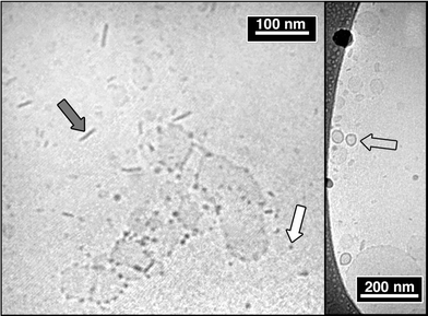 | ||
| Fig. 4 Cryo-TEM micrographs of the BTHA1 sample. Both micellar (white arrow) and disc-like (grey arrow) structures are present. Vesicles are indicated by the transparent arrow. | ||
Therefore the hypothesis of micelles made of cis-BTHA (whose azobenzene/quaternary nitrogen distance is about 15 Å) and vesicle bilayers formed by trans-BTHA (the distance between two quaternary nitrogen is about 29 Å) appears to be substantiated.
Ternary systems
Four different BTHA–SDS–water samples were investigated (see Table 1). The photosurfactant concentration was kept constant, while SDS concentration was varied around its cmc (8.3 mM at 25 °C).24Fig. 5 reports the UV–vis measurements for the photoconversion of SDS4.8 sample. The SDS concentration for this sample is below its cmc.
 | ||
| Fig. 5 UV–vis absorption spectra of SDS4.8 sample as a function of the irradiation time at 365 nm (a) and 430 nm (b). | ||
Before discussing photoconversion, we would like to stress the effect of the local chemical environment on spectral properties of the chromophore. In particular, the molar absorption of the trans species shows a strong increase in the presence of SDS (see Fig. 6, dark adapted samples), while the cis band is practically unaffected by the presence of SDS (irradiated samples). This observation is indicative of higher affinity of trans-BTHA for the anionic surfactant and of the formation of mixed aggregates.
 | ||
| Fig. 6 UV–vis absorption spectra of BTHA as a function of SDS concentration for dark-adapted (a), lab-adapted (b) and fully irradiated (c) samples. | ||
A careful comparison of ternary samples' spectra shows that UV–vis absorption of BTHA depends on SDS concentration, especially for the dark and the lab adapted samples, as Fig. 6 shows. The differences are smaller for low SDS/BTHA ratios and level off when the mole ratio exceeds 8. This evidence can be taken as an indication of a structural evolution of the self-assembled structures that implies rearrangement of the BTHA environment. Moreover, the fact that the dark and the lab adapted samples present such striking differences again indicates that the trans isomer has a preferential affinity for SDS.
This can be qualitatively understood by taking into account the chemical structure of SDS and comparing it to those of the two isomers. Trans-BTHA matches with its fully extended length two SDS molecules arranged in a tail-to-tail fashion. We can therefore hypothesize that BTHA in a trans conformation and SDS cooperate in the formation of bilayered structures. This arrangement prevents photoconversion imposing conformational constraints on the molecule that disfavor the trans-to-cis isomerization.
It is interesting to note that the SDS4.8 sample exhibits almost no difference between dark and lab adapted spectra. For this sample composition the conformational freedom of BTHA must be particularly restricted, and probably most of the BTHA molecules are hosted in bilayered structures.
A quantitative evaluation of the isomerization provides both phototransformation rates and yields as a function of the sample composition. According to the literature,22 we can assume that the cis isomer is present only in negligible amounts in dark-adapted samples. For all the other samples, even if the absorptions corresponding to the two isomers are partially overlapped, it is reasonable to assume that 95% of the absorption at 365 nm is due to the trans form. On this basis it is possible to calculate the trans isomer percentage in solution by simply weighting the absorbance values at the maximum trans absorbance wavelength with respect to the dark sample:
 | (13) |
In Fig. 7 the trans percentage of BTHA is reported as a function of the irradiation time for binary and ternary systems. Both trans-to-cis and cis-to-trans isomerization data have been interpreted according to a single exponential decay, using respectively the eqns. (14) and (15):
 | (14) |
 | (15) |
 | ||
| Fig. 7 trans-BTHA percentage as a function of irradiation time for the trans-to-cis (a) and cis-to-trans (b) isomerizations. Dotted lines represent the best fitting according to a single exponential law. | ||
The values of τ and %transmax/min obtained by non-linear least squares fitting of data according to eqns. (14) and (15) are reported in Fig. 8.
 | ||
| Fig. 8 (a): Characteristic time of the process τ as a function of SDS/BTHA ratio. (b): Minimum and maximum trans-BTHA percentages obtained respectively for the trans-to-cis and cis-to-trans isomerizations. | ||
When SDS/BTHA ratio is 4.8, the characteristic time of the trans-to-cis process τ is the lowest, while %transmin is the highest. For the reverse process the characteristic rates are closer to each other and no straightforward dependence on the mole ratio can be traced, while the value of the %transmax in the case of SDS4.8 is again maximum. Increasing both BTHA and SDS concentrations, but keeping constant the molar ratio (4.8), we observe the same behavior, suggesting that the photoionization dominant factor is the ratio between BTHA and SDS, and not the overall concentration.
Fig. 9 shows results obtained by means of dynamic light scattering in terms of mean diameter and scattered intensity as a function of [SDS]/[BTHA] composition. As in the case of the binary system, the autocorrelation functions are not characterized by a single exponential, suggesting the presence of different populations. The averaged diameter reported in Fig. 9 and deduced from a second order cumulant suffers of this limitation. SDS4.8 shows for both aggregate size and scattered intensity the strongest irradiation effect. Therefore, this sample represents the most promising candidate to devise a light-responsive system and deserves a more detailed analysis of its microstructure.
 | ||
| Fig. 9 Mean diameters (a) and scattered intensities (b) as obtained by dynamic light scattering. | ||
The cryo-TEM on binary system evidenced the simultaneous presence of disks and vesicles. For the ternary system cryo-TEM shows the presence of vesicles and no disks could be observed (see Fig. 10). Interestingly, an accurate analysis of the micrographs shows the coexistence of two vesicle populations: the first constituted by small vesicles with a mean diameter of about 50 nm, and the other constituted by more polydisperse vesicles with diameters up to 350 nm. Within the experimental resolution, both vesicle populations are characterized by the same bilayer thickness obtained for the binary system (ca. 3 nm). Micelles are still present, even if it is again very difficult to accurately evaluate their size and shape.
 | ||
| Fig. 10 Cryo-TEM micrographs of the SDS4.8 sample in lab-adapted conditions. | ||
The auto-correlation functions of the samples SDS4.8 and SDS8 both in lab-adapted and fully irradiated conditions were Laplace-inverted by using the CONTIN routine.25 The mean hydrodynamic radii and the weight by volume of both vesicle populations are reported in Table 3, showing a good agreement with cryo-TEM results.
| SDS4.8 | SDS8 | |||||
|---|---|---|---|---|---|---|
| Lab-adapted | Fully irradiated | Lab-adapted | Fully irradiated | |||
| a The volume percentage accounts for the whole scattering objects, i.e., the sum of inner water core, bilayer and water of hydration volumes.b The volume fraction accounts only for the volume occupied by the surfactant molecules, i.e., the micelle or bilayer volume. | ||||||
| DLS | Small Vesicles | %volumea | 87.7 | 40.8 | 88.5 | 39.0 |
| rhd/Å | 228 | 244 | 222 | 255 | ||
| Large Vesicles | %volumea | 12.3 | 59.2 | 11.5 | 61.0 | |
| rhd/Å | 2018 | 1400 | 1357 | 1417 | ||
| SANS | Ellipsoids | ϕmb | 3.5 × 10⁻⁵ | 1.378 × 10⁻³ | 1.429 × 10⁻³ | 1.843 × 10⁻³ |
| rmin/Å | 18.5 | 18.3 | 18.5 | 17.3 | ||
| rmax/Å | 27.0 | 27.2 | 27.9 | 24.2 | ||
| %volumea | <0.1 | 0.1 | 0.1 | 0.3 | ||
| Small Vesicles | ϕsvb | 1.391 × 10⁻³ | 1.11 × 10⁻⁴ | 5.96 × 10⁻⁴ | 5.4 × 10⁻⁵ | |
| rc/Å | 240 | 241 | 235 | 252 | ||
| σc | 0.24 | 0.23 | 0.23 | 0.23 | ||
| t/Å | 29.0 | 29.0 | 29.0 | 29.0 | ||
| %volumea | 55.9 | 18.6 | 58.8 | 18.1 | ||
| Large Vesicles | ϕlvb | 2.5 × 10⁻⁶ | 3.3 × 10⁻⁶ | 2.9 × 10⁻⁶ | 1.8 × 10⁻⁶ | |
| rc/Å | 2015 | 1397 | 1354 | 1414 | ||
| σc | 0.52 | 0.35 | 0.51 | 0.63 | ||
| t/Å | 29.0 | 29.0 | 29.0 | 29.0 | ||
| %volumea | 44.1 | 81.3 | 41.1 | 81.6 | ||
Further insights can be obtained from SANS. SANS measurements have then been performed on SDS4.8 and SDS8 samples. As mentioned in the experimental section, only lab-adapted and fully-irradiated samples have been investigated.
Fig. 11 reports SANS curves for non-irradiated samples. In the case of the SDS4.8 samples, a Q−2 scattering power law, typical of bilayers, is present for a remarkable scattering vector range.
 | ||
| Fig. 11 Small angle neutron scattering curves for lab adapted samples. The line represents the Q−2 power law. | ||
Assuming an asymptotic law for bilayers,26I(Q) ∝ exp(−q2Rt2), the bilayer thickness t = 2.9 nm can be evaluated (Rt2 = t2/12). This thickness is in excellent agreement with cryo-TEM results.
All the experimental evidences gathered so far support that SDS4.8 and SDS8 are constituted by mixture of micellar and bilayered structures.
It is worth noting that BTHA can be strucurally assimilated to two dodecyltrimethylammonium bromide (DTAB) molecules with a flexible covalent connection at the end of hydrophobic tails. In particular, two DTAB molecules assembled in a side-to-side fashion (as in DTAB micelles) resemble cis-BTHA, while the tail-to-tail assembly (DTAB bilayers) looks similar to trans-BTHA. This parallelism turns out to be very useful in the analysis of SANS data. The self assembly properties in water of catanionic systems constituted by dodecylsulfate and dodecyltrimethylammonium have been extensively studied in the last years.17,27–32 According to SANS results, DTAB and SDS form micelles and bilayered aggregates in solution.32 Bergström and Pedersen found that small unilamellar vesicles (350 Å < R < 650 Å) form in very dilute samples when one of the two surfactants is at a concentration lower than 1 : 3 molar ratio, while larger vesicles are predominant near the equimolar composition.30
Lab-adapted and fully irradiated SANS spectra of SDS4.8 and SDS8 samples were therefore interpreted according to the indications inferred from cryo-TEM and DLS results: the total scattered intensity was simulated as arising from micelles, small vesicles and large vesicles (see Fig. 12), neglecting possible contributions from interference effects, as explained in the experimental section. The introduction of multiple form factors in scattering data analysis should generally be taken with due caution, due to a risk of misinterpretation connected to the high number of variational parameters. However, in the present case, SANS analysis is not stand-alone and is compared to results obtained with different and complementary techniques.
 | ||
| Fig. 12 Sketch of the model used for SANS data fitting. Micelles were simulated according to a prolate ellipsoid (bottom-left), while vesicles (right) were simulated according to a core–shell model, with an aqueous polydisperse core and a monodisperse shell. | ||
SANS experimental and simulated curves of SDS4.8 and SDS8 samples are shown in Figs. 13 and 14, respectively, while the best-fit parameters are reported in Table 3. The SDS4.8 sample in lab-adapted conditions is almost entirely constituted by vesicles, with the small vesicle population representing the majority of the scattering objects, in good agreement with DLS results. When the sample is fully irradiated, the small vesicle population volume fraction is reduced by a factor of 10, while micelles become the prevalent aggregate.
 | ||
| Fig. 13 SANS experimental (markers) and fitted (lines) curves of the SDS4.8 sample in lab-adapted (○) and fully irradiated conditions (●). | ||
 | ||
| Fig. 14 SANS experimental (markers) and fitted (lines) curves of the SDS8 sample in lab-adapted (□) and fully irradiated conditions (■). | ||
In the case of the SDS8 sample, ellipsoidal micelles are the prevailing objects both for lab-adapted and fully irradiated conditions. Similarly to the SDS4.8 case, the vesicle amount diminishes when the sample is irradiated.
With regards to the large vesicle population, the Q range investigated by SANS is not ideally suited for the determination of both size and volume fraction of the large vesicle population. Therefore, we have used the radius determined from DLS to extract the vesicle thickness from SANS analysis.
SANS analysis results confirm the tendency of trans-BTHA to form bilayered structures, while cis-BTHA preferentially form micelles. Moreover, the peculiarity of the SDS4.8 sample indicate that the [SDS]/[BTHA] 4.8 ratio is the ideal ratio for stabilizing these bilayered structures: in fact, UV irradiation causes a dramatic change in the microstructure of this sample, consisting of the break up of vesicles to form ellipsoidal micelles. Eastoe et al.4 have previously reported a similar behaviour for a stilbene-containing gemini photosurfactant (SGP). They have shown that this vesicle-to-micelle mechanism can be successfully used to effectively modulate the interfacial properties of the photoactive system. Unfortunately, the investigated SGP shows a poor reversibility, being the major drawback in the design of optical devices based on this photosurfactant. On the contrary, the BTHA/SDS system is fully reversible and can be indefinitely photoconverted between each photostationary state.
Conclusions
We have reported a structural investigation of the aggregates formed in aqueous solutions by a di-cationic bolaform photosurfactant in binary (BTHA–water) and ternary (BTHA–SDS–water) systems. The chemical structure of the surfactant is such that the photoresponsive moiety connects two identical hexyl chains, terminated by two quaternary ammonium groups. UV irradiation shifts the cis/trans thermal equilibrium towards the cis form, with a dramatic change in surfactant packing preference. Photochemical properties (i.e., isomerization kinetics and equilibria) have been determined as a function of photosurfactant concentration for binary systems and sample composition for ternary systems. Photoconversion is strongly dependent on sample composition, irradiation time, and storage conditions of the aqueous solutions (i.e., laboratory light or dark conditions). For each sample a photoconversion cycle has been determined and reproducible light responsiveness established. Isomerization is affected by packing constraints of the photosurfactant, which are meaningfully different for the binary and the ternary systems. Photoconversion has a profound effect on the kind and on the relative abundance of the aggregates in solution, as deduced by QELS and SANS results, both supported by cryo-TEM analysis. We have observed the presence of vesicles that form for SDS/BTHA 4.8 ratio; this observation is fully consistent with geometrical and packing considerations of surfactant molecules. The opposite charges present on the surfactant headgroups (SDS and BTHA) makes this system similar to classical catanionic systems. Upon UV irradiation, major reversible changes have been found for this particular mole ratio that are consistent with a vesicle to micelle transition imposed by trans–cis isomerization. This feature makes this sample composition an excellent candidate for a photosensitive nanodevice with tunable external control on encapsulation and release properties.Acknowledgements
Massimo Bonini, Debora Berti and Piero Baglioni acknowledge CSGI, MIUR (PRIN-2003) and Programma Galileo (CRUI) for financial support. Göran Karlsson (Department of Physical Chemistry, University of Uppsala) is gratefully acknowledged for cryo-TEM measurements. The authors would like to thank Dr Pierluigi Barbaro for NMR measurements.References
- H. Rau, in Photochemistry and Photophysics, ed. J. K. Rabek, Boca Raton, 1990 Search PubMed.
- A. A. Blevins and G. C. Blanchard, J. Phys. Chem. B, 2004, 108, 4962 CrossRef CAS.
- A. Natansohn and P. Rochon, Chem. Rev., 2002, 102, 4139 CrossRef CAS.
- J. Eastoe, M. S. Dominguez, P. Wyatt, A. Beeby and R. K. Heenan, Langmuir, 2002, 18, 7837 CrossRef CAS.
- J. Eastoe, M. Sanchez-Dominguez, H. Cumber, G. Burnett, P. Wyatt and R. K. Heenan, Langmuir, 2003, 19, 6579 CrossRef CAS.
- J. Eastoe, M. Sanchez-Dominguez, P. Wyatt and R. K. Heenan, Chem. Commun., 2004, 2608 RSC.
- J. Eastoe, P. Wyatt, M. Sanchez-Dominguez, A. Vesperinas, A. Paul, R. K. Heenan and I. Grillo, Chem. Commun., 2005, 2785 RSC.
- C. Rosslee and N. L. Abbott, Curr. Opin. Colloid Interface Sci., 2000, 5, 81 Search PubMed.
- S. Abbott, J. Ralston, G. Reynolds and R. Hayes, Langmuir, 1999, 15, 8923 CrossRef CAS.
- K. Ichimura, S. K. Oh and M. Nakagawa, Science, 2000, 288, 1624 CrossRef CAS.
- J. Y. Shin and N. L. Abbott, Langmuir, 1999, 15, 4404 CrossRef CAS.
- B. M. Bogoslowsky, J. Gen. Chem., 1946, 16, 193 Search PubMed.
- J. P. Hansen and J. B. Hayter, Mol. Phys., 1982, 46, 651.
- J. B. Hayter and J. Penfold, Mol. Phys., 1981, 42, 109 CAS.
- http://www.ncnr.nist.gov/programs/sans..
- L. A. Feigin and D. I. Svergun, Structure Analysis by Small-Angle X-Ray and Neutron Scattering, Plenum Press, New York, 1987 Search PubMed.
- M. Bergstrom and J. S. Pedersen, J. Phys. Chem. B, 1999, 103, 8502 CrossRef.
- P. Bartlett and R. H. Ottewill, J. Chem. Phys., 1992, 96, 3306 CrossRef CAS.
- J. Hayter, in Physics of Amphiphiles: Micelles, Vesicles and Microemulsions, ed. V. DeGiorgio and M. Corti, North Holland, Amsterdam, 1983 Search PubMed.
- M. Bergstrom and J. S. Pedersen, Phys. Chem. Chem. Phys., 1999, 1, 4437 RSC.
- M. Almgren, K. Edwards and G. Karlsson, Colloids Surf., A, 2000, 174, 3 CrossRef CAS.
- W. R. Brode, J. H. Gould and G. M. Wyman, J. Am. Chem. Soc., 1952, 74, 4641 CrossRef CAS.
- J. Israelachvili, in Intermolecular and Surface Forces, Academic Press, London, 1985 Search PubMed.
- N. M. van Os, J. R. Haak and L. A. M. Rupert, in Physico-Chemical Properties of Selected Anionic, Cationic and Nonionic Surfactants, Elsevier, Amsterdam, 1993 Search PubMed.
- S. W. Provencher, Comput. Phys. Commun., 1982, 27, 213 CrossRef.
- O. Glatter and O. Kratky, Small Angle X-Ray Scattering, Academic Press, London, 1982 Search PubMed.
- P. Baglioni, L. Dei, L. Kevan and E. Rivara-Minten, ACS Symp. Ser., 1992, 501, 180 CAS.
- P. Baglioni, L. G. Dei, E. Rivara-Minten and L. Kevan, J. Am. Chem. Soc., 1993, 115, 4286 CrossRef CAS.
- N. Kamenka, M. Chorro, Y. Talmon and R. Zana, Colloids Surf., 1992, 67, 213 CrossRef CAS.
- M. Bergstrom and J. S. Pedersen, Langmuir, 1998, 14, 3754 CrossRef.
- M. Bergstrom and J. S. Pedersen, Langmuir, 1999, 15, 2250 CrossRef.
- M. Bergstrom, J. S. Pedersen, P. Schurtenberger and S. U. Egelhaaf, J. Phys. Chem. B, 1999, 103, 9888 CrossRef.
Footnote |
| † Electronic supplementary information (ESI) available: Absorbance calibration curves of BTHA in water, 1H-NMR spectra of 4,4′-dihydroxyazobenzene and BTHA, dynamic surface tension of BTHA in water and autocorrelation functions of the scattered light of BTHA1. See DOI: 10.1039/b511544b |
| This journal is © The Royal Society of Chemistry 2005 |