Rapid in situ detection of alkaloids in plant tissue under ambient conditions using desorption electrospray ionization
Nari Talaty, Zoltán Takáts and R. Graham Cooks*
Department of Chemistry, Purdue University, West Lafayette, IN 47907. E-mail: cooks@purdue.edu; Fax: +1-(765) 494-9421; Tel: +1-(765) 494-5262
First published on 19th October 2005
Abstract
Desorption electrospray ionization (DESI) mass spectrometry is applied to the in situ detection of alkaloids in the tissue of poison hemlock (Conium maculatum), jimsonweed (Datura stramonium) and deadly nightshade (Atropa belladonna). The experiment is carried out by electrospraying micro-droplets of solvent onto native or freshly-cut plant tissue surfaces. No sample preparation is required and the mass spectra are recorded under ambient conditions, in times of a few seconds. The impact of the sprayed droplets on the surface produces gaseous ions from organic compounds originally present in the plant tissue. The effects of operating parameters, including the electrospray high voltage, heated capillary temperature, the solvent infusion rate and the carrier gas pressure on analytical performance are evaluated and optimized. Different types of plant material are analyzed including seeds, stems, leaves, roots and flowers. All the previously reported alkaloids have been detected in C. maculatum, while fifteen out of nineteen known alkaloids for D. stramonium and the principal alkaloids of A. belladonna were also identified. All identifications were confirmed by tandem mass spectrometry. Results obtained show similar mass spectra, number of alkaloids, and signal intensities to those obtained when extraction and separation processes are performed prior to mass spectrometric analysis. Evidence is provided that DESI ionization occurs by both a gas-phase ionization process and by a droplet pick-up mechanism. Quantitative precision of DESI is compared with conventional electrospray ionization mass spectrometry (after sample workup) and the RSD values for the same set of 25 dicotyledonous C. maculatum seeds (one half of each seed analyzed by ESI and the other by DESI) are 9.8% and 5.2%, respectively.
Introduction
The study of natural products has played a major role in the development of organic and medicinal chemistry, including fundamental aspects of stereochemistry, mechanistic chemistry, biosynthesis and mechanisms of biological action, as well as providing medically useful compounds. Amongst natural products, it is the secondary metabolites which give a particular species its characteristic features. Unlike primary metabolites, these compounds are neither ubiquitous in the living organisms that produce them nor are they expressed continuously. Plants are the best known sources of secondary metabolites which are classified by their chemical structure and/or physical properties into one or more of the following groups: alkaloids, terpenoids, polyketides, steroids, saponins, peptides, ethereal oils, resins and balsams. The alkaloids are organic compounds with basic chemical properties, containing at least one nitrogen atom normally in a heterocyclic ring, occurring chiefly in vascular plants and some fungi. Many alkaloids, such as nicotine, coniine, hyoscyamine, scopolamine, quinine, cocaine, and morphine are known for their medicinal value. Alkaloid analysis is of interest for pharmaceutical quality control (detection of byproducts or degradation), drug discovery and development, clinical drug monitoring (complex biological fluid or forensic analysis), and in biosynthetic studies in plants.1–7 Standard separation and analysis methods are commonly used in alkaloid identification, with hyphenated methods such as HPLC-MS and GC-MS having particular value. Carbon-13 and proton NMR methods are useful in structural elucidation and in biosynthetic studies.8 These methods are sometimes coupled with solid-phase extraction or liquid–liquid extraction prior to the chemical analysis process. Alkaloid isolation and structural elucidation are often laborious and there is room for analytical methods that provide rapid results by eliminating some of the separation and isolation steps. Methods that require little or no sample preparation and can be used in the field or on-site by allowing direct analysis of plant surfaces would be particularly useful. Applications would include cases where new alkaloids are being sought or when plant material is being processed to extract known compounds as in the case of semi-synthetic drug production. A well-known example of this is the isolation from the common yew of the constituent deacetylbaccatin III, which serves as a precursor in taxol (paclitaxel) synthesis.9Direct analysis of biological tissues is one of the newer applications of mass spectrometry.10,11 Direct mass spectrometric analysis is advantageous in cases where the analyte is otherwise destroyed by sample preparation, or in high throughput applications. However, the direct ionization and mass spectrometric detection of constituents of biological tissues faces two serious problems. Firstly, the complexity of such systems usually leads to poor quality spectra and poor sensitivity, in part due to suppression of analyte ionization by the matrix. Secondly, the intact native samples are not compatible with mass spectrometric methods of ionization other than the desorption ionization (DI) techniques.12 Two such methods, matrix assisted laser desorption ionization (MALDI) and secondary ion mass spectrometry (SIMS) have seen some use for the interrogation of tissue samples,13 however until now there have been no reports of the application of these methods to the direct analysis of plant tissues. In the case of animal tissues, imaging of the spatial distributions of particular compounds14 is an emerging area of application. The characteristic features of MALDI and SIMS are somewhat different: while SIMS does not require chemical pretreatment of the sample surface and can give a spatial resolution in the low nm range, MALDI requires the deposition of a matrix compound and the resolution is in the micron range.12 Both methods normally are implemented under high vacuum conditions, although atmospheric pressure MALDI15 is a newer method that avoids this complication although it is still to be used for tissue imaging.
Direct interrogation of plant tissues was demonstrated as early as the 1970's by tandem mass spectrometry in the form of mass-analyzed ion kinetic energy mass spectrometry.16 In this experiment the alkaloids and other constituents of interest were released thermally. However, neither this nor the previously mentioned ionization methods are able to perform tissue analysis under ambient conditions or on chemically unmodified samples. Desorption electrospray ionization (DESI) was developed specifically for such applications.17
DESI is carried out by directing electrosprayed droplets and ions of solvent onto the surface of a complex sample of interest. The impact of the spray on the surface produces gaseous ions from compounds originally present at or near the surface; these gaseous ions are transferred into the vacuum system and mass spectra are recorded. Compound identifications are confirmed by MS/MS analysis. The plant samples include seed, stem, leaf, root or the flower of the species of interest. It will be seen that the method provides a way to obtain both quick and efficient qualitative and semi-quantitative chemical profiling from plant surfaces under ambient conditions.
The plants analyzed, poison hemlock (Conium maculatum), jimsonweed (Datura stramonium) and deadly nightshade (Atropa belladonna), are rich in alkaloids. A weed belonging to the family Apiaceae, C. maculatum is one of the most common poisonous plants found in the northern hemisphere. It was once used as a drug, however its medicinal importance is now very limited due to the small difference between its therapeutic and toxic dosages.18 The principal alkaloids in the plant are the piperidine alkaloids, coniine and γ-coniceine, which are present in ∼100–1000 µg g−1 quantities and N-methyl coniine, conhydrine, pseudoconhydrine and conhydrinone, which are secondary alkaloids present in ∼10–100 µg g−1 quantities.19 The plant is extremely toxic to mammals, affecting the central nervous system.20D. stramonium and A. belladonna are rich in tropane alkaloids, primarily atropine and scopolamine.21,22 Hyoscyamine, a pure enantiomer of atropine, is used to control symptoms associated with disorders of the gastrointestinal (GI) tract while scopolamine is used to prevent nausea and vomiting caused by motion sickness.23 DESI has the potential to provide survey-type chemical analysis information on whole plant material for constituents of interest.
Experimental
Materials
The plant material (C. maculatum) used in these studies originated from a natural population in West Lafayette, Indiana. Roots and seeds of Datura stramonium and Atropa belladonna were purchased from Bouncing Bear Botanicals, Lawrence, KS. Acetonitrile, methanol, formaldehyde and methylene dichloride and coniine were purchased from Aldrich Chemicals, Milwaukee, WI, ammonium hydroxide (1 M) from Fisher Scientific, Pittsburgh, PA, and trifluoroacetic acid from Fluka, Allentown, PA. All the chemicals were used as obtained and all dilutions requiring the use of deionized water were carried out using Barnstead Mega-Pure system D1 (Barnstead/Thermolyne, Dubuque, IA).Instrumentation
Experiments were carried out using a commercial Thermo Finnigan (San Jose, CA) linear ion trap mass spectrometer (LTQ) equipped with a DESI ion source. The DESI source was home-built, as detailed elsewhere.17 Physical parameters of the source, instrument control parameters and chemical parameters were investigated and optimized.Physical parameters of the DESI source were carefully optimized to enhance the signal intensity. The angle the DESI source capillary makes with the sample surface was ∼45°; the angle the heated capillary of the LTQ makes with respect to the sample was ∼30°; the spray tip was 5 mm from the surface of the sample; the front end of the heated capillary of the LTQ was 4 and 5 mm from the sample; the nebulizing nitrogen gas pressure was maintained in the range 150–175 psi; the spray outer capillary dimensions were OD 0.4 mm and ID 0.25 mm while the inner capillary dimensions were OD 0.15 mm and ID 0.1 mm.
In seeking optimum values of the instrument control parameters, solvent flow-rates were varied over the range 0.5–10 µL min−1, heated capillary temperatures over the range 100–300 °C, and spray voltage was varied in the range 0.5 kV–6.5 kV. The optimum instrumental parameters are summarized in Table 1.
| Parameter | Optimal value |
|---|---|
| Spray angle | 45° |
| Take off angle | 30° |
| Nitrogen carrier gas pressure | 150–175 psi |
| Solvent flow-rate | 3–5 µL min−1 |
| Capillary temperature | 275 °C |
| Spray voltage | 4–5 kV |
| Distance from sample to tip | 5 mm |
| Distance from sample to analyzer | 4–5 mm |
For all the experiments, tissues of different parts of the plant were cut into small sections (surface area ∼4 mm2) and analyzed without any further treatment. Different parts of the plant (roots, stems, leaves, flower or seed) and four different spray solvents were used (methanol![[thin space (1/6-em)]](https://www.rsc.org/images/entities/char_2009.gif) ∶water (1∶1), 1 M ammonium hydroxide, acetonitrile∶water (1∶1)
+ 0.5% trifluoroacetic acid and 0.2% formaldehyde).
∶water (1∶1), 1 M ammonium hydroxide, acetonitrile∶water (1∶1)
+ 0.5% trifluoroacetic acid and 0.2% formaldehyde).
Precision determination
The reproducibility of the DESI data was investigated using twenty-five C. maculatum seeds. Each dicotyledonous seed was cut axially into its two halves. One half of each seed was subjected to DESI and the intensity of the signal for γ-coniceine (m/z = 126) was monitored. The other half of the seed was subjected to solvent extraction using methanol:dichloromethane (1∶2) using 2 mL of solvent at ambient temperature. The extracted solvent was then filtered and subjected to ESI-MS analysis. The ESI and DESI spectrum were then compared.Results and discussion
Optimum conditions for DESI
Seeds of C. maculatum were used for the systematic characterization and optimization of the DESI method as they are rich in a single alkaloid and exhibited a strong signal corresponding to protonated γ-coniceine (m/z = 126) in the positive ion mode. A seed was placed on a solid support (PTFE) and an electrospray generated using methanol∶water (1∶1) was directed at this seed. Three measurements were made. For each measurement, three scans were recorded, making a total of nine scans for each seed. The entire procedure was repeated, still using the same seed and hence a total of eighteen scans were averaged to record each data-point shown on the graphs (Fig. 1). This figure shows how the intensity of the peak for protonated γ-coniceine varied with the conditions used for DESI. | ||
| Fig. 1 Variation of (a) spray angle (b) nitrogen carrier gas pressure (c) solvent flow-rate (d) capillary temperature and (e) spray voltage with the logarithmic intensity of peak 126 of the C. maculatum seed. | ||
The signal intensity for m/z 126 as a function of the spray angle (Fig. 1a) showed the maximum signal intensity to occur when the spray angle was ∼45°. The optimal distance of the plant seed from the tip of the spray was about 5 mm and approximately the same distance was found to be optimal for the distance from the heated capillary tip of the mass spectrometer. High gas flow velocity (equivalently, the pressure of the nitrogen gas supplied to the outer capillary of the DESI) was important in achieving a good signal. Best responses were obtained in the range of pressures from 150–200 psi (Fig. 1b). The instrumental parameters were then varied and the signal intensity recorded (Fig. 1 c, d and e) as a function of the three remaining parameters, solvent flow-rate (the spray solvent which travels in the inner capillary of the DESI source and interacts with the sample surface), capillary temperature (temperature of the outer capillary of the LTQ which is the transfer tube) and the spray voltage (applied to the DESI spray tip). From the results, it can be seen that the best signal was obtained for a solvent flow-rate between 3 and 5 µL min−1, although values as low as 0.5 µL min−1 can be used. The signal dropped by two orders of magnitude when the temperature was below 150 °C relative to its optimum. The best performance was obtained for a temperature in the range 250 and 300 °C for this particular system (Fig. 1d). The optimum spray voltages are in the kilovolt range, and 4 kV was selected for subsequent work but similar results were obtained using 3 and 5 kV (Fig. 1e). A summary of the best operating parameters which were used for further analysis is given in Table 1.
DESI-MS/MS of Conium maculatum, Datura stramonium and Atropa belladonna
Fig. 2 shows the optimized positive ion DESI-MS spectrum of a C. maculatum seed, examined without any sample preparation and using 1 M ammonium hydroxide as the spray solvent. (The effect of changing solvent is detailed in a later section.) The spectrum obtained from the surface of the seed shows the presence of a major peak at m/z 126 which was identified as γ-coniceine (M + H)+. The insets in the figure show product ion MS/MS spectra obtained for the principal alkaloids γ-coniceine and coniine. These spectra matched those reported in the literature or recorded for standard compounds (data not shown). One interesting observation is that coniine was not detectable in the DESI mass spectrum but could be detected by recording the MS/MS spectrum of m/z 128. This result is consistent with the very low concentrations of this alkaloid which leads the signal in the mass spectrum to be obscured by chemical noise. The low concentration of coniine is in agreement with other results for plants obtained from Illinois and Indiana which are also much richer in γ-coniceine, the parent alkaloid.19 Several other alkaloids (N-methyl coniine, conhydrine, pseudoconhydrine and conhydrinone) were identified by analogous procedures from the other parts of the plant. The DESI experiments on C. maculatum account for all the alkaloids that have been reported so far from this plant and a list of all the alkaloids, their characteristic molecular weights and their key fragments is presented in Table 2. | ||
| Fig. 2 DESI mass spectrum of C. maculatum seed in the positive ion mode using 1 M ammonium hydroxide as solvent showing the (M + H)+ peak for γ-coniceine; insets show MS/MS spectra for protonated γ-coniceine and coniine. | ||
| Plants analyzed | Alkaloids identified | Molecular weight | (M + H)+ and main fragments |
|---|---|---|---|
| Conium maculatum | γ-Coniceine | 125 | 126, 98, 84, 70 |
| Coniine | 127 | 128, 111, 100, 83, 69 | |
| N-Methyl coniine | 141 | 142, 127, 69 | |
| Conhydrinone | 141 | 142, 124, 98 | |
| Conhydrine | 143 | 144, 129, 124, 98, 84 | |
| N-Methyl pseudoconhydrine | 157 | 158, 98 | |
| Datura stramonium | |||
| (Root) | 3-Acetoxy-6-hydroxytropane | 199 | 200, 94 |
| 3-Tygloyloxytropane | 223 | 224, 124 | |
| 3-Hydroxy-6-tygloyloxytropane | 239 | 240, 113 | |
| 3-Tygloyloxy-6-hydroxytropane | 239 | 240, 94 | |
| 3-Tygloyloxy-6,7-dihydroxytropane | 255 | 256, 94 | |
| Hyoscyamine | 289 | 290, 124 | |
| Scopolamine | 303 | 304, 138 | |
| 3-(2′-Hydroxytropoyloxy)tropane | 305 | 306, 124 | |
| 3-Tropoyloxy-6-hydroxytropane | 305 | 306, 94 | |
| Alkaloid 325 | 325 | 326, 94 | |
| 3α,6β-Ditygloyloxy-7β-hydroxytropane | 337 | 338, 94 | |
| 3β,6β-Ditygloyloxy-7β-hydroxytropane | 337 | 338, 94 | |
| 3α-Tygloyloxy-6-isovaleroyloxy-7-hydroxytropane | 339 | 340, 124 | |
| 3β-Tygloyloxy-6-isovaleroyloxy-7-hydroxytropane | 339 | 340, 94 | |
| 3-Tropoyloxy-6-tygloyloxytropane | 387 | 388, 94 | |
| (Seed) | 3α-Phenylacetoxytropane | 259 | 260, 124 |
| 3α-Apotropoyloxytropane | 271 | 272, 124 | |
| Hyoscyamine | 289 | 290, 124 | |
| Scopolamine | 303 | 304, 138 | |
| Atropa belladonna | Hyoscyamine | 289 | 290, 124 |
| (Seed) | Scopolamine | 303 | 304, 138 |
| 3α-Tygloyloxy-6-isovaleroyloxy-7-hydroxytropane | 339 | 340, 124 | |
| 3-Tropoyloxy-6-tygloyloxytropane | 387 | 388, 94 |
Fig. 3 shows the DESI mass spectrum of the Datura stramonium root. Several alkaloids were identified and cross-checked with those observed by Philipov and Berkov22 who used a multi-step extraction process followed by GC-MS. Twenty-eight alkaloids have been reported for this species, nineteen of which are known (from previous GC-MS studies) to be present in the intact plant. Of these nineteen alkaloids, we identified fifteen alkaloids from Datura stramonium root using DESI in combination with tandem mass spectrometry with methanol∶water (1∶1) as the spray solvent. The principal alkaloids identified in the Datura stramonium root were atropine and scopolamine. Atropine (m/z 290, after protonation) and scopolamine (m/z 304, after protonation) are known to exist in the Datura stramonium seed at concentrations of 1.69–2.71 mg g−1 and 0.36–0.69 mg g−1, respectively.24 Several of the other alkaloids identified were accompanied by positional isomers which were also identified by their different fragmentation patterns, as indicated in Table 2.
 | ||
| Fig. 3 DESI mass spectrum of Datura stramonium root using methanol∶water (1∶1) as spray solvent showing the presence of several alkaloids (fifteen out of nineteen alkaloids were identified and confirmed by MS/MS experiments, cf. Table 2). | ||
Fig. 4 presents the DESI mass spectrum of an Atropa belladonna seed. The analysis was carried out primarily for the identification of the principal alkaloids atropine and scopolamine. Other abundant alkaloids that were identified are reported in Table 2. The inset in the figure shows MS/MS spectra recording the products of the ions of m/z 290 and 304, which were identified and confirmed as corresponding to protonated atropine and scopolamine, respectively. The ability to use DESI-MS/MS to successfully identify structural isomers is also noted. Littorine is a positional isomer of atropine which was found by Mateus et al.25 to be very difficult to isolate from atropine, even when using CZE-ESI-MS for analysis of the plant extract. The MS/MS experiment readily distinguishes the two isomers: littorine, if present, gives rise to a peak at m/z 142 and the minor peak in the MS/MS product ion spectrum of m/z 290 is associated with the presence of this compound in the sample. These conclusions are identical with those of Friedman,24 Berkov and Pavlov26 and Drager1 who also found atropine and scopolamine as the principal alkaloids in the Atropa belladonna seed. The m/z 304 peak was confirmed as scopolamine as the MS/MS spectrum was found to match that of the standard alkaloid.
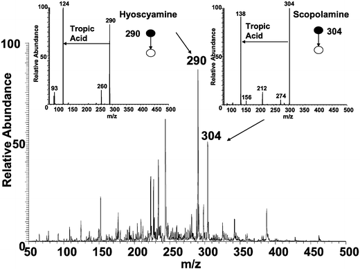 | ||
| Fig. 4 DESI mass spectrum of Atropa belladonna seed using methanol∶water (1∶1) as spray solvent; insets show MS/MS of protonated hyoscyamine and scopolamine showing the loss of tropic acid in both cases. | ||
Distribution of alkaloids in different plant parts
The seeds, stem, leaves, skin of stem, flower and roots were investigated for their alkaloid content. Different parts of the C. maculatum plant give different patterns of alkaloids, as shown in Fig. 5. The occurrence of alkaloids in the different plant parts of C. maculatum has been investigated thoroughly using standard isolation and spectroscopic methods by Lopez et al.,19 Corsi and Biasci27 and Leete and Olson.28 The present results are in good agreement with this literature data, with the single exception of coniine. This compound has repeatedly been found to be one of the main alkaloids in C. maculatum, however it was only found in trace amounts in our study. Tissue samples spiked with coniine at ng cm−2 levels gave intense signals due to coniine, so it can be concluded that C. maculatum in Northwestern Indiana contains unusually low amounts of coniine.19 γ-Coniceine is the parent alkaloid and is the precursor in the biosynthesis of all other alkaloids. The seeds are the plant parts that are richest in alkaloid content, as judged by the signal intensity in the DESI spectra, while the root contains smaller amounts. Note that this procedure can only be semi-quantitative, given the changes in matrix and the absence of internal standards in the plant material. The leaf, stem (cross-section) and the skin of the stem contain N-methyl coniine and conhydrine appearing at m/z values 142 and 144, respectively. The flower is particularly rich in N-methyl coniine. DESI spectra of C. maculatum are unusually easy to interpret as they show piperidine alkaloids almost exclusively. | ||
| Fig. 5 Alkaloid content of different tissues of the C. maculatum plant using 1 M NH4OH as the DESI spray solvent; m/z 126, 142 and 144 correspond to protonated γ-coniceine, N-methyl coniine and conhydrine, respectively. | ||
DESI mass spectra of Datura and Atropa tissues are more complex, each showing the presence of almost a hundred different compounds. Fifteen out of nineteen known alkaloids were identified for the Datura stramonium root. Some of these alkaloids are evident in the mass spectrum shown in Fig. 3, and all are listed in Table 2. The alkaloids present in the Datura stramonium seed were also examined by DESI MS/MS in order to differentiate the alkaloid content in the two different parts of the plant. The seed was found to contain a smaller number of alkaloids and lower concentrations (as judged by signal intensities under identical mass spectral conditions) than the root. The alkaloids identified in the seed are listed in Table 2.
The marked difference in the alkaloid distributions seen in the DESI spectra of C. maculatumvs. the other species could simply mean that C. maculatum has a smaller number of higher concentration alkaloids or it could be associated with ionization suppression effects. In some cases, the matrix is known to suppress ionization to such an extent that analytes are almost completely undetectable by MS although such cases normally involve different analytes and are judged unlikely to occur as a result of the minor differences in plant matrix material encountered in a case like this.
Nothing is yet known of the depth of sampling of DESI and in order to make a preliminary examination of this factor, spectra were obtained for tissue cross sections and compared with those taken from the surface of the same tissue. In most cases the spectra recorded from the surface were found to show very similar characteristics to those from the interior. This indicates that the alkaloid distribution over the tissues was approximately uniform, at least on the scale of this experiment.
Composition of spray solution and DESI mechanism
When the spray solvent was changed, the spectra and alkaloid distributions changed significantly. The DESI mass spectra shown in Fig. 6 illustrate spray solvent associated spectral changes in the particular case of C. maculatum stem tissue. The mass spectra are shown for four different solvent compositions: (a) acetonitrile∶water (1∶1)
+ 0.5% trifluoroacetic acid, (b) methanol∶water (1∶1), (c) 1 M ammonium hydroxide and (d) 0.2% formaldehyde. Fig. 6 clearly shows that the acidic solvent system gives lower signal intensity and that the acidity/alkalinity of the solvent also has an effect on the relative peak ratios. It is important to note that all the alkaloids detected from C. maculatum have considerable vapor pressure, which might facilitate the gas-phase ionization of these compounds. It is not unreasonable to suppose that the solvent can modify the pH of the liquid present on the surface and suppress or enhance the evaporation of different species, depending on their acidic/basic character. | ||
| Fig. 6 C. maculatum stem examined using different solvents (acetonitrile∶water (1∶1)
+ 0.5% trifluoroacetic acid, methanol∶water (1∶1), 1 M ammonium hydroxide and 0.2% formaldehyde). | ||
Assuming that m/z 126 is due to protonated coniceine and m/z 142 is mainly protonated N-methyl coniine; these two alkaloids represent compounds with comparable volatility but different basicity (hydrolytic dissociation constants) in aqueous solution. While the absolute intensity of protonated coniceine does not change dramatically when the solvent is changed, N-methyl coniine gives considerably higher signal intensity in the case of neutral methanol–water solvent which further increases in the case of ammonium hydroxide. N-methyl coniine is a tertiary aliphatic amine, thus it has a high gas-phase basicity and also is highly volatile in its neutral form. One explanation of the observed data is that spraying acidic solvent onto the surface shifts the N-methyl coniine protonation equilibrium towards the ionic species, suppressing evaporation and hence ionization by a gas-phase mechanism (Fig. 6). Another factor that may influence the signal intensity could be possible signal suppression by TFA as observed by Mallet et al.29 By contrast, ammonium hydroxide deprotonates the species on the surface, enhancing evaporation and hence gas-phase ionization. The best overall result obtained in terms of the highest signal intensity (counts) and minimum noise is when 1 M ammonium hydroxide is the solvent. This explanation assumes that these compounds are ionized in the gas phase rather than at the interface. It is known, as now discussed, that this is not the ionization mechanism for some other types of compounds, including large proteins and highly non-volatile explosives like RDX.17,30,31 Droplet pick-up, a known DESI mechanism, may also occur for alkaloids.
Semi-quantitative information and statistical evaluation of DESI data
Quantification of components present in complex materials like plant tissues involves serious problems in almost any method of direct molecular analysis. Since the surface characteristics are usually non-reproducible and surfaces cannot be spiked with internal standards, these methods are semi-quantitative at best. The feasibility of quantitative measurements and the reproducibility of DESI spectra were studied by recording DESI spectra of twenty-five C. maculatum seeds. One half of each seed was interrogated by DESI while the other was extracted, filtered and analyzed by ESI-MS. One of the seeds was investigated in greater detail and DESI spectra were obtained at 10 positions on the same seed. ESI spectra were also obtained by extracting the alkaloids from the same seed and then examining 10 aliquots of the extracted sample. This was done to compare the relative standard deviation of ESI with that of DESI as well as that associated with a data set consisting of twenty-five different seeds vs. one seed. We also compared the ratio of the intensity for the two peaks (m/z 126 and 154) for ESI with that for DESI. The plot of seed number vs. the logarithmic intensity for peak m/z 126 is shown in Fig. 7a; and a plot of the seed number vs. peak ratio is shown in Fig. 7b. Reproducibility of DESI is considerably poorer than that of the extraction/ESI experiment, at least in part because the smaller quantity of alkaloid sampled gives lower signal intensities. For a single ion (m/z 126) the relative standard deviation of DESI is found to be about 30.5% for twenty-five seeds and 10.1% for one seed. The greater value in the case where more data were recorded is an indication that sample-to-sample variability makes a significant contribution to the precision of this experiment. On the other hand, the reproducibility of peak ratios is comparable in the ESI and DESI cases. The relative standard deviation for the peak ratios is 9.8%. The results for the relative standard deviations for both DESI and ESI are summarized in Table 3. From this rough comparison it is concluded that DESI gives semi-quantitative results for the alkaloid content of plant tissues. DESI also gives mass spectral peak patterns with higher reproducibility and this feature allows the differentiation of different types of tissues which is a key feature for imaging applications. | ||
| Fig. 7 Semi-quantitative direct seed analysis using DESI. (a) Seed number vs. logarithmic intensity for peak m/z 126 γ-coniceine (b) seed number vs. peak ratio for peaks m/z 126:m/z 154. | ||
| ESI | DESI | |
|---|---|---|
| Highest observed intensity of 126 | 8.30 E + 6 | 7.00 E + 5 |
| Average intensity of 126 | 5.92 E + 6 | 3.41 E + 5 |
| Relative standard deviation of 126 for 25 seeds | 12.5% | 30.8% |
| Relative standard deviation of 126 for 1 seed | 3.4% | 10.1% |
| Relative standard deviation for ratio of peaks 126∶154 | 5.2% | 9.8% |
Conclusions
Application of DESI for the direct interrogation of plant tissues is demonstrated. All the alkaloids from C. maculatum and fifteen out of nineteen known alkaloids from Datura stramonium were successfully identified. In a narrower sense, this result is important from the point of view of natural product analysis or toxicological applications with special regard to forensic toxicology. However, in a broader sense this study suggests that the DESI methodology can probably be applied to animal or human tissues with appropriate modifications and indeed such studies are underway.11 It has been demonstrated that DESI can be used to differentiate between different types of tissues of the same plant, so it has the potential to be used to differentiate animal tissues in the same way, e.g. tumor cells from healthy tissue. One should also note that the precision data suggest that DESI can probably be used for fingerprinting of plant species.The main advantage of DESI compared to other DI methods is that it can be used for in vivo analysis and presumably for in vivo imaging of tissues. Although imaging has been demonstrated17 on the mm scale, by application of droplet-on-demand sources producing micrometre sized droplets, the spatial resolution of the method must now be improved. High-throughput analysis by DESI has already been demonstrated,32,33 it can also be used for screening applications and when coupled to a fieldable mass spectrometer will enable onsite plant analysis with minimal sample destruction.
Acknowledgements
This work was supported by Inproteo, LLC, Prosolia, Inc. (Indianapolis, IN) and the Office of Naval Research (N00014-05-1-0454).References
- B. Drager, J. Chromatogr., A, 2002, 978, 1–35 Search PubMed.
- I. N. Papadoyannis, Instrum. Sci. Technol., 1994, 22, 241–258 CrossRef CAS.
- A. Baerheim Svendsen and R. Verpoorte, Chromatography of Alkaloids, Part A: TLC, Elsevier, Amsterdam, 1983, p. 91 Search PubMed.
- M. Popl, J. Faehnrich and V. Tatar, Chromatographic Analysis of Alkaloids, Marcel Dekker, New York, 1990 Search PubMed.
- D. Dagnino and R. E. Verpoorte, Modern Methods of Plant Analysis—Alkaloids, Springer, Berlin, Heidelberg, New York, 1994, p. 113 Search PubMed.
- R. Verpoorte and W. M. A. Niessen, Phytochem. Anal., 1994, 5, 217–232 CrossRef.
- R. B. van Breemen, B. M. Johnson and J. L. Bolton, Chem. Res. Toxicol., 2001, 14, 1546–1551 CrossRef.
- G. A. Cordell, Introduction To Alkaloids. A Biogenetic Approach, Wiley, New York, 1981, p. 80 Search PubMed.
- R. N. Patel, Annu. Rev. Microbiol., 1998, 52, 361–395 CrossRef CAS.
- M. L. Reyzer, Y. S. Hsieh, K. Ng, W. A. Korfmacher and R. M. Caprioli, J. Mass Spectrom., 2003, 38, 1081–1092 CrossRef CAS.
- J. M. Wiseman, S. Puolitaival, Z. Takats, R. M. Caprioli and R. G. Cooks, Angew. Chem., Int. Ed., 2005 DOI:10.1002/anie.200502362.
- S. L. Luxembourg, T. H. Mize, L. A. McDonnell and R. M. A. Heeren, Anal. Chem., 2004, 76, 5339–5344 CrossRef CAS.
- P. J. Todd, T. G. Schaaff, P. Chaurand and R. M. Caprioli, J. Mass Spectrom., 2001, 36, 355–369 CrossRef CAS.
- S. A. Schwartz, M. L. Reyzer and R. M. Caprioli, J. Mass Spectrom., 2003, 38, 699–708 CrossRef CAS.
- V. V. Laiko, M. A. Baldwin and A. L. Burlingame, Anal. Chem., 2000, 72, 652–657 CrossRef CAS.
- R. W. Kondrat and R. G. Cooks, Science, 1978, 199, 978–980 CrossRef CAS.
- Z. Takats, J. M. Wiseman, B. Gologan and R. G. Cooks, Science, 2004, 306, 471–473 CrossRef CAS.
- J. Vetter, Food Chem. Toxicol., 2004, 42, 1373–1382 CrossRef CAS.
- T. A. Lopez, M. S. Cid and M. L. Bianchini, Toxicon, 1999, 37, 841–865 CrossRef CAS.
- Y. Gaillard and G. Pepin, J. Chromatogr., B: Biomed. Appl., 1999, 733, 181–229 CrossRef CAS.
- M. Friedman and C. E. Levin, J. Agric. Food Chem., 1989, 37, 998–1005 CrossRef CAS.
- S. Philipov and S. Berkov, Z. Naturforsch., C: Biosci., 2002, 57, 559–561 CAS.
- D. O'Hagan, Nat. Prod. Rep., 2000, 17, 435–446 RSC.
- M. Friedman, J. Chromatogr., A, 2004, 1054, 143–155 Search PubMed.
- L. Mateus, S. Cherkaoui, P. Christen and J. L. Veuthey, Electrophoresis, 1999, 20, 3402–3409 CrossRef CAS.
- S. Berkov and A. Pavlov, Phytochem. Anal., 2004, 15, 141–145 CrossRef CAS.
- G. Corsi and D. Biasci, Ann. Bot. London, 1998, 81, 157–162 Search PubMed.
- E. Leete and J. O. Olson, J. Am. Chem. Soc., 1972, 94, 5472–5477 CrossRef CAS.
- C. R. Mallet, Z. L. Lu and J. R. Mazzeo, Rapid Commun. Mass Spectrom., 2004, 18, 49–58 CrossRef CAS.
- Z. Takats, I. Cotte-Rodriguez, N. Talaty, H. W. Chen and R. G. Cooks, Chem. Commun., 2005, 1950–1952 RSC.
- Z. Takats, J. M. Wiseman and R. G. Cooks, J. Mass Spectrom., 2005 DOI:10.1002/JMS.922.
- H. W. Chen, N. Talaty, Z. Takats and R. G. Cooks, Anal. Chem., 2005 DOI:10.1021/ac050989d.
- D. J. Weston, C. S. Creaser, R. Bateman, T. R. Wood and I. D. Wilson, 53rd ASMS Conference on Mass Spectrometry, Poster 188, San Antonio, Texas, June 5–9, 2005 Search PubMed.
| This journal is © The Royal Society of Chemistry 2005 |