Immunoassay for B. globigii spores as a model for detecting B. anthracis spores in finished water
Svetlana
Farrell
,
H. Brian
Halsall
* and
William R.
Heineman
*
Department of Chemistry, University of Cincinnati, P.O. Box 210172, Cincinnati, OH 45221-0172, USA
First published on 10th February 2005
Abstract
The 2001 anthrax alarm in the US raised concerns about the Nation's preparedness to the threat of bioterrorism, and the demand for early warning systems that might be used in the case of a biological attack continues to grow. Here we develop an ultra-sensitive rapid detection method for B. globigii (BG) spores, the simulant of B. anthracis (BA) spores. BG spores were detected by a bead-based sandwich immunoassay with fluorescence detection. Paramagnetic Dynal® beads were used as a solid support, primary antibody was attached to the beads by streptavidin-biotin coupling and the secondary antibody had an alkaline phosphatase (AP) enzyme label. Enzymatic conversion of fluorescein diphosphate (FDP) to fluorescein by AP was measured in real time with λex = 490 nm and λem = 520 nm. The assay was linear from 2.6 × 10³–5.6 × 10⁵ BG spores mL−1, and the detection limit was 2.6 × 10³ spores mL−1 or 78 spores. All reagent concentrations and incubation times were optimized. The assay time from the moment the spores were introduced to the system was 30 min, and real-time fluorescence detection was done in less than 1 min. Formation of the BG spores–capture beads complex was confirmed by environmental scanning electron microscopy (ESEM). BG spores were detected successfully when doped into Cincinnati tap water to demonstrate the applicability of the developed method to detect the spores in non-buffered media.
Introduction
Terrorism in the United States was not considered a serious threat even a few years ago. Recent international and domestic terrorist attacks, however, showed the vulnerability of the nation to such threats, and forced the government to increase the effort in making the homeland security defense system more efficient.The Nation's critical infrastructure, such as air, drinking water, food and energy sources is particularly susceptible to terrorist attacks. These sources are also the most difficult ones to protect due to their wide accessibility to the public and because their wide distribution makes constant monitoring very challenging. The vulnerability of the public water supply system to physical attack, and biological and chemical terrorism, has becoming increasingly apparent.1 The possibility of public water contamination with biological warfare agents was first recognized by Berger in 1955.2 He observed that water can be used as a way to transmit diseases, and that any person experienced in water works systems can put a biological agent into the water flow. Even though there are many potential microorganisms and their toxins that can be used in biological warfare, B. anthracis (BA) is of particular interest. BA causes anthrax, a highly infectious disease of hoofed animals that can be readily transmitted to humans. There is evidence that BA has been weaponized for aerosol application and food contamination in biological warfare.3–5 Under unfavorable conditions, such as lack of nutrients, drought, heat and radiation, BA forms spores—a dormant form of a bacterium that helps it survive harsh environmental conditions. BA spores are very stable in the environment, surviving for two years in pond water, and for 40 years or more in soil.6 They are heat resistant and can only be inactivated by boiling in water for 25 min.6 Even though BA is readily destroyed by chlorine treatment, the spores are highly resistant to water chlorination.6 The infectious dose for gastrointestinal anthrax is not known. In animal models (guinea pigs, rabbits, and rhesus monkeys), investigators failed to induce infection following oral challenge with 108 spores.7 These animals were thought to be more susceptible to the disease than humans. For inhalation anthrax the infectious dose is estimated to be 6,000 spores.8 This corresponds to a drinking water concentration of 57 spores L−1 for a consumption of 15 L day−1 for 7 days or 171 spores L−1 for consumption of 5 L day−1.6 The long lifetime of BA spores, their resistance to chlorination, low infective dose, the high anthrax mortality rate and relatively wide accessibility of the water distribution system to the public, make the water supply a particularly vulnerable target for contaminating with BA spores.
When developing a device to detect biopathogens, exposure to the highly infectious agent in the initial stages of method development is hazardous to the personnel and requires specialized facilities, proper decontamination and controlled disposal of biological material. These difficulties are circumvented by using simulants, with B. globigii (BG) spores being recommended as a non-pathogenic simulant for BA spores.9 BG (also called B. subtilis variant niger) is a spore-forming microorganism whose spores resemble those of BA in size, shape and surface morphology. This microorganism has been studied extensively, and its genome, proteome, sporulation mechanisms and spore coat morphology are well understood.10,11 Similarly to BA, the spore coat of BG helps maintain the dehydrated state of the spore and provides resistance against organic solvents,11 solar and artificial radiation12 and damage from free-radicals.11
Our group has developed bead-based immunoassays for various types of biological agents.13–15 Para-magnetic beads function as both a solid support for the immunoassay and also as a pre-concentration device, as they can be homogeneously dispersed in large sample volumes, pulled out of the solution with a magnet and re-dispersed in a smaller volume. These assay formats might be suitable for detecting BG spores in finished water (i.e. water received by the consumers). However, all of the previous immunoassays were done in a buffered system, where pH and ionic strength were controlled. In comparison, finished water from different sources is variable in composition, pH and ionic strength making detection of biological agents in finished water more challenging than in a controlled buffered system. Since the proposed detection method is based on immuno-recognition, it was thought that water composition might cause conformational or chemical changes in the antigenic (Ag) surface of the spores, rendering them unrecognizable by the antibodies (Ab). Here, we develop an optimized bead-based enzyme-labeled immunoassay for BG spores in a controlled, buffered system, and then validate the developed method for detecting bacterial spores in finished water.
Materials and methods
Apparatus
All fluorescence measurements were done in an LS 45 luminescence spectrometer (Perkin Elmer Life and Analytical Science, Boston, MA, USA). A Lambda 45 UV/Vis System (Perkin Elmer Life and Analytical Science, Boston, MA, USA) was used in the rabbit anti-BG biotinylation procedures. A 0.4 cm path length fluorescence cuvette equipped with an adaptor (McCarthy Scientific Co., Fullerton, CA, USA) was used for all fluorescence and spectroscopic measurements.An Olympus BX-40 microscope with a Hitachi HV-C20 camera and Scion CG-7 frame grabber software on a Macintosh G3 platform were used to count spores. The obtained images were processed with Scion Image Software.
The interactions between the spores and the capture beads were imaged using an XL 30 Environmental Scanning Electron Microscope (ESEM) (Philips Electron Optics, Hillsboro, OR, USA).
Origin 7.0 (OriginLab, Northampton, MA, USA) and SigmaPlot 4.0 (Jandel Scientific, Corte Madera, CA, USA) software were used for data processing.
Buffers and reagents
Streptavidin-coated M-280 Dynabeads® were from Dynal (Lake Success, NY, USA). BG spores were supplied as a dry powder by Aberdeen Proving Ground (Aberdeen, MD, USA). Rabbit anti-BG IgG was from Tetracore Inc. (Gaithersburg, MD, USA). Biotin-SP-conjugated donkey anti-mouse IgG (H+L) and mouse IgG (whole molecule) were from Jackson Immunoresearch Laboratories, Inc. (West Grove, PA, USA). Folin-Ciocalteau's phenol reagent, and sodium azide were from Sigma (St. Louis, MO, USA). EZ-Link sNHS-LC-biotinylation kit (containing s-NHS-LC-biotin, D-Salt® Dextran desalting column, 10 mM HABA in 0.01 M NaOH and avidin) was from Pierce (Rockford, IL, USA). Fluorescein diphosphate (FDP) was from Molecular Probes (Eugene, OR, USA). A Neubauer hemocytometer, tris (hydroxymethyl) aminomethane (THAM), bovine serum albumin (BSA, biotech grade), sodium chloride, sodium carbonate, sodium hydroxide, sodium tartrate, copper sulfate and 12 × 75 mm borosilicate culture tubes were from Fisher Scientific (Fair Lawn, NJ, USA). Glycine, potassium phosphate monobasic and potassium phosphate dibasic were from Matheson Coleman & Bell Manufacturing Chemists (Norwood, OH, USA). All salts were ACS grade. Tween-20 was from Aldrich (Milwaukee, WI, USA). The neodymium-iron-boron rare earth magnets were from Radio Shack (Fort Worth, TX, USA). All chemicals were used without further purification.The following aqueous buffer solutions were used. Phosphate buffer (PBS): 44 mM KH2PO4, 56 mM K2HPO4, 0.1 M NaCl, 1% (w/v) BSA, 0.02% (w/v) NaN3, 0.02% (v/v) Tween-20, pH 7.0 was used in the immunoassay preparation step. BSA and Tween-20 help to reduce nonspecific adsorption. Concentrated PBS buffer (2× PBS): 88 mM KH2PO4, 112 mM K2HPO4, 0.2 M NaCl, 2% (w/v) BSA, 0.04% (w/v) NaN3, 0.04% (v/v) Tween-20, pH 7.0 was used in detecting BG spores in finished water. Tris buffer: 0.1 M THAM, 10 mM MgCl2, 0.02% (w/v) NaN3, 10 mM glycine, pH 9.0 was used with fluorescence detection.
Reagent preparation
Purified and preconcentrated biotinylated rabbit anti-BG IgG was prepared using the EZ-link® sNHS-LC biotinylation kit.16 The final concentration of the biotinylated Ab was 1.2 mg mL−1 (53% yield) determined by the Folin-Ciocalteu assay17,18 with mouse IgG as a protein standard. The molar ratio of incorporated biotin to IgG (determined by a HABA displacement assay19) was 2.5 on average, close to the optimal 3 ∶ 1 ratio.20Custom alkaline phosphatase (AP) conjugation to rabbit anti-BG IgG (secondary Ab) was done by American Qualex (San Clemente, CA, USA). Primary and secondary Ab solutions were stored at 4 °C.
Experimental
Determination of the Dynabeads capacity for rabbit anti-BG IgG
First, 5 µL aliquots of 6.7 × 10⁸ beads mL−1 were transferred into 9 fresh test tubes. One tube was set aside as a non-specific adsorption (NSA) control. Biotinylated rabbit anti-BG IgG was added to the rest of the tubes to a concentration of 0.5–40 µg mL−1 in a final volume of 30 µL adjusted with PBS. The tubes were incubated for 10 min at RT on a vortex and washed 5 times with 50 µL of PBS. After that, 30 µL of 40 µg mL−1 goat anti-rabbit IgG–AP conjugate were added to each tube including a control, and the tubes were incubated for 30 min at room temperature (RT) on a vortex. The tubes were then washed 5 times with 50 µL of Tris buffer, pH 9.0 and resuspended in 30 µL of Tris.Fluorescence detection was done in real time with λex = 490 nm and λem = 520 nm, and the detection cell contained 293 µL of Tris, 5 µL of 1 mM FDP and 2.5 µL of beads. The beads were added last, the cuvette was inverted a few times and placed in the instrument. Each IgG concentration was run in triplicate, and the reaction velocity was calculated as the slope of the most linear portion of the reaction progress curve for each run. The average velocity was plotted vs. IgG concentration and the curve fitting was done using SigmaPlot 4.0.
Determination of the BG working concentration range
Paramagnetic capture beads were prepared in one batch by adding 300 µL of 12 µg mL−1 biotinylated rabbit anti-BG IgG to 60 µL of 6.7 × 10⁸ beads mL−1. The concentration of primary Ab in the final volume was 10 µg mL−1, which is above the determined saturation concentration. The beads were incubated for 10 min at RT on a vortex, washed 5 times with 200 µL PBS and resuspended in 60 µL of PBS (a final bead concentration of 6.7 × 10⁸ beads mL−1).A suspension of BG spores was prepared by adding 4.6 mg of the spores to 1 mL of PBS. Spores were washed by centrifugation prior to analysis to remove soluble proteins. The spore concentration was 6.7 × 10⁸ spores mL−1 as determined with a Neubauer hemocytometer under the Olympus BX-40 optical microscope. Capture beads (2.5 µl aliquots) were transferred into nineteen fresh test tubes. One tube was set aside as the NSA control. BG spores were then added to the rest of the tubes to concentrations between 2.0 × 10³ and 5.6 × 10⁷ spores mL−1 in the final volume of 30 µL adjusted with PBS. The tubes were incubated for 30 min at RT on a vortex and washed 5 times with 50 µL of PBS. Enzyme conjugate working solution was prepared by diluting the 500 µg mL−1 stock solution to 50 µg mL−1 with PBS. Thirty µL of the diluted enzyme conjugate were added to each tube, including the control, and the tubes were agitated for 30 min at RT on a vortex. The tubes were then washed 5 times with 50 µL of Tris buffer, pH 9.0 and resuspended in 30 µL of the same buffer. One mM FDP in Tris buffer was prepared from the 10 mM stock FDP solution to be used as the enzyme substrate during detection.
Fluorescence detection was done in real time as described in the previous section. All BG concentrations were run in triplicate. The reaction velocity was calculated as the most linear portion of the reaction progress curve for all BG concentrations. The average velocity was plotted versus BG concentration, and the limit of detection was calculated as 3× standard deviation of the lowest detected concentration/slope of the calibration curve.21
Imaging interactions between the BG spores and the capture beads by ESEM
Four sample tubes were prepared prior to the ESEM analysis. Tube # 1 contained 5 µL of 6.7 × 10⁸ anti-BG capture beads. Tube # 2 contained 5 µL of 6.7 × 10⁷ BG spores mL−1, and tube # 3 contained 5 µL of the BG-capture beads complex with a BG concentration of 5.6 × 10⁷ spores mL−1. The contents of tubes 1–3 were washed 5 times with 30 µL diH2O and resuspended in 30 µL diH2O. Tube # 4 was prepared for evaluating NSA by first mixing 5 µL of 6.7 × 10⁸ streptavidin-coated Dynabeads with 25 µL of 12 µg mL−1 biotinylated donkey anti-mouse IgG. The tube was incubated for 10 min at RT on a vortex and washed 5 times with 50 µL PBS. BG spores were then added to give a concentration of 5.58 × 10⁷ spores mL−1 in a final volume of 30 µL adjusted with PBS. The tube was incubated for 30 min at RT on a vortex, washed 5 times with 50 µL diH2O and resuspended in 30 µL diH2O. Samples 1–4 were then imaged with the ESEM using a secondary electron detector, and multiple images of each sample were taken.Optimizing the BG immunoassay
Assay preparation, fluorescence detection and data analysis for all optimization experiments were done as described in the “BG working concentration range” section with the following exceptions.Three sets of test tubes were prepared to optimize the BG spores–capture beads incubation time. The BG concentration varied between 1.7 × 10⁴ and 5.6 × 10⁵ spores mL−1 in each set. BG spores were incubated with the capture beads for 30 min (set 1), 10 min (set 2) and 5 min (set 3). All BG concentrations were run in triplicate for each incubation time.
The concentration of the secondary Ab–conjugate complex was optimized by keeping the BG concentration constant at 5.6 × 10⁵ spores mL−1, and varying the AP–anti BG Ab conjugate concentration between 1 and 60 µg mL−1. Each conjugate concentration was run in triplicate.
The conjugate incubation time was optimized using two sets of test tubes in which BG concentration varied between 1.7 × 10⁴ and 5.6 × 10⁵ spores mL−1 in each set. The conjugate concentration was 30 µg mL−1. Conjugate was incubated with the capture beads–BG complex for 30 min (set 1) and 5 min (set 2). All BG concentrations were run in triplicate for each incubation time.
Detecting B. globigii spores in finished water
BG spores were suspended in Cincinnati tap water (tubes 1 and 2) and in PBS (tube 3) to give 6.7 × 10⁷ spores mL−1 in each. All tubes were incubated for 7 days at RT. After that, an equal volume of Cincinnati tap water, 2× PBS and PBS was added to tubes 1, 2 and 3, respectively. The pH in tube 2 was adjusted to 7.0.Three sets of six tubes each were set up. For each set of tubes, a standard BG assay protocol (“Determination of BG working concentration range”) was used with the following exceptions:
1. The BG concentration range in all three sets was between 1.7 × 10⁴ and 3.1 × 10⁵ spores mL−1.
2. BG spores were incubated with the capture beads for 10 min.
3. The AP–anti-BG IgG conjugate concentration was 30 µg mL−1. The conjugate incubation time was 5 min.
4. For set # 1, BG spores incubated in PBS were used as the Ag.
5. For set # 2, BG spores incubated in Cincinnati tap water were used as the Ag.
6. For set # 3, BG spores incubated in Cincinnati tap water and then adjusted with PBS were used as the Ag.
Fluorescence detection and data analysis were done as described. All BG concentrations were run in triplicate for each tube set.
Results and discussion
Developing an immunoassay for B. globigii spores
When developing an immunoassay for a new Ag, various parameters in each assay preparation step need to be considered. Optimal reagent concentrations and incubation times are of particular interest. Under “optimal conditions” we assume the lowest reagent concentration or the shortest incubation time that gives the highest practical assay sensitivity. Operating under optimal conditions allows the overall cost of the assay and the total assay time to be reduced. Another important parameter is the working concentration range for the particular Ag, which is usually the linear response portion of a calibration plot and depends on the nature of the Ag, its size and shape, on the properties of the Abs, and on the detection method used.22In this study, we developed and optimized the immunoassay for BG spores with fluorescence detection. The assay schematic and the optimization required for each assay step are shown in Fig. 1, and it can be seen that the following steps must be addressed individually, and the following conditions need to be optimized:
• In step # 1 (capture beads preparation): saturation concentration of the primary Ab and optimal incubation time.
• In step # 2 (BG capture): BG working concentration range and optimal incubation time.
• In step # 3 (conjugate attachment): optimal conjugate concentration and incubation time.
Optimization of these steps is discussed below.
AP-FDP was chosen as the enzyme–substrate couple for fluorescence detection for its low detection limit,23 low background signal and high quantum yield of the reaction product, fluorescein.24
 | ||
| Fig. 1 Schematic of an immunoassay for BG spores. Dynal® capture bead is the solid support, biotinylated (B) rabbit anti BG IgG is the primary Ab, BG spore is the antigen, rabbit anti-BG IgG–AP is the secondary antibody. Fluorescein diphosphate (FDP) is a fluorogenic substrate of AP; fluorescein is the reaction product. Each assay step and optimization procedures are identified. | ||
Determination of the Dynabeads capacity for rabbit anti-B. globigii IgG
The first step of the assay involves attaching the primary Ab to the surface of paramagnetic capture beads, and is done by the biotin–streptavidin interaction. The biotin–streptavidin bond forms very rapidly19 and is not affected by the source of biotinylated Ab. Therefore, capture beads were incubated with rabbit anti-BG IgG for 10 min. The saturation concentration of a biotinylated primary Ab, however, is a variable that depends on the degree of biotinylation and on the bead size. It changes when going from one Ab to another, and must be determined when developing a new immunoassay.In a bead-based immunoassay, it is desirable for the capture beads to be completely coated (saturated) with primary Ab. Having saturated beads increases the active surface area and the number of binding sites for the epitopes of the Ag, thereby increasing the sensitivity and lowering the detection limit. Thus, it is important to determine the concentration of a primary Ab that results in complete bead coverage.
Fig. 2 (insert) shows an instrumental output obtained for the saturation studies. When beads are added to a solution of FDP, AP hydrolyses FDP, and the concentration of the reaction product, fluorescein, increases, which can be seen as the increase of fluorescence intensity with time. Initially the slopes of the reaction progress curves are proportional to the IgG concentration, however, at some point they become concentration-independent. When the reaction velocity (determined as the slope of each reaction progress curve from 5–30 s) is plotted versus IgG concentration (Fig. 2), the resulting plot has the shape of a saturation isotherm where the initial increase is followed by a plateau. The latter indicates that the beads have reached saturation and cannot accommodate more Ab. The saturation concentration was determined to be 4 µg mL−1 from the plot. The number of IgG molecules per bead (bead capacity) was calculated to be 1.4 × 10⁵ molecules bead−1 assuming the molecular weight of IgG to be 150,000.25 This number was compared with the 2.8 µm Dynal bead capacity of 6.8 × 10⁵ molecules bead−1 obtained for commercially biotinylated donkey anti-mouse IgG.14 A value of the same order of magnitude indicates successful in-lab biotinylation of anti-BG IgG.
 | ||
| Fig. 2 Dynal® beads capacity for biotinylated rabbit anti-BG IgG detected by fluorescence (λex = 490 nm, λem = 520 nm). Saturation plot and fluorescence intensity (Int) vs. time (S) reaction progress curves (insert). Concentration of biotinylated rabbit anti BG IgG: 0.5–40 µg mL−1. Saturation concentration: 4 µg mL−1. Each error bar represents standard deviation from triplicate measurements. | ||
Determination of the BG working concentration range
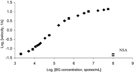
When designing a detection method for a pathogen, it is important to establish the form of the response curve and the range of the pathogen's concentration that gives a linear response so that the Ag in a contaminated sample can be detected and quantitated. Another parameter to be considered is the infectious dose, the minimum amount of a pathogen required to cause an infection. The infectious dose depends on the type of agent, the route of exposure and the health status of the host. For example, for inhalation anthrax the infectious dose is between 8,000 and 10,000 spores.26 Here, we establish a working concentration range and determine the limit of detection for BG spores.The instrumental outputs resembled those shown in Fig. 2 (insert) with steeper reaction progress curves observed at higher BG concentrations. Fig. 3 is a calibration plot for BG spores. The plot has a sigmoid shape with a linear range between 5.0 × 10³ and 5.6 × 10⁵ spores mL−1. The velocity value for 2.0 × 10³ spores mL−1 is within the value of NSA. Above 5.6 × 10⁵ spores mL−1 the plot begins to plateau as the capture beads are being saturated by BG spores. The determined range of BG spores concentration is a “true” linear range since it is limited by the NSA and the capture beads capacity for the spores. It was also confirmed in a control experiment for BG spore detection in finished water done under completely optimized conditions. From the plot, the detection limit was calculated to be 2.6 × 10³ spores mL−1, or 78 spores in 30 µL used for analysis. This detection limit is 10 times lower than the published value obtained by an immunoassay-based fiber optics biosensor.27 Excellent method sensitivity was attributed to the use of the paramagnetic bead solid support and enzyme labels. The beads provide a higher surface area compared to fiber optic probes, wells and capillaries, enhancing Ag capture. Enzyme labels amplify the signal by continuously generating detectable product. With an assumption that the BG spores mimic B. anthracis spores in size, shape and surface morphology, and that the binding constants of the capture Abs are equivalent, one can speculate that an assay for B. anthracis spores with the same format could easily detect infective concentrations of the pathogen.
 | ||
| Fig. 3 Sandwich immunoassay for BG spores, fluorescence detection (λex = 490 nm, λem = 520 nm). BG calibration plot. BG spore concentration: 2.0 × 10³ to 5.6 × 10⁷ spores mL−1. Each error bar represents standard deviation from three replicates. NSA—nonspecific adsorption of the enzyme conjugate to the capture beads (control). | ||
Imaging interactions between the BG spores and the capture beads by ESEM
Visualizing the interactions between the Ag and the capture beads by ESEM can provide very useful information when designing a bead-based immunoassay. Not only can the BG capture by the Dynal beads be visualized, but also the specificity of the interactions between the two particles can be indicated. When developing a bead-based immunoassay for a new Ag, one should prove that the interactions between the beads and the Ag are specifically due to Ab–Ag recognition. All other types of particle–particle interactions are non-specific and, therefore, are not desired. Two samples were prepared to evaluate the BG–capture bead interactions. In the first sample, BG spores were incubated with the capture beads coated with the anti-BG Ab. Anti-BG Abs bind to the complementary BG spore coat Ags (specific interaction), and the bead–spore complex was expected to be seen under the ESEM. In the second sample, BG spores were introduced to the capture beads covered with the anti-mouse IgG, which doesn't form immunocomplexes with the BG spore coat proteins. If the BG spores are seen on the surface of the capture beads in this case, these interactions will be non-specific. Two controls were used to confirm the size and shape of the spores and the capture beads.Fig. 4 shows images obtained for the samples. The capture beads and BG spores were homogeneous in shape and size distribution (Fig. 4A, B). The formation of the bead–spores complex can be seen in Fig. 4C, where the spores were incubated with the anti-BG capture beads. When the spores were incubated with the non-complementary anti-mouse IgG capture beads, no spores were detected on the surface of the beads by the ESEM (Fig. 4D). Spore capture by the specific anti-BG capture beads in the absence of interaction with the non-specific beads indicates that the formation of a bead-spore complex is due to the Ag–Ab recognition and not to NSA of the spores to the beads.
 | ||
| Fig. 4 Images of interactions between the BG spores and the capture beads. (A): control capture beads; (B): control BG spores; (C): specific interactions between the spores and the capture beads; (D): non-specific interactions between the spores and the capture beads. | ||
Optimizing the BG–capture beads incubation time
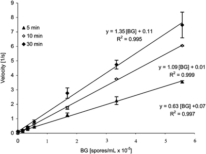
The optimal Ag incubation time depends primarily on the nature of the Ag, its size and shape, on the quality of the Abs and on the number of capture beads. This study was conducted using beads completely saturated with the primary antibody. Ideally, operating under optimal conditions allows one to obtain the highest sensitivity in the shortest time and at the lowest cost. Here we compare the results obtained with 5, 10 and 30 min BG spores–capture beads incubation times.The instrumental outputs for each trial resembled those shown in Fig. 2. Steeper reaction progress curves were observed at higher spore concentrations. Overlapped BG calibration plots obtained with various BG incubation times are shown in Fig. 5, where the highest sensitivity can be seen after 30 min incubation (y = 1.35 × 10⁻⁵ [BG] + 0.10). About 15% of the maximum sensitivity was lost when going from 30 to 10 min incubation (y = 1.09 × 10⁻⁵ [BG] + 0.01). A significant decrease in sensitivity, about 52% loss, was observed at 5 min incubation (y = 0.63 × 10⁻⁵ [BG] + 0.07). The detection limits for 5, 10 and 30 min incubations were 2.1 × 10⁴, 3.3 × 10³ and 5.3 × 10³ spores mL−1, respectively. The large difference in the sensitivity and detection limits between 5 and 30 min incubations can be attributed to the relatively large size of both beads and spores. It is assumed that in a spore–bead complex, a spore can be effectively held by a bead via multiple attachment sites, rather than by just a single attachment point. The formation of multiple attachments may be accompanied with some spore–bead positional readjustment and also requires a certain time. Ten minutes incubation time resulted in slightly lower sensitivity and comparable detection limits to the 30 min incubation time. Based on that, 10 min BG–capture beads incubation time was chosen as optimal, and it was used in the subsequent immunoassays.
 | ||
| Fig. 5 Optimizing BG spores capture time. BG spores–capture beads incubation time: 5, 10 and 30 min. BG concentration range: 1.7 × 10⁴–5.6 × 10⁵ spores mL−1 Fluorescence detection (λex = 490 nm, λem = 520 nm). | ||
Optimizing the concentration of the secondary Ab–conjugate complex
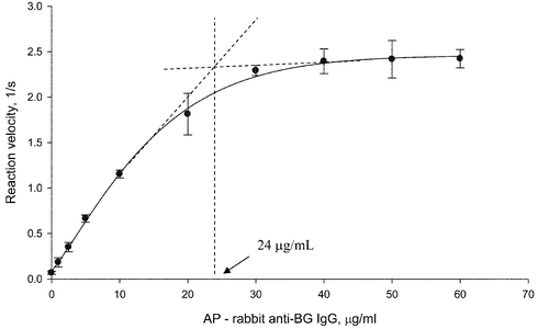
The optimal conjugate concentration was determined in order to minimize the use of the reagents and the assay's overall cost. The optimal conjugate concentration is the lowest concentration at which all accessible epitopes of the spore surface have bound with the secondary Ab–enzyme conjugate. The optimal conjugate concentration gives the lowest detection limit with the shortest incubation time. It is, therefore, a balance of conjugate concentration and NSA. Using concentrations below the optimal can result in higher detection limits; operating above the optimal concentration will waste the reagent.The optimal conjugate concentration was determined by preparing the spore–capture beads complex using the highest BG concentration that gave a linear response on the calibration curve. The instrumental output resembled that shown in Fig. 2. The average velocity was plotted vs. the conjugate concentration; the resulting graph had the form of a saturation isotherm (Fig. 6), where the initial increase in velocity was followed by a plateau. The saturation concentration was calculated from the saturation isotherm to be 24 µg mL−1. Concentrations of the plateau region slightly above the saturation concentration are traditionally used in bead-based immunoassays.13–15 This improves the assay to assay reproducibility, since the assay outcome is conjugate concentration-independent in the plateau region. Therefore, 30 µg mL−1 was assumed to be the optimal conjugate concentration.
 | ||
| Fig. 6 Optimizing concentration of rabbit anti-BG IgG–AP conjugate. Saturation plot. Conjugate concentration: 1–60 µg mL−1. Saturation concentration: 24 µg mL−1. Fluorescence detection (λex = 490 nm, λem = 520 nm). | ||
Optimizing the conjugate incubation time
The benefits of lowering the total assay time are discussed in the previous sections. Here, the effect of reducing the conjugate incubation time from 30 to 5 min is evaluated. The instrumental outputs obtained in this study resembled those shown in Fig. 2. Calibration plots for BG spores were constructed for both incubation times and are shown in Fig. 7. It can be seen that the assay sensitivity is the same for 30 and 5 min incubation, with slope equations of y = 1.09 × 10⁻⁵ [conj] + 0.01 and y = 1.06 × 10⁻⁵ [conj] − 0.03, respectively. Compared to the results obtained for BG spores, where 52% loss in sensitivity was observed between 30 and 5 min BG spores incubation, no loss in sensitivity was seen with change in conjugate incubation time. This result was attributed to the smaller size of the conjugate compared to the spores. Since the formation of the BG–anti BG IgG–AP complex involves interactions between two single protein molecules (as compared to the multiple Ab–Ag interactions involved in the formation of the bead–BG complex), a shorter period of time is required for complexation. Five minutes was found to be a sufficient time for the secondary Ab attachment, and it was used in the subsequent assays. | ||
| Fig. 7 Optimizing conjugate incubation time with the BG–capture beads complex. BG concentration range: 1.7 × 10⁴ – 5.6 × 10⁵ spores mL−1. Conjugate incubation time: 5 and 30 min. Fluorescence detection (λex = 490 nm, λem = 520 nm). | ||
Immunoassay for B. globigii spores in Cincinnati tap water
Until now, the immunoassay for BG spores was done in a buffered system of a known composition, pH and ionic strength. However, the goal of this project is to detect the spores in the Nation's water supply. Even though the composition of the finished water is somewhat regulated by the EPA, its pH, ionic strength, organic and metal content vary greatly throughout the Nation. Here, we compare three optimized standard assays in which the BG spores are exposed to three different matrices prior to detection. In the first assay control, the spores are incubated in PBS for 7 days. In the second assay, the spores are exposed to Cincinnati tap water for 7 days, and PBS is added to the spores immediately prior to detection to control the pH and ionic strength. In the third assay, the spores are incubated and detected in Cincinnati tap water. According to the Cincinnati Water Works, finished water in the Greater Cincinnati area typically circulates in the distribution system for a maximum of 7 days after leaving the treatment plant. For this reason, a 7 day incubation was chosen for the BG exposure time to reflect a pathogen's residence time in the distribution system.Fig. 8 shows calibration plots obtained for the BG spores exposed to different conditions. It can be seen that the highest sensitivity was obtained for the control (BG in PBS) with the slope equation of y = 0.232[BG] + 0.008 and the detection limit of 2.6 × 10³ spores mL−1. The assay in which the pH and ionic strength were adjusted following the spores' exposure to tap water had the second highest sensitivity (y = 0.201[BG] + 0.013) with only 13% loss in sensitivity and a detection limit of 3.0 × 10³ spores mL−1. The assay was the least sensitive when incubation and detection were done in tap water (y = 0.159[BG] + 0.009), with the loss in sensitivity being 31% and a detection limit of 3.8 × 10³ spores mL−1. These findings suggest that the water matrix has an effect on the spore capture by the beads and, possibly, on the spores' morphology. Adjusting the pH and the ionic strength of tap water with PBS helps to significantly improve the assay sensitivity and brings the detection limit closer to that of the control.
 | ||
| Fig. 8 Sandwich immunoassay for BG spores in PBS buffer, Cincinnati tap water and Cincinnati tap water buffered with PBS. BG spores were suspended in PBS and Cincinnati tap water for 7 days prior to analysis. Spore concentration range: 1.7 × 10⁴ – 3.0 × 10⁵ spores mL−1. Each data point is the average of three replicates. | ||
Conclusions
The work presented is the first step towards developing a detection method for anthrax spores. A bead-based immunoassay for B. globigii spores—B. anthracis spore simulant—was successfully developed. Reagent concentrations, and incubation times were optimized. The optimal assay conditions are given in Table 1. The data shown are determined for 5.6 × 10⁷ capture beads mL−1 or 1.7 × 10⁶ beads in the final volume of 30 µL.| Immunoassay step | Concentration/range | Incubation time |
|---|---|---|
| Capture bead preparation | Primary Ab: 10 µg mL−1 | 10 min |
| Capturing BG spores | Spores working range: 2.6 × 10³–5.58 × 10⁵ spores mL−1 | 10 min |
| Attaching the secondary Ab | Secondary Ab: 30 µg mL−1 | 5 min |
The total incubation time for the assay is 25 min, and an additional 25 min is spent on the bead washing procedures, giving a total assay of 50 min. Before optimization, the assay could be completed in an average of 150 min. Therefore, optimizing the incubation time for each step of the immunoassay reduced the total assay time by 3×. Considering that the capture beads can be prepared in advance, the total assay time from the moment the spores are introduced to the system was 30 min. The assay time can be reduced even further by minimizing the reaction volumes and/or by automation. Once the immunoassay sandwich is complete the spores can be detected by fluorescence in less than a minute.
The optimized BG immunoassay had a linear working range of two orders of magnitude (2.6 × 10³ – 5.58 × 10⁵ spores mL−1). The assay's experimental detection limit was 2.6 × 10³ spores mL−1 or 78 spores in the 30 µL sample used for analysis; below this value the assay was limited by the NSA. With an assumption that the BG spores morphologically resemble B. anthracis spores, we can speculate that similar detection limits can be obtained for B. anthracis spores. The infectious dose for anthrax (8,000–10,000 spores) will then be above the limit of detection, and the assay will be reliable in detecting pathogenic quantities of the agent. Undoubtedly, additional optimization will be required when the developed method is applied to detecting the anthrax spores.
The assay for BG spores was successful in finished water. It was demonstrated that adjusting the pH and the ionic strength of the spores' suspension is necessary to maintain the assay's high sensitivity and low detection limits. The loss in sensitivity was attributed to the effect of the individual components of finished water (possibly chlorine) on the immunoassay for BG, and needs further investigation. The effect, however, was not as profound as it was for a virus simulant.28. The difference can be attributed to the difference in composition of the outer coat between these classes of microorganisms. The outer coat of a virus consists primarily of proteins, while the bacterial spore coat has proteins, phospholipids and lipopolysaccharides that are very densely packed in a laminate-like structure. This dense packing reduces the accessibility of the spore-coat proteins to harmful agents and is a basis for spore resistance to the various environmental factors.11,29
Overall, the developed method showed very good assay-to-assay reproducibility without any major problems or drawbacks. Current efforts are directed towards achieving lower detection limits by sample pre-concentration, incorporating the developed assay into a microchip device and validating the method with the various water samples collected throughout the Nation.
Acknowledgements
This project was funded by the American Water Works Association Research Foundation (AWWARF) and Yellow Springs Instruments. The authors would also like to acknowledge Jack DeMarco and David Hartman from the Cincinnati Water Works for helpful discussions.References
- R. M. Clark and R. A. Deininger, J. Contingencies Crisis Manage., 2000, 8, 73–80 Search PubMed.
- B. B. Berger and A. H. Stevenson, Am. Water Works Assoc., 1955, 2, 101–110 Search PubMed.
- E. Marshall, Science, 1997, 275, 745 CrossRef CAS.
- R. A. Zilinskas, J. Am. Med. Assoc., 1997, 278, 418–424 Search PubMed.
- G. W. Christopher, T. J. Cieslak, J. A. Pavlin and E. M. Eitzen, Jr, J. Am. Med. Assoc., 1997, 278, 412–417 Search PubMed.
- W. D. Burrows and S. E. Renner, Environ. Health Persp., 1999, 107, 975–984 CAS.
- M. E. Beatty, D. A. Ashford, P. M. Griffin, R. V. Tauxe and J. Sobel, Arch. Intern. Med., 2003, 163, 2527–2531 Search PubMed.
- A. Watson and D. Keir, Epidemiol. Infect., 1994, 113, 479–490 CrossRef CAS.
- Under Secretary for Health's Information Letter, IL 10-200-012. Dec. 1, 2000; www.va.gov/shad/docs/IL_10-2000-012.pdf.
- Y. Hiragi, J. Gen. Microbiol, 1972, 72, 87–99 CAS.
- A. Driks, Microbiol. Mol. Biol. Rev., 1999, 63, 1–20 CAS.
- P. J. Riesenman and W. L. Nicholson, Appl. Environ. Microbiol., 2000, 66, 620–626 CrossRef CAS.
- S. Purushothama, S. Kradtap, C. A. Wijayawardhana, H. B. Halsall and W. R. Heineman, Analyst, 2001, 126, 337–341 RSC.
- S. Kradtap, C. A. Wijayawardhana, K. T. Schlueter, H. B. Halsall and W. R. Heineman, Anal. Chim. Acta., 2001, 444, 13–26 CrossRef CAS.
- C. A. Wijayawardhana, S. Purushothama, M. A. Cousino, H. B. Halsall and W. R. Heineman, J. Electroanal. Chem., 1999, 468, 2–8 CrossRef CAS.
- “EZ-Link Sulfo-NHS-LC Biotinylation Kit, Instructions”, Cat. # 21430, Pierce Biotechnology; http://www.piercenet.com/files/0215-js4.pdf.
- R. L. Switzer and L. F. Garrity, Experimental Biochemistry.Theory and Exercises in Fundamental Methods, W. H. Freeman Publishing, 3rd edn., 1999 Search PubMed.
- O. H. Lowry, N. J. Rosebrough, R. L. Farr and R. J. Randall, J. Biol. Chem., 1951, 193, 265–275 CAS.
- N. M. Green, Adv. Protein Chem., 1975, 29, 85–133 CAS.
- D. J. Hnatowich, F. Virzi and M. Rusckowski, J. Nucl. Med., 1987, 28, 1294–1302 CAS.
- R. Q. Thompson, G. S. Barone, III, H. B. Halsall and W. R. Heineman, Anal. Biochem., 1991, 192, 90–95 CrossRef CAS.
- P. Tijssen, ”Practice and Theory of Enzyme Immunoassays”, Laboratory Techniques in Biochemistry and Molecular Biology, 1985, vol. 15 Search PubMed.
- J.-S. Yu, S.-H. Chang, W.-H. Chan and H.-C. Chen, J. Biochem., 2001, 129, 243–251 CAS.
- R. P. Haugland, Handbook of Fluorescent Probes and Research Products, Molecular Probes, OR, USA, 9th edn., 2002, ch. 10 Search PubMed.
- C. K. Mathews, K. E. van Holde and K. G. Ahern, Biochemistry, Addison Wesley Longman, Inc., 3rd edn., 2000 Search PubMed.
- New York State Department of Health, “Communicable Disease Fact Sheet”; http://www.health.state.ny.us/nysdoh/communicable_diseases/en/anthrax.htm.
- G. P. Anderson, C. A. Rowe-Taitt and F. S. Ligler, Proceedings of the First Conference on Point Detection for Chemical and Biological Defense, October, 2002 Search PubMed.
- J. H. Thomas, S. K. Kim, P. J. Hesketh, H. B. Halsall and W. R. Heineman, Anal. Chem., 2004, 76, 2700–2707 CrossRef CAS.
- E. W. Koneman, S. D. Allen, W. M. Janda, P. C. Schreckenberger and W. C. Winn, Color Atlas and Textbook of Diagnostic Microbiology, Lippincott, Philadelphia, 5th edn., 1997 Search PubMed.
| This journal is © The Royal Society of Chemistry 2005 |
