A preliminary comparison of radial and axial excitation fluorescence in the ICP using non-laser sources†
Anita
Young
a,
Les
Pitts
b,
Stanley
Greenfield
b and
Mike
Foulkes
*b
aAstraZeneca, Brixham Enivronmental Laboratory, Freshwater Quarry, Brixham, Devon, UK TQ5 8BA
bSchool of Environmental Sciences, University of Plymouth, Drake Circus, Plymouth, Devon, UK PL4 8AA
First published on 21st November 2002
Abstract
This paper describes a preliminary comparison of radial and axial excitation fluorescence within the ICP utilising hollow cathode lamps (HCLs) and narrow band-width light emitting diodes (LEDs) as sources. Univariate searches were used to optimise various plasma parameters, i.e., forward power, viewing height ALC, plasma, nebuliser and auxiliary gas flow rates, and various excitation source parameters, i.e., modulation frequency, duty cycle and peak current for each of the elements studied (Ba, Li, Mg and Na). Calibrations showed excellent linearity over five orders of magnitude (R2 = 0.99995–1.0000) with precision better than 5% RSD. Vertical profiles of the plasma, using radial excitation, were obtained for Ba and Mg using HCLs and for Li and Na using LEDs. Limits of detection were found to be 27.6, 0.51, 0.43 and 0.20 µg l−1 for Ba, Li, Mg and Na, respectively. Using the optimum conditions for Li and Na, vertical profiles of the plasma were obtained using axial excitation with the more intense LEDs. Two different excitation lens systems were investigated in the axial mode: a focusing lens and a collimating lens. The relatively sharp profiles and optimum viewing heights ALC obtained for each analyte studied were identical using both radial and axial excitation fluorescence (circa 60–80 mm at forward powers of 475–750 W and injector flow rates of 2.0–2.5 l min−1). The axial LODs obtained under these conditions were found to be 1.12 and 0.70 µg L−1, and 5.3 and 3.1 µg L−1 for Li and Na, using a focusing and collimating lens system, respectively. The potential for increased sensitivity and lower limits of detection is discussed using the novel axial excitation system. ‘Temperature’ measurements (Texc and Trot) determined under fluorescence conditions both gave values in the region 2500–3000 K, indicating a plasma operating closer to LTE.
Introduction
The last few decades have seen much progress in atomic fluorescence spectrometry (AFS) with the introduction of intense sources such as the boosted discharge hollow cathode lamp (BDHCL),1 the inductively coupled plasma (ICP)2–4 and various types of laser.5,6 The driving force for this is that the intensity of the atomic fluorescence signal is proportional to the intensity of the incident light. This has been in addition to the benefits that AFS offers, i.e. long linear dynamic range (like atomic emission spectrometry (AES)), being well suited to the development of simple and low-cost instrumentation and the fact that AF spectra are relatively simple, like atomic absorption (AA) spectra. Moreover, when the modulated excitation source is synchronous with the lock-in detector, the effects of baseline drift associated with any continuous emission from, for example, a plasma, are effectively removed. The potential for improved measurement characteristics is therefore offered. Finally, if an ICP is operated as an atomisation cell, the possibility arises that certain matrix effects may be reduced compared with alternative atomisation sources, e.g. flames, ETV, etc., and scattered radiation interferences may be eliminated for all practical purposes.5–7Although the characteristics of ICP-AFS are attractive, the technique has rarely been used for routine analysis, primarily because of the absence of commercially available instrumentation.4,8
The important operating conditions used in ICP-AFS are low forward powers (ca. 600–900 W), high nebuliser gas flows (1.5–2.5 L min−1) and viewing heights of 50–120 mm above load coil (ALC). These reduce the plasma background and allow suitable fluorescence signals to be obtained.1,4–9 For comparison, ICP-AES uses forward powers that are typically in the range 1000–1200 W with a nebuliser gas flow generally below 1.0 L min−1.
To date, all fluorescence studies have ‘focused’ on a radial excitation arrangement in plasmas1,4–9 and no in-depth study using axial excitation has been published.4,5,9,10 In the radial mode, collection of fluorescence is restricted by the position of the source radiation. When irradiating the plasma axially, a larger number of atoms lie within the excitation sources’ emission and fluorescence can be collected from a larger cell volume. This offers the potential for increased fluorescence and detection efficiency. In addition, while the use of HCLs has been previously reported, the use of narrow band-width light emitting diodes (LEDs) as an excitation source has yet to be fully investigated. This paper describes a preliminary comparison of radial and axial excitation fluorescence within the ICP utilising HCLs and LEDs as sources.
Experimental
Instrumentation
A plasma, produced by a PlasmaQuad PQ2 ICP (Thermo Elemental, Winsford, Cheshire, UK) was used as the atom cell for both radial and axial excitation fluorescence experiments. The instrument was equipped with an ICP-MS demountable plasma torch fitted with a glass injector (1.5 mm id), a ‘Scott-type’ double pass spray chamber, and an Ebdon high solids, high gas flow nebuliser (P.S. Analytical, Orpington, Kent, UK). Hollow cathode lamps (Ba and Mg, Starna, Romford, Essex, UK) and LEDs (Toshiba part nos. TLYH190P and TLRH190P for Na and Li, respectively, TOSBright, RS Components, Corby, Northamptonshire, UK), powered by a custom-built (in-house) modulation system, were used as excitation sources. The modulation power generator allowed the excitation sources to be driven at higher peak currents, dependent on the mark–space ratios (on–off periods in a duty cycle). This facilitated the production of increased excitation-source intensities. The effect of varying the modulation frequency (167–1067 Hz) and mark–space ratios (10–50%) upon the fluorescence signal was investigated. This was achieved by a univariate search technique under optimum/near optimum plasma conditions. The detection system comprised a fibre optic (1000 µm core diameter HPSUV1000P; 97% transmission efficiency; Oxford Electronics Ltd., Four Marks, Hampshire, UK), optical filters (Model No. 5325, λmax 280 nm for Mg and Model No. 5370, λmax 460, 680 and 580 nm for Ba, Li and Na, respectively; LOT Oriel, Leatherhead, Surrey, UK), a PMT (IP 28, Hamamatsu Photonics UK Ltd., Welwyn Garden City, Hertfordshire, UK) operated by a high voltage power supply (Model 456, EG & G Ortec, Oak Ridge, TN, USA), a 50 Hz notch filter, a low noise instrumentation amplifier (both designed and built in-house) and a lock-in amplifier (Model 9503, EG & G Brookdeal, Bracknell, Berkshire, UK). Schematics of the two experimental arrangements used for the fluorescence experiments are shown in Fig. 1 (a) and (b). Operating conditions and parameters for analytes of interest are presented in Tables 1 and 2.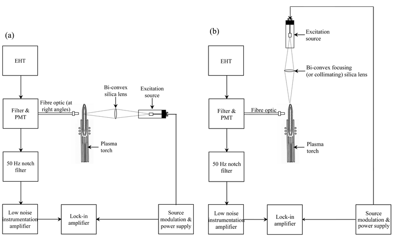 | ||
| Fig. 1 Schematic of experimental arrangements used for (a) radial excitation and (b) axial excitation fluorescence experiments. | ||
| Operating parameter | |
| HCL primary current | 6–15 mA |
| LED operating current | 20 mA |
| Modulation frequency | 167–1042 Hz |
| Duty cycle | 10–50% |
| RF generator | |
| Frequency | 27.12 MHz, crystal controlled |
| Power | 300–900 W |
| Sample introduction system | |
| Nebuliser | Ebdon, high solids, high gas flow |
| Torch | Demountable with 1.5 mm id silica injector |
| Spray chamber | Scott Double Pass, water cooled |
| Peristaltic pump | Gilson Minipuls 3, computer controlled |
| Sample flow rate | 2.0 ml min−1 |
| Argon flow rate | |
| Plasma | 8–15 L min−1 |
| Auxiliary | 0.0–1.0 L min−1 |
| Nebuliser | 1.50–3.00 L min−1 |
| Excitation source | Current/mA | Element | Wavelength/nm | Forward power/W | Viewing height/mm | Gas flow rate/L min−1 | ||
|---|---|---|---|---|---|---|---|---|
| Nebuliser | Plasma | Auxiliary | ||||||
| HCL | 15 | Ba II | 455.403 | 750 | 70 | 2.25 | 10 | 0.20 |
| 6 | Mg I | 285.213 | 700 | 60 | 2.00 | 10 | 0.80 | |
| LED | 20 | Li I | 670.800 | 500 | 80 | 2.50 | 14 | 0.50 |
| 20 | Na I | 589.000 | 475 | 80 | 2.00 | 9 | 0.20 | |
For radial profiling experiments, the emission from a HCL or LED (6 and 15 mA primary current for Mg and Ba, respectively, 20 mA primary current for Li and Na, 667 Hz modulation frequency, 50% duty cycle) was focused at a range of viewing heights ALC (60–120 mm) and the fibre optic (1000 µm core diameter HPSUV1000P; Oxford Electronics Ltd.) used for detection aligned at a 90° angle. Axial excitation fluorescence profiles of the plasma were obtained using the fibre optic for detection aligned at a 90° angle at a range of viewing heights ALC (60–120 mm) to collect the fluorescence. For axial profiling experiments, two silica lens systems were investigated.
A focusing lens (50 mm diameter; 50 mm focal length; LOT Oriel) was used to focus the excitation source emission from a HCL/LED at particular viewing heights ALC.
A collimating lens (18 mm diameter; 100 mm focal length; Speirs Robertson Ltd., Bromham, Bedford, UK) was used to illuminate the whole length of the tailflame at once.
Using the optimum plasma conditions given in Table 2, vertical profiles of fluorescence from the plasma were obtained at viewing heights of 60–120 mm ALC (10 mm increments) while aspirating Ba, Li, Mg and Na analytes. The signal to background ratio was used as the criterion of merit and all the points (n = 3) were corrected by measuring a blank and a 100 µg ml−1 standard.
Temperature measurements
Excitation (Texc) and rotational (Trot) temperature measurements were obtained using a PlasmaQuad PQ2 Thermo Elemental ICP (Winsford, Cheshire, UK) source unit under fluorescence conditions and a fibre optic (1000 µm core diameter HPSUV1000P; Oxford Electronics Ltd.) held at right angles to the plasma tailflame. Measurements were taken using a Datascan 2 (ISA Instruments S.A. (UK) Ltd., Middlesex, UK) interfaced with a current/phase stepper drive and a monochromator (1700 Series; SPEX Industries Inc., Metuchen, NJ, USA). An IEEE488 communications port provided standardised electronic protocols to receive commands and sent data to a host computer. All readings were automated by constructing a program within the instrument software (SpectRad Version 2 for Windows (ISA Instruments SA (UK) Ltd.). Operating parameters are presented in Tables 1 and 2. Manual background correction points were used for all measurements. ‘Excitation’ temperatures were determined from a Boltzmann distribution using 50 Fe I lines,11,12 whilst ‘rotational’ temperatures were determined using OH (0–0) Q and R branch lines (see Table 3).13,14| SPEX monochromator | ||
| Entrance and exit slit widths | 25 µm | |
| PMT voltage | 1200 V | |
| Viewing height ALC | 10–100 mm (in 10 mm increments) | |
| Lateral position | 0.0 mm (centre of the plasma) | |
| Fe I Boltzmann distribution measurements | ||
| Scan range | 360–400 nm | |
| Step size | 0.1 nm | |
| Scan speed | 0.1 nm s−1 | |
| OH molecular species | ||
| Scan range | 306.5–310.0 nm | |
| Step size | 0.01 nm | |
| Scan speed | 0.01 nm s−1 | |
Reagents
The reagents used in this work were of ‘AnalaR’ reagent grade and all solutions were prepared using doubly deionised water (Milli-Q, Millipore, Harrow, Middlesex, UK). Calibration standards were prepared by serial dilution of commercially available stock standard solutions (Ba, Fe, Li, Mg and Na, 1000 or 10000 µg ml−1, Merck, Poole, Dorset, UK).Results and discussion
The preliminary optimum plasma excitation source and modulation/power parameters derived from the univariate searches using radial fluorescence are presented in Table 2 for the various analyte lines. The range of conditions determined in this study agrees closely with those found in the literature.1,4,7,9 This includes the higher power (750 W) and nebuliser gas flow rate (2.25 L min−1) required for Ba, which in this case is taking advantage of fluorescence from the ground state ion line (455.403 nm). In contrast, the lower powers (450–500 W) and greater viewing heights (75–85 mm) used for the alkali metal (Li and Na) atomic lines follow the trends expected for these ‘easy-to-excite’ analytes. The requirement of relatively higher power (700 W) and lower viewing heights (60 mm) for Mg may reflect the refractory nature of this element in competition with the higher energy, shorter wavelength, atomic line utilised for its fluorescence (285.213 nm).The effect of varying the modulation frequency and mark–space ratio upon the fluorescence signal is presented in Fig. 2 for the Mg I (285.213 nm) line. The modulation range 460–670 Hz at 30–50% duty cycle produces the greater fluorescence under the low power plasma conditions stated.
 | ||
| Fig. 2 Effect of varying modulation frequency and duty cycle on Mg I 285.213 nm radial excitation fluorescence signal utilising a Thermo Elemental ICP (forward power 700 W; plasma gas flow rate 10 L min−1; nebuliser gas flow rate 2.00 L min−1; auxiliary gas flow rate 0.80 L min−1; viewing height 60 mm ALC; n = 5; RSDs < 5%). | ||
The fluorescence results from the viewing height profiling studies using radial excitation are presented in Fig. 3. This shows that there is a relatively sharp optimum for viewing height ALC and that the fluorescence signal observed was much greater for Li and Na than for Ba and Mg. This may be attributed to the source intensity of the LEDs being greater than that of the HCLs. Optimum viewing heights for the ICP atomiser and ICP source atomic fluorescence spectrometry system (ASIA) have been reported as 70, 72 and 84 mm ALC for the same Ba, Li and Na lines, respectively.4 This study agrees closely with these viewing height values which, as stated, also compare well with the power ranges found for each of the analytes.
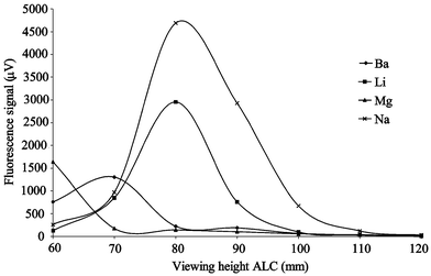 | ||
| Fig. 3 Results from a profiling study using radial excitation fluorescence signal for Ba, Li, Mg and Na (n = 3, RSDs < 5%). Operating conditions are as shown in Table 2. | ||
Using the optimum parameters given in Table 2, the working linear ranges were determined for each of the elements studied. Excellent linearity is seen up to at least 100 mg L−1, covering 5 orders of magnitude of concentration (R2 values range from 0.99995 to 1.0000) with the precision on each data point better than 5%.
Limits of detection (LODs) were calculated using the mean blank signal and three standard deviations of the blank and were found to be 27.6, 0.51, 0.43 and 0.20 µg L−1 for Ba, Li, Mg and Na, respectively. A comparison of the LODs obtained for this system with the LODs presented in the literature for HCL-ICP-AFS (25, <0.1, <0.1 and <0.1 µg l−1 for Ba, Li, Mg and Na, respectively) show that the values obtained are approaching those of a previously available commercial system.1,4 This was despite the fact that the experimental arrangement used was still being refined and improvements in terms of collection efficiency and optical coupling should be possible.
It was found that axial profiles of the plasma could not be obtained for Ba and Mg using focused or collimated excitation–source HCLs. This was because the line emission was not sufficiently intense to produce a measurable fluorescence signal. However, using the optimum conditions determined for Li and Na (Table 2), vertical profiles of the plasma, using axial excitation, were obtained using the more intense LEDs. These results are presented in Fig. 4.
 | ||
| Fig. 4 Results from a profiling study using axial excitation fluorescence signal for Li and Na (n = 3, RSDs < 5%). Operating conditions are as shown in Table 2. | ||
Fig. 4 shows that there was a relatively sharp optimum of the fluorescence signal, with respect to the viewing height ALC. The optimum viewing height (ALC) was identical to that determined from the vertical profiles of the plasma using radial excitation fluorescence. It was also independent of the use of focusing or collimating lens conditions. Again, this suggests that, irrespective of the excitation arrangement employed, only particular operating conditions produced in the plasma give the optimum conditions for fluorescence, and that these are spatially dependent.
Two different excitation lens systems were investigated in the axial mode. Initially, a ‘focusing’ lens was used to focus the emission from the excitation source at particular viewing heights (ALC) in the plasma tailflame and the fluorescence radiation measured. These results are shown in Fig. 4. The LODs obtained under these conditions were determined to be 1.12 and 0.70 µg L−1 for Li and Na, respectively. The axial profile (Fig. 4) obtained also shows that, despite an optimum (maximum) fluorescence signal being observed at a particular viewing height (ALC), there was a detectable but decreasing fluorescence signal at other viewing heights on either side of the optimum, approximately 20–30 mm wide along the plasma. If the fluorescence radiation were to be collected simultaneously along and around the region of the tailflame where this emission is effective at any one time then improvements in detection parameters may be possible. One way to achieve this broad (in spatial terms) excitation of the central channel of the tailflame is to use a collimating lens to produce a narrow beam of light of approximately equal intensity along the length of the plasma tailflame. Using this second axial arrangement, the fluorescence radiation was measured under the same operating conditions and a profile plotted. As Fig. 4 shows, the intensity of the fluorescence signal produced using the collimating lens system was less than that produced using a focusing lens (approximately one third in the case of Na and one sixth in the case of Li). The LODs obtained under these conditions were determined to be 5.3 and 3.1 µg L−1 for Li and Na, respectively. Under these source excitation conditions, and considering the relationship between source intensity and efficiency of fluorescence together with any ‘inner filter’ effect, it is seen that a collimating lens system in the axial mode does not offer any advantage over the focused axial arrangement when only one position in the plasma is measured. However, it is possible that integration of the fluorescence signal, collected from and around the plasma (360°) and along its length may improve both sensitivity and limits of detection, theoretically for both focusing and collimating lens arrangements. This is only possible using an axial excitation arrangement which effectively extends the ‘fluorescence cell’ available for measurement. In practice, this improvement may only be realised if a sufficiently intense source is used, particularly when using the collimating lens arrangement. This may be achieved with a laser, which can be used to create saturated fluorescence conditions to overcome axial absorption (“inner filter”) effects.6,16
Temperature measurements
A suitable population of ground state atoms (or ions) is required for the more intense production of fluorescence. This is facilitated by the optimum conditions determined, i.e., lower forward plasma powers (475–700 W), higher nebuliser gas flow rates (2.0–2.5 L min−1) and viewing heights (60–90 mm ALC). Under these conditions, the plasma would be expected to operate as an efficient ‘atomiser’ rather than ‘exciter’ of the analytes of interest. In order to verify this, viewing height ‘temperature’ profiles were determined under the optimum conditions using suitable diagnostic markers. Iron (Fe I) lines were chosen for Texc measurements and OH (0–0) rotational emission for the Trot measurements because of the accuracy and precision of the well-documented data available.11–14 At least four fundamental characteristics (trends) would be expected if, under fluorescence conditions, the plasma was operating more as an ‘atomiser’ and less as an ‘exciter’. Firstly, the emission profile from Fe I and OH excited species would decrease, with an increase in viewing height, in contrast with the fluorescence emission profile (an inverse matching, which shows a signal above 50 mm ALC). Secondly, the ‘temperatures’ obtained would decrease with viewing height. Thirdly, the ‘temperatures’ measured would be lower than expected, especially for Texc compared with a plasma operated under conventional higher power conditions. Fourthly, the plasma might be expected to approach conditions closer to local thermodynamic equilibrium (LTE) and therefore, in both energy and spatial terms, the temperatures of Texc and Trot would be similar in value and profile.Excitation temperatures (Texc, Fe I species) obtained using a Boltzmann distribution and rotational temperature measurements (Trot) determined using the OH (0–0) rotational band spectra are presented in Table 4. The ‘temperatures’ obtained are quite similar both in value (2700–3000 K at 50 mm ALC) and profile, suggesting that the plasma is approaching closer to LTE when operated under ‘fluorescence’ conditions. The decrease in ‘temperature’ values with viewing height and loss of line and band emission, where fluorescence begins, is also seen. An example plot of the Boltzmann distribution for Fe I is given in Fig. 5. It is interesting to note that while Fig. 5 shows that the Boltzmann plot is curved (and not a straight line), the ‘degree’ of curvature is smaller, compared with similar profiles for Fe I but under conventional emission conditions.12,14,15 This suggests that the plasma is indeed closer to LTE when operated under fluorescence conditions. It is important to note that all the Fe I lines used to plot the Boltzmann distribution had relatively low transition probabilities but were also checked for evidence of self-absorption.
 | ||
| Fig. 5 Plot of the Fe I Boltzmann distribution for the Thermo Elemental plasma operated under fluorescence conditions (forward power 900 W; plasma gas flow rate 10 L min−1; nebuliser gas flow rate 2.00 L min−1; auxiliary gas flow rate 0.20 L min−1; viewing height 20 mm ALC; n=3, RSDs < 5%). | ||
| Forward power/W | ||||||||
|---|---|---|---|---|---|---|---|---|
| 900 | 700 | 500 | 300 | 900 | 700 | 500 | 300 | |
| Viewing height ALC/mm | Excitation temperature/K | Rotational temperature/K | ||||||
| a n.d. = not detected due to low/absent emission intensity. | ||||||||
| 10 | n.d. | n.d. | n.d. | n.d. | n.d. | n.d. | n.d. | n.d. |
| 20 | 3740 | 3330 | 3220 | 3140 | 3200 | 3230 | 2760 | 2820 |
| 30 | 3210 | 3270 | 3260 | n.d. | 3370 | 2870 | 2680 | n.d. |
| 40 | 3050 | 3140 | n.d. | n.d. | 3080 | 2860 | n.d. | n.d. |
| 50 | 3080 | n.d. | n.d. | n.d. | 2720 | 2670 | n.d. | n.d. |
| 60 | n.d. | n.d. | n.d. | n.d. | 2500 | n.d. | n.d. | n.d. |
| 70 | n.d. | n.d. | n.d. | n.d. | n.d. | n.d. | n.d. | n.d. |
Conclusions
An excitation source-driver system was designed and built in-house to operate HCLs, BDHCLs and LEDs with variable modulation frequencies and duty cycle capabilities. Studies have indicated that a lamp operated with a low modulation frequency range (460–670 Hz) and higher duty cycles (30–50%) provided the preferred intense excitation conditions for the production of fluorescence in the ICP.Vertical profiles of the plasma were obtained utilising radial and axial excitation. The LODs obtained using radial excitation were superior to those obtained using both focusing and collimating lens systems for axial excitation. However, the axial profile obtained showed that, despite a maximum fluorescence signal being observed at a particular viewing height, there was a detectable but decreasing fluorescence signal at other viewing heights. If the fluorescence signal was collected from and around the plasma (360°) and along its length then both sensitivity and limits of detection should be improved. This is only possible using an axial excitation arrangement, which effectively extends the ‘fluorescence cell’ available for measurement. The inner filter effect, possibly present during axial excitation, may be addressed using a suitably intense source, e.g. a laser.6,16
Plasma excitation temperatures (Texc) and OH rotational temperatures (Trot) show similar values in the range 2500–3000 K at these low power and high viewing heights (ca. 700 W, 50–60 mm ALC). The low emission intensities obtained for these species, under these conditions, is indicative of the higher population of ground state species required for fluorescence. The values indicate a plasma operating closer to LTE compared with a plasma operated under conventional conditions.
References
- D. R. Demers, Am. Lab., 1987, 19, 30 Search PubMed.
- M. A. Kosinski, H. Uchida and J. D. Winefordner, Anal. Chem., 1983, 55, 688 CrossRef CAS.
- S. Greenfield, Anal. Proc., 1984, 21, 61 Search PubMed.
- S. Greenfield, J. Anal. At. Spectrom., 1995, 10, 183 RSC.
- H. G. C. Human, N. Omenetto, P. Cavalli and G. Rossi, Spectrochim. Acta, 1984, 39B, 1345 Search PubMed.
- P. Stchur, K. X. Yang, X. D. Hou, T. Sun and R. G. Michel, Spectrochim. Acta, 2001, 56B, 1565 Search PubMed.
- D. R. Demers and C. D. Allemand, Anal. Chem., 1981, 53, 1915 CrossRef CAS.
- D. R. Demers, D. A. Busch and C. D. Allemand, Am. Lab., 1982, 14, 167 Search PubMed.
- D. R. Demers, Spectrochim. Acta,, 1985, 40B, 93 Search PubMed.
- X. R. Liu and G. Horlick, Spectrochim. Acta, 1994, 50B, 537 Search PubMed.
- National Institute of Standards and Technology, ‘DAS’ database, available from http://physics.nist.gov/asd., 2002.
- S. Nakamura, J. Anal. At. Spectrom., 1995, 10, 467 RSC.
- G. H. Dieke and H. M. Crosswhite, J. Quant. Spectrosc. Radiat. Transfer, 1962, 2, 97 CrossRef CAS.
- J. M. Mermet, in Inductively Coupled Plasma and its Applications, ed. S. J. Hill, Sheffield Academic Press, Sheffield, 1999 Search PubMed.
- J. F. Alder, R. M. Bombelka and G. F. Kirkbright, Spectrochim. Acta, 1980, 35B, 163 CAS.
- K. Niemax, A. Zybin, C. Schnurer Patschan and H. Groll, Anal. Chem., 1996, 68, A351.
Footnote |
| † Presented at the Eleventh British National Atomic Spectroscopy Symposium (BNASS), Loughborough University, UK, July 8–10, 2002. |
| This journal is © The Royal Society of Chemistry 2003 |