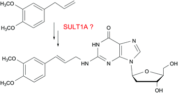
The influence of the SULT1A status – wild-type, knockout or humanized – on the DNA adduct formation by methyleugenol in extrahepatic tissues of mice
Abstract
Methyleugenol, present in herbs and spices, has demonstrated carcinogenic activity in the liver and, to a lesser extent, in extrahepatic tissues of rats and mice. It forms DNA adducts after hydroxylation and sulphation. As previously reported, hepatic DNA adduct formation by methyleugenol in mice is strongly affected by their sulphotransferase (SULT) 1A status. Now, we analysed the adduct formation in extrahepatic tissues. The time course of the adduct levels was determined in transgenic (tg) mice, expressing human SULT1A1/2, after oral administration of methyleugenol (50 mg per kg body mass). Nearly maximal adduct levels were observed 6 h after treatment. They followed the order: liver > caecum > kidney > colon > stomach > small intestine > lung > spleen. We then selected liver, caecum, kidney and stomach for the main study, in which four mouse lines [wild-type (wt), Sult1a1-knockout (ko), tg, and humanized (ko-tg)] were treated with methyleugenol at varying dose levels. In the liver, caecum and kidney, adduct formation was nearly completely dependent on the expression of SULT1A enzymes. In the liver, human SULT1A1/2 led to higher adduct levels than mouse Sult1a1, and the effects of both enzymes were approximately additive. In the caecum, human SULT1A1/2 and mouse Sult1a1 were nearly equally effective, again with additive effects in tg mice. In the kidney, only human SULT1A1/2 played a role: no adducts were detected in wt and ko mice even at the highest dose tested and the adduct levels were similar in tg and ko-tg mice. In the stomach, adduct formation was unaffected by the SULT1A status. In conclusion: (i) the SULT1A enzymes only affected adduct formation in those tissues in which they are highly expressed (mouse Sult1a1 in the liver and caecum, but not in the kidney and stomach; human SULT1A1/2 in the liver, caecum and kidney, not in the stomach of tg mice and humans), indicating a dominating role of local bioactivation; (ii) the additivity of the effects of both enzymes in the liver and caecum implies that the enzyme level was limiting in the adduct formation; (iii) SULT1A forms dominated the activation of methyleugenol in several tissues, but non-Sult1a1 forms or SULT-independent mechanisms were involved in its adduct formation in the stomach.


 Please wait while we load your content...