
Biomolecular recognition at the cellular level: geometrical and chemical functionality dependence of a low phototoxic cationic probe for DNA imaging†
Abstract
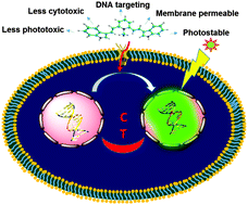
A combined approach was adopted to understand the impact of structural geometry as well as suitable chemical functionality of a molecular probe on its efficient binding in the minor groove of DNA. The development of a small chemical library of different lutidinium conjugates (P1–P5) and molecular simulations using DFT calculations clearly demonstrated that the semilunar conformation of a molecular probe equipped with requisite chemical functionality is the key parameter for its proper binding in the minor groove of DNA. The comparative optical responses of these probes (P1–P5) coupled with the theoretical studies illustrated that only P3 displayed considerable fluorescence enhancement in the presence of DNA because of its semilunar geometry and special chemical architecture. Furthermore, the bioassays clearly revealed that the probe can penetrate the cell membrane of live as well as dead cells without the help of any permeabilization agent. Microscopic cellular imaging established that probe P3 can stain the nuclear region of the cells with high contrast and negligible cytoplasmic spillage without causing any cellular deterioration. The specificity and binding efficiency of P3 toward DNA were established by performing DNase/RNase digest tests and gel electrophoresis experiments. Most importantly, P3 exhibited minimum phototoxicity and high photobleaching resistance in cellular medium under continuous exposure to a light source, which are highly desirable for real time monitoring of many biological events. Altogether, the investigated properties of P3 shed light on its admirable and persuasive standing as a cell-compatible, bright and photostable molecular probe for nuclear imaging in various bio-medical applications.
 Please wait while we load your content...

