Cell response on the biomimetic scaffold of silicon nano- and micro-topography
Abstract
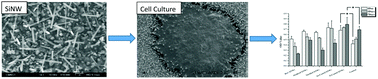
Silicon scaffolds were synthesized in a low-pressure furnace via a vapor–liquid–solid (VLS) mechanism. Structural dimensions of silicon scaffolds were tunable in the synthesis to satisfy diverse requirements for cell culture applications. Cylindraceous SiNWs structurally resemble fibrous proteins in connective tissue and the extracellular matrix (ECM), which are main cell adhesion substrata in vivo. Hemispherical silicon microbroccolis (SiMBs) possess large contact area with microscale topology for cell contact and attachment. Mouse 3T3 fibroblasts were cultured on microscale and nanoscale silicon structures with different surface modifications. Silicon scaffolds were functionalized by several physical and chemical vapor deposition methods to modify scaffold surface physical and chemical properties. Metal-coated SiNWs and SiMBs had been demonstrated and compared for their ability to provide mechanical support sites for cell adhesion and promote cell proliferation and maintain normal cell functionality. Scanning electron microscopy (SEM) micrographs at high magnification show cell–scaffold interactions, and immunofluorescence images reveal nuclear DNAs and actin cytoskeleton distribution on nanostructure covered substrates and selected biomarker expression was analyzed by enzyme-linked immunosorbent assay (ELISA).
 Please wait while we load your content...

