Quantification of the mesh structure of bundled actin filaments†
Abstract
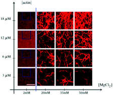
Biopolymer networks are essential for a wide variety of cellular functions. The biopolymer actin is known to self-assemble into a variety of spatial structures in response to physiological and physical mechanisms. So far, the mechanics of networks of single actin filaments and bundles has previously been described. However, the spatial structure of actin bundles remains poorly understood. Here, we investigate this question by bundling actin filaments with systematically varied concentrations of known physical bundling agents (MgCl2 and PEG) and physiological bundling agents (α-actinin and fascin). We image bundled actin networks with confocal microscopy and perform analysis to describe their mesh size and the nearest-distance distribution, which we call “mesh structure”. We find that the mesh size ξ scales universally with actin concentration as ξ ∼ [actin]−1/2. However, the dependence of ξ on the concentration of the bundling agent depends on the agent used. Finally, we find that nearest-distance distributions are best fit by Weibull and Gamma distributions. A complete understanding of the mesh structure of biopolymer networks leads to a more mechanistic understanding of the structure of the cytoskeleton, and can be exploited to design filters with variable porosity for microfluidic devices.
 Please wait while we load your content...

