Issue 12, 2013

Mapping the mechanical stiffness of live cells with the scanning ion conductance microscope

Abstract

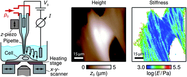
Mapping the mechanical properties of living cells with high spatial and temporal resolution is important for the exploration of cell function. Widely used imaging techniques such as the atomic force microscope are generally based on direct mechanical contact between the probe and the cell, thereby involving the risk of damaging the cell. Here, we present a noncontact method for fast and quantitative stiffness mapping of living cells with sub-micrometer lateral resolution. This was achieved by repeatedly moving a pressurized nanopipette toward and away from the sample in a scanning ion conductance microscope (SICM). The pressure-induced microfluidic flow through the nanopipette produced a time-varying force on the sample surface, thereby locally indenting it without direct mechanical contact. Maps of sample stiffness (quantified by the Young's modulus) were then determined from ion current approach curves using a finite element model. To demonstrate the capability of the method we visualized the dynamics of individual cytoskeleton fibers in living cells over several hours. Additionally, we found that spreading extensions of migrating fibroblast cells tend to be softer than their lamellum, which is consistent with a mechanism of cell migration by osmotic swelling.

Graphical abstract: Mapping the mechanical stiffness of live cells with the scanning ion conductance microscope

Supplementary files

Article information

Article type
Paper
Submitted
18 Oct 2012
Accepted
20 Dec 2012
First published
17 Jan 2013

Soft Matter, 2013,9, 3230-3236

Mapping the mechanical stiffness of live cells with the scanning ion conductance microscope

J. Rheinlaender and T. E. Schäffer, Soft Matter, 2013, 9, 3230 DOI: 10.1039/C2SM27412D

To request permission to reproduce material from this article, please go to the Copyright Clearance Center request page.

If you are an author contributing to an RSC publication, you do not need to request permission provided correct acknowledgement is given.

If you are the author of this article, you do not need to request permission to reproduce figures and diagrams provided correct acknowledgement is given. If you want to reproduce the whole article in a third-party publication (excluding your thesis/dissertation for which permission is not required) please go to the Copyright Clearance Center request page.

Read more about how to correctly acknowledge RSC content.

Social activity

Spotlight

Advertisements