Issue 21, 2011

Morphology and organization of tissue cells in 3D microenvironment of monodisperse foam scaffolds

Abstract

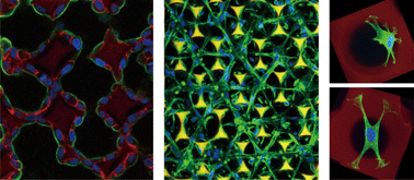
Here we demonstrate an efficient method to fabricate large-domain monodisperse foam scaffolds made of gelatin for 3D cell culture. We tested three distinct tissue cell types cultured in foam scaffolds composed of uniform spherical pores. The cells displayed appropriate morphological and physiological characteristics: epithelial cells formed cyst-like structures and were polarized inside pores, myoblasts adopted a tubular structure and fused into myotubes, and fibroblasts exhibited a wide variety of morphologies. Scaffolds with uniform pores can thus provide a platform for systematic study of 3D cell–matrix interactions.

Graphical abstract: Morphology and organization of tissue cells in 3D microenvironment of monodisperse foam scaffolds

Supplementary files

Article information

Article type
Paper
Submitted
03 Mar 2011
Accepted
04 Aug 2011
First published
01 Sep 2011

Soft Matter, 2011,7, 10010-10016

Morphology and organization of tissue cells in 3D microenvironment of monodisperse foam scaffolds

J. Lin, W. Lin, W. Hong, W. Hung, S. H. Nowotarski, S. M. Gouveia, I. Cristo and K. Lin, Soft Matter, 2011, 7, 10010 DOI: 10.1039/C1SM05371J

To request permission to reproduce material from this article, please go to the Copyright Clearance Center request page.

If you are an author contributing to an RSC publication, you do not need to request permission provided correct acknowledgement is given.

If you are the author of this article, you do not need to request permission to reproduce figures and diagrams provided correct acknowledgement is given. If you want to reproduce the whole article in a third-party publication (excluding your thesis/dissertation for which permission is not required) please go to the Copyright Clearance Center request page.

Read more about how to correctly acknowledge RSC content.

Social activity

Spotlight

Advertisements