
Blisters on graphite surface: a scanning microwave microscopy investigation
 *a
Rossella
Yivlialin,
†b
Christopher
Hardly Joseph,
a
Gianluca
Fabi,
a
Davide
Mencarelli,
a
Luca
Pierantoni,
a
Gianlorenzo
Bussetti
b
and
Marco
Farina
a
*a
Rossella
Yivlialin,
†b
Christopher
Hardly Joseph,
a
Gianluca
Fabi,
a
Davide
Mencarelli,
a
Luca
Pierantoni,
a
Gianlorenzo
Bussetti
b
and
Marco
Farina
a
Abstract
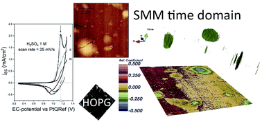
Scanning microwave microscopy (SMM) is based on the interaction between a sample and an electromagnetic evanescent field, in the microwave frequency range. SMM is usually coupled with a scanning probe microscopy (SPM) technique such as in our case, a scanning tunneling microscope (STM). In this way, the STM tip is used to control the distance between the probe and the sample while acting as an antenna for the microwave field. Thanks to the peculiarity of our home-made setup, the SMM is a suitable method to study blisters formed on HOPG surface as consequence of an electrochemical treatment. Our system has a “broad-band” approach that opens the way to perform local microwave spectroscopy over a broad frequency range. Moreover, microwaves have the ability to penetrate into the sample allowing the sub-surface characterization of materials. The application of the SMM to characterize blisters formed on the HOPG surface provides information on the sub-layer structures.
 Please wait while we load your content...

