Peak force tapping atomic force microscopy for advancing cell and molecular biology
Abstract
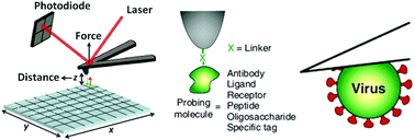
The advent of atomic force microscopy (AFM) provides an exciting tool to detect molecular and cellular behaviors under aqueous conditions. AFM is able to not only visualize the surface topography of the specimens, but also can quantify the mechanical properties of the specimens by force spectroscopy assay. Nevertheless, integrating AFM topographic imaging with force spectroscopy assay has long been limited due to the low spatiotemporal resolution. In recent years, the appearance of a new AFM imaging mode called peak force tapping (PFT) has shattered this limit. PFT allows AFM to simultaneously acquire the topography and mechanical properties of biological samples with unprecedented spatiotemporal resolution. The practical applications of PFT in the field of life sciences in the past decade have demonstrated the excellent capabilities of PFT in characterizing the fine structures and mechanics of living biological systems in their native states, offering novel possibilities to reveal the underlying mechanisms guiding physiological/pathological activities. In this paper, the recent progress in cell and molecular biology that has been made with the utilization of PFT is summarized, and future perspectives for further progression and biomedical applications of PFT are provided.

- This article is part of the themed collection: Recent Review Articles


 Please wait while we load your content...