Issue 16, 2018

Visualizing a core–shell structure of heavily doped silicon quantum dots by electron microscopy using an atomically thin support film

Abstract

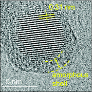
We successfully visualize a core–shell structure of a heavily B and P codoped Si quantum dot (QD) by transmission electron microscopy using an ultra-thin graphene oxide support film. The enhanced contrast reveals that a codoped Si QD has a highly crystalline Si core and an amorphous shell composed of Si, B and P.

Graphical abstract: Visualizing a core–shell structure of heavily doped silicon quantum dots by electron microscopy using an atomically thin support film

Supplementary files

Article information

Article type
Communication
Submitted
20 Dec 2017
Accepted
19 Mar 2018
First published
21 Mar 2018

Nanoscale, 2018,10, 7357-7362

Author version available

Visualizing a core–shell structure of heavily doped silicon quantum dots by electron microscopy using an atomically thin support film

H. Sugimoto, M. Yamamura, M. Sakiyama and M. Fujii, Nanoscale, 2018, 10, 7357 DOI: 10.1039/C7NR09474D

To request permission to reproduce material from this article, please go to the Copyright Clearance Center request page.

If you are an author contributing to an RSC publication, you do not need to request permission provided correct acknowledgement is given.

If you are the author of this article, you do not need to request permission to reproduce figures and diagrams provided correct acknowledgement is given. If you want to reproduce the whole article in a third-party publication (excluding your thesis/dissertation for which permission is not required) please go to the Copyright Clearance Center request page.

Read more about how to correctly acknowledge RSC content.

Social activity

Spotlight

Advertisements